Anatomy lect 1 : - L1 transyloric plane -> hilum
advertisement
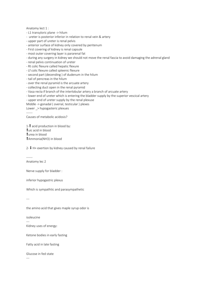
Anatomy lect 1 : - L1 transyloric plane -> hilum - ureter is posterior inferior in relation to renal vein & artery - upper part of ureter is renal pelvis - anterior surface of kidney only covered by peritenum - First covering of kidney is renal capsule - most outer covering layer is pararenal fat - during any surgery in kidney we should not move the renal fascia to avoid damaging the adrenal gland - renal pelvis continuation of ureter - Rt colic flexure called hepatic flexure - Lf colic flexure called spleenic flexure - second part (decending ) of dudenum in the hilum - tail of pencreas in the hilum - over the renal pyramid is the arcuate artery - collecting duct open in the renal pyramid - Vasa recta if branch of the interlobular artery a branch of arcuate artery - lower end of ureter which is entering the bladder supply by the superior vescical artery - upper end of ureter supply by the renal plexuse Middle -> gonadal ( overial, testicular ) plexes Lower _> hypogasteric plexues -----Causes of metabolic acidosis? 1-⬆ acid production in blood by: ⬆uic acid in blood ⬆urea in blood ⬆Ammonia(NH3) in blood 2- ⬇ H+ exertion by kidney caused by renal failure -----Anatomy lec 2 Nerve supply for bladder : inferior hypogastric plexus Which is sympathtic and parasympathetic --the amino acid that gives maple syrup odor is isoleucine --Kidney uses of energy: Ketone bodies in early fasting Fatty acid in late fasting Glucose in fed state --- Synthesis of ketone bodies : In liver BUT do not use it for energy , they use FA ( in starvation) ----Synthesis of glucose (gluconeogenesis) : -liver . -kidney in starvation (prolonged fasting) after 60 hrs . Thus renal failure patients will be affected more in prolonged fasting ---Cause of metabolic acidosis is Not enough produce NH3 and increase in H --Phsl6 Chatecolamines works most: Afferent arterioles Angiotensin II works on : efferent arterioles كلهمvasoconstriction ---Interstetial cystitis without bacterial infection ---Ureteric bud gives: 1-ureter 2-Renal pelvis 3-major calyces 4-minor calyces 5-collecting tubules ---Cap of the metanephric mesenchyme gives: 1-bowmans calpsule 2-PCT 3-DCT 4-ascending &descending limb of henle loop ---Overflow protienurea Make sure to know the 3 causes of them ---- Urea recirculation 1_the concentration of urea if h2o is low or deficient 2_ the diluation of urea if h2o is excess لو الموية قليل اليوريا تركيزها بيعلى وبيصير اليورن مركز ... العكس لو الموية زادت ---Triphasic pattern in wilms tumor: 1-blastema* cells 2-stromal cells 3-epithelial cells --Extrophy of cloca include : 1-extrophy of bladder 2-spinal defect 3-imperforate anus 4-omphalocele --2 function of ADH ? :1-anti diuretic 2-vasoconstriction 3- increase Aquaporin-2 water channel protein --Cystic change in Clear cell Collecting duct Cystic formation in Wilms tumor --Clear cell RCC common in familia cases At 67 years of age, Mr. H underwent a transurethral prostatectomy For cancer of the prostate . Because of concern about postoperative bleeding from straining during urination, he had a catheter placed into the bladder. Three days later, Mr. H developed a urinary tract infection with low-grade fever, some pain, and pyuria . Quantitative urine culture counts 3*105 CFU/ml of urine. The microbe was Gram’s negative lactose fermenter bacilli with indole positive reaction. Physicians were able to control the infection with gentamicin therapy. --- التنسون في الباث Best prognosis is chromophobe RCC --صور االنات اللي صورناها https://www.dropbox.com/sh/xc6v1nhw29x3p6s/AACwtJv7Yu-cuWZCTYwvCDYka?dl=0 الpoint's of Identification https://www.dropbox.com/sh/2nxha880d40gcxk/AAA17J5flmvSQZ_P-hlCCndga?dl=0 صور ثانية لالنات https://www.dropbox.com/sh/s010s083o1hb0mw/AADDubwmVMVvGFn3_2F0stDpa?dl=0 --OSPE Anat Penile urethra Membranous urethra Detrouse muscle Ureter Renal vein Parymid (medulla)


