9 - The Biology Corner
advertisement
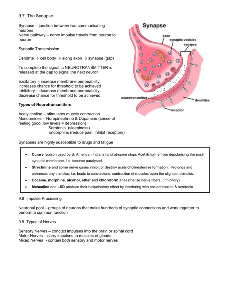
9.7 The Synapse Synapse – junction between two communicating neurons Nerve pathway – nerve impulse travels from neuron to neuron Synaptic Transmission Dendrite cell body along axon synapse (gap) To complete the signal, a NEUROTRANSMITTER is released at the gap to signal the next neuron Excitatory – increase membrane permeability, increases chance for threshold to be achieved Inhibitory – decrease membrane permeability, decrease chance for threshold to be achieved Types of Neurotransmitters Acetylcholine – stimulates muscle contraction Monoamines – Norepinephrine & Dopamine (sense of feeling good, low levels = depression) Serotonin (sleepiness) Endorphins (reduce pain, inhibit receptors) Synapses are highly susceptible to drugs and fatigue Curare (poison used by S. American Indians) and atropine stops Acetylcholine from depolarising the postsynaptic membrane, i.e. become paralysed. Strychnine and some nerve gases inhibit or destroy acetylcholinesterase formation. Prolongs and enhances any stimulus, i.e. leads to convulsions, contraction of muscles upon the slightest stimulus. Cocaine, morphine, alcohol, ether and chloroform anaesthetise nerve fibers. (inhibitory) Mescaline and LSD produce their hallucinatory effect by interfering with nor-adrenaline & serotonin 9.8 Impulse Processing Neuronal pool – groups of neurons that make hundreds of synaptic connections and work together to perform a common function 9.9 Types of Nerves Sensory Nerves – conduct impulses into the brain or spinal cord Motor Nerves – carry impulses to muscles of glands Mixed Nerves - contain both sensory and motor nerves 9.10 Nerve Pathways Reflex arc – simple pathway, includes only a few neurons (reflexes) Reflex Behavior – automatic, subconscious responses to stimulu Knee-jerk reflex (patellar tendon reflex) stimulus knee sensory nerve spinal cord motor nerve Withdrawal reflex – occurs when you touch something painful 9.11 Meninges = membranes located between bone and soft tissues of the nervous system Dura mater = outmost layer, blood vessels, nerves Arachnoid mater = no blood vessels, located between Pia mater = contains many nerves and blood vessels to nourish cells of brain and spinal cord *Cerebrospinal fluid = between arachnoid and pia maters 9.12 Spinal Cord - nerve column, passes from brain down through the vertebral canal - has 31 segments, each with a pair of spinal nerves Cervical enlargement = supplies nerves to upper limbs (neck) Lumbar enlargement = supplies nerves to the lower limbs (lower back) FUNCTION: conducting nerve impulses, serves as a center for spinal reflexes Ascending tracts = carry sensory info to the brain Descending tracts = carry motor impulses from the brain to the muscles Spinal reflexes – reflex arcs pass through the spinal cord 9.13 Brain Three Major Parts: Cerebrum – largest, sensory and motor functions, higher mental function (memory, reasoning) Cerebellum – coordinate voluntary muscles Brain stem – regulate visceral functions DESCRIBE THE FUNCTIONS: 1. Cerebral Hemispheres 2. Corpus Callosum 3. Convolutions / Sulcus / Gyrus 4. Transverse / Lateral / Longitudinal Fissures Lobes of the Brain 5. Frontal Lobe 6. Parietal Lobe 7. Temporal Lobe 8. Occipital Lobe 9. Cerebral Cortex 10. Ventricles 11. Cerebrospinal Fluid Functional Regions: 12. Motor 13. Sensory 14. Association DIENCEPHALON & BRAIN STEM 1. Diencephalon 2. Thalamus 3. Hypothalamus 4. Optic Tract / Chiasma 5. Midbrain 6. Pons 7. Medulla 8. Pituitary Gland 9. Hippocampus 10. Limbic System TYPES OF MEMORY: Episodic | Procedural | Semantic | Working
