'Science of Psychology' Class Notes (Milgram and
advertisement
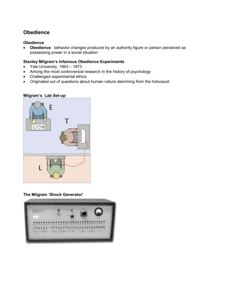
Obedience Obedience Obedience: behavior changes produced by an authority figure or person perceived as possessing power in a social situation Stanley Milgram’s Infamous Obedience Experiments Yale University, 1963 – 1973 Among the most controversial research in the history of psychology Challenged experimental ethics Originated out of questions about human nature stemming from the holocaust Milgram’s Lab Set-up The Milgram ‘Shock Generator’ Milgram Results: Original Experiment: 63% shocked to 450 volts Heart Condition: 65% shocked to 450 volts No Audio Group: 93% shocked to 450 volts Same Room Group: 40% shocked to 450 volts Forced Shock Group: 30% shocked to 450 volts Phone Command: 21% shocked to 450 volts Non-Status Authority: 20% shocked to 450 volts Three Teacher Groups: 10% shocked to 450 volts No Association with Yale Group: 50% shocked to 450 volts Criticism of Milgram’s Research Criticism: The experiment put the subjects through needless, torturous anxiety Response: Only 1% of the subjects over the years regretted participating, while 80+% were glad they had participated Conclusions Drawn from the Milgram studies A powerful situation can produce high obedience and surprising behavior Separating the ‘teacher’ (or ‘obedient aggressor’) from his victim increases obedience Getting the ‘teacher’ (or ‘obedient aggressor’) alone increases obedience The physical presence of an authority figure increases obedience Functional Specificity in the Brain Phrenology Franz Joseph Gall (1796) Brain has specific areas of functioning Highly developed brain areas force bumps on the cranial bone Discounted as pseudoscience fMRI ‘Functional Magnetic Resonance Imaging’ MRI vs. fRMI –MRI = reveals anatomy (15 images per 45 minutes) –fMRI = reveals brain function (15 per second) Non-invasive, no radiation, widely available How fMRI Works Neural activity takes energy Blood flow increases in active areas Hemoglobin is magnetic Difference in ratio of oxyhemoglobin vs. deoxyhemoglobin = BOLD signal (blood-oxygen-leveldependence) Changes in blood flow are reflected in fMRI images as bright areas Questions About Brain Organization Generalized or Specialized? What do specific brain regions do? Analyze only? Provide consciousness? How do specific brain regions get there? Genes? Or Experience? Is most of the brain highly specified or more generalized? Are specified areas rare exceptions? Research On Faces vs. Objects Showed subjects photos of faces – dot -- objects – dot – and so on… Observed brain activity in occipital / fusiform region of the brain Results: one specific region showed considerably higher activity when exposed to faces than objects The area is not exclusive to faces, though First results to support ‘Fusiform Face Area’ [FFA] Research on Places vs. Objects Russell Epstein, Kanwisher and others Exposed subjects to images of ‘places’ Used photos of places like ‘Florida,’ empty rooms, or actual lego layouts Results: high activity in a bilateral, medial area in front of the FFA ‘Parahippocampus Place Area’ (PPA) Research on Body Images vs. Objects Showed subjects photos of body parts (feet, arms, legs, knees) vs. objects Stick figures vs. random lines = response cut in half ‘Extrastriate Body Area’ (EBA) Bilateral, occipital, external location Photo Stimuli vs. Mental Imagery Is a physical visual stimulus needed to activate the FFA or PPA? Famous faces, famous places photos were shown Then subjects asked to form a mental image of the face / place without an external stimulus Face / place areas light up, but weaker Autism and Brain Function Robert Schultz [Yale University] Schultz: FFA = ‘storing social knowledge’ and facial recognition Autistic subjects did not show significant FFA activity when shown faces FFA and amygdala [emotional center] activity remain dark / appear impaired Frontal brain regions needed for empathizing are impaired ‘Superior temporal sulcus’ (STS) light up when another person (or animal) looks at a healthy subject but not in autistic subjects The Political Brain [1] Drew Westen (2006) Exposed voters to contradictory remarks from their preferred candidate Both conservatives and liberals – Showed little activity in ‘reasoning’ area – Emotional circuits became active • Regulation of emotion circuits active – Conflict resolution, moral judgment areas are active • Increased activity in reward circuits • Illustrated circuits used in confirmation bias The Political Brain [2] Other research: – Conservative brains oriented toward higher amygdala activity • Amygdala = primary fear emotion area – Liberal brains oriented toward anterior cingulate cortex (ACC) and left posterior insula • • Anterior Cingulate Cortex = reward anticipation, decision making, empathy, emotion Left Posterior Insula = processes deep, visceral emotions Social Rejection in the Brain Naomi Eisenberger, Kipling Williams, et al Cyberball to cause rejection Social rejection linked with activity in the dorsal anterior cingulate and the anterior insula -two areas that show increased activity in response to physical pain The Brain in Love Helen Fisher, et al Studied subjects experiencing intense, passionate love Procedure: – subjects looked at a photo of their beloved • then countback task – then looked at a photo of an acquaintance • [countback task, repeat]… Researchers compared brain activity related to long-term spousal relations (‘beloved person’) and casual acquaintances Right ventral tegmental area, right caudate nucleus are activated for intense, passionate love Both are dopamine-rich areas that are associated with reward and motivation Fisher’s hypothesis / claim: passionate love is a primary motivational system rather than simply an emotion ‘Thoughts of Other People’ Area Rebecca Saxe, MIT Temporoparietal junction lights up when people think about other people's thoughts Area does not activate when thinking about different aspects of other people Used often as we try to figure out why others behave as they do
![milgram[1].](http://s2.studylib.net/store/data/005452941_1-ff2d7fd220b66c9ac44050e2aa493bc7-300x300.png)
