Interactive Spine:
By Primal Pictures
EDITOR'S NOTE: The following is a small sample of the Interactive Spine
CD in the ground breaking Primal Pictures 3-D Anatomy CD ROM series.
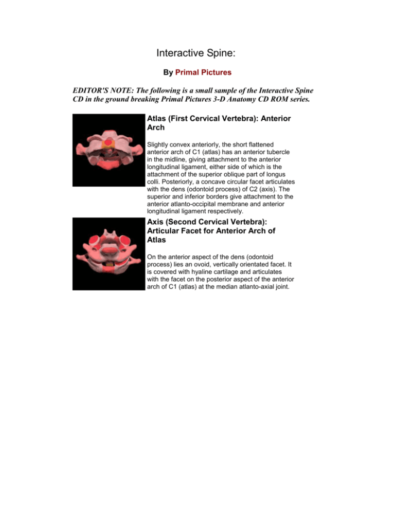
Atlas (First Cervical Vertebra): Anterior
Arch
Slightly convex anteriorly, the short flattened
anterior arch of C1 (atlas) has an anterior tubercle
in the midline, giving attachment to the anterior
longitudinal ligament, either side of which is the
attachment of the superior oblique part of longus
colli. Posteriorly, a concave circular facet articulates
with the dens (odontoid process) of C2 (axis). The
superior and inferior borders give attachment to the
anterior atlanto-occipital membrane and anterior
longitudinal ligament respectively.
Axis (Second Cervical Vertebra):
Articular Facet for Anterior Arch of
Atlas
On the anterior aspect of the dens (odontoid
process) lies an ovoid, vertically orientated facet. It
is covered with hyaline cartilage and articulates
with the facet on the posterior aspect of the anterior
arch of C1 (atlas) at the median atlanto-axial joint.
Clavicle
The clavicle is a curved subcutaneous bone
extending almost horizontally from the manubrium
of the sternum to the acromion and acts as a strut
to hold the scapula laterally. The flat lateral end
articulates with the medial aspect of the acromion
and the enlarged medial, sternal end with the
manubrium and first costal cartilage. The medial
two-thirds of the shaft is convex anteriorly and
approximately circular in cross-section. The lateral
third is concave anteriorly and flattened from above
downwards. With the scapula it forms the pectoral
(shoulder) girdle transmitting the weight of the
upper limb to the axial skeleton and facilitating the
wide range of movement of the upper limb. A direct
blow or indirect force may fracture the clavicle at
the junction of the two curvatures. In females, the
clavicle is shorter, thinner, less curved and
smoother.
The lateral third has superior and inferior surfaces
and anterior and posterior borders. The palpable
superior border is smooth except at its margins.
Posteriorly on the inferior surface at the junction of
the two curvatures is the rounded conoid tubercle
for the conoid part of the coracoclavicular ligament.
The rough trapezoid line runs antero-laterally. The
anterior border is thin, rough and concave and has
a small deltoid tubercle; the posterior border is
convex. A small oval articular facet for articulation
with the medial aspect of the acromion faces
laterally. The lateral third provides attachment for
the deltoid anteriorly and trapezius posteriorly.
The medial two-thirds has anterior, superior,
posterior and inferior surfaces. The posterior
surface is smooth but has a roughened depression
(rhomboideus fossa) medially for the
costoclavicular ligament. The inferior surface is
grooved laterally for the attachment of the
subclavius muscle. The medial end faces medially
and slightly antero-inferiorly. The inferior threequarters is beveled, sometimes extending onto the
inferior surface, for articulation with the manubrium
above, which it projects.
Ossification
The clavicle undergoes intramembranous
ossification and is the first bone in the body to
begin ossification. Medial and lateral centers
appear in the shaft in the fifth and sixth weeks inutero, and fuse with each other one week later. The
medial end contributes more to growth than the
lateral end. An ossification center appears in the
sternal end of the clavicle between the ages of 14
and 18 years, usually two years earlier in females,
fusing with the shaft between the ages of 18 and 23
years. A small inconstant lateral center appears
between 18 and 20 years, this soon fuses with the
shaft.
Sternoclavicular joint
The sternoclavicular joint is a shallow, saddleshaped joint between the manubrium and the first
costal cartilage medially and the medial end of the
clavicle laterally. The sternal articular surface of the
clavicle is larger than that of the sternum, and is
convex vertically and slightly concave in the sagittal
plane. The clavicular notch of the sternum is
reciprocally curved, although the surfaces are not
completely congruent. The joint is lined with
fibrocartilage and a fibrocartilaginous disc divides it
into two synovial cavities, each lined with its own
synovial membrane. The fibrous capsule surrounds
the articular surfaces, thickened in front and behind
but thin above and below.
The brachiocephalic vein is formed posterior to the
sternoclavicular joint.
Acromioclavicular joint
The acromioclavicular (AC) joint is a planar
synovial joint between the small oval facet on the
lateral end of the clavicle and the facet at the
medial aspect of the acromion. The articular
surfaces are lined with fibrocartilage.
A wedge-shaped articular disc is often found in the
AC joint, partially separating the joint surfaces. The
capsule is attached to the articular margins and
reinforced by superior and inferior
acromioclavicular (AC) ligaments. The
coracoclavicular ligament, with its conoid and
trapezoid components provides major stability to
the joint.
Coccyx
The coccyx is formed by the fusion of three to five
(usually four) coccygeal vertebrae to form a small
triangular bone. The coccygeal vertebrae are
usually fused and consist of rudimentary vertebral
bodies, possibly with traces of transverse
processes and pedicles to the level of the second
vertebra. The second, third and fourth coccygeal
vertebrae decrease in size as they descend.
The coccyx descends antero-inferiorly, its upper
end articulating with the sacral apex at the sacrococcygeal joint. Its orientation varies with its
mobility, but generally the pelvic surface faces
supero-anteriorly and the dorsal surface posteroinferiorly. It gives attachment to the sacrotuberous
and sacrospinous ligaments and coccygeus, levator
ani and gluteus maximus muscles.
The base is formed by the upper surface of the first
coccygeal vertebral body and exhibits an oval
articular facet for the sacrococcygeal disc. Posterolateral to this facet are two processes referred to as
the coccygeal cornua. The coccygeal cornua
project upwards to articulate with the sacral cornua.
Rudimentary transverse processes project superolaterally from each side of the body. They may
articulate or fuse with the infero-lateral sacral angle
to complete the fifth sacral foramina. The interval
between the body of the fifth sacral vertebra and
the sacral and coccygeal cornua on each side
forms an intervertebral foramen, transmitting the
fifth sacral spinal nerve.
Ossification
Coccygeal segments each ossify from a single
primary center with that of the first ossifying at birth
and the others at various intervals until the age of
20 years. The coccygeal cornua may ossify from
separate centers soon after birth. The segments
gradually fuse; however, fusion between the first
and second segments is commonly delayed until
the 30th year. In females, the coccyx often fuses
with the sacrum later in life.
Eighth Thoracic Vertebra
The eighth thoracic vertebra (T8) is a typical
thoracic vertebra with a heart-shaped body and a
small, circular vertebral foramen; it has a larger
body than that of the thoracic vertebrae above. On
each side of the body are two costal demifacets for
articulation with the heads of the eighth (superior
facet) and ninth (inferior facet) ribs.
From the vertebral body pedicles project posteriorly
and the short, thick laminae project posteromedially, overlapping the laminae of the vertebra
below. A spinous process projects inferiorly from
the junction of the laminae, overlapping that of T9.
Anteriorly and infero-medially facing inferior
articular facets are located on the inferior articular
processes that project from the laminae. The
pediculo-laminar junctions give rise to thin, flat
superior articular processes, which face posteriorly
and supero-laterally and support the oval superior
articular facets. Also projecting postero-laterally
from the pediculo-laminar junctions are the
transverse processes, which display concave oval
facets on their anterior surfaces for articulation with
the tubercle of the eighth ribs.
Ossification
The centrum and each half of the vertebral arch
ossify from single centers that appear in-utero in
the seventh week and fourth month, respectively.
The arches unite during the first year and then join
with the vertebral body by the fourth year. The ring
apophyses (or epiphyseal rings) form secondary
ossification centers at approximately 12 years and
fuse with the vertebral body between the ages of 14
and 25 years. At 16 years centers also appear at
the tips of the transverse and spinous processes
and fuse with the rest of the vertebra by 25 years.
Secondary ossification centers may remain unfused
at these sites and are known as accessory
ossification centers. They must be differentiated
from fracture on plain-film radiographs.
Vertebral Column
The normal vertebral column is made up of 29
vertebrae (7 cervical, 12 thoracic, 5 lumbar and 5
sacral) and the coccyx. Anteriorly, the vertebrae are
connected via secondary cartilaginous joints, which
form the intervertebral discs. Posteriorly, the neural
arch has paired synovial joints, known as facets or
zygapophyseal joints.
Contours of the Spine
The overall contour of the spine in the coronal
plane is straight. However, in the sagittal plane the
contour changes with development. At birth, there
is a kyphotic posture to the whole spine (primary
curves). With development of the erect posture,
lordotic (secondary) curves develop in the cervical
and lumbar spines.
Overall, spine alignment is altered in many
conditions. Scoliosis, which is a descriptive term for
lateral curvature, is usually accompanied by
rotational abnormality as well. This can be due to
congenital deformity, degeneration or associated
with numerous neuro-muscular conditions. The
most common type, however, is idiopathic.
One way to quantify the degree of curvature is to
use the Cobb Measurement Method. The curvature
is measured by drawing a line along the upper and
lower end plates of the respective upper and lower
vertebrae that are most tilted. The angle between
these lines is then measured, usually by drawing
additional lines at perpendicular angles to the endplates .
Sagittal plane alignment can also be altered by
disease and injury. This is manifested clinically with
abnormal kyphosis or lordosis.
Tumors
Primary spinal tumors may result from primary
tumors of bone, spinal cord and nerve roots, or the
meninges. The vertebral column may also be
involved with secondary disease via local spread
from paraspinous soft tissues, or distant spread via
metastatic disease.
Location
The majority of malignant tumors, both primary and
metastatic, will originate anteriorly and involve the
vertebral body and possibly one or both pedicles.
Strictly posterior localization, even when more than
one level is involved, is far more typical of benign
lesions.
Tumors have a tendency to be located in certain
parts of the vertebrae:
-Vertebral body: chordoma, giant cell tumor,
hemangioma, eosinophilic granuloma, metastatic
disease and multiple myeloma
-Posterior elements: aneurysmal bone cyst ,
osteoblastoma, osteoid osteoma and
osteochondroma
-Adjacent vertebrae: aneurysmal bone cyst,
chondrosarcoma and chordoma
-Multiple vertebrae: eosinophilic granuloma,
metastases and myeloma
CLICK HERE for more information on the Primal Pictures 3-D Anatomy
series and for details on exclusive PTontheNET.com member discounts
Disclaimer
No warranty is given as to the accuracy of the information on any of the pages in this website. No responsibility is accepted for
any loss or damage suffered as a result of the use of that information or reliance on it. It is a matter for users to satisfy themselves
as to their or their client’s medical and physical condition to adopt the information or recommendations made. Notwithstanding a
users medical or physical condition, no responsibility or liability is accepted for any loss or damage suffered by any person as a
result of adopting the information or recommendations.
© Copyright Personal Training on the Net 1998 2003 All rights reserved


