Surface Anatomy of the Nose
advertisement
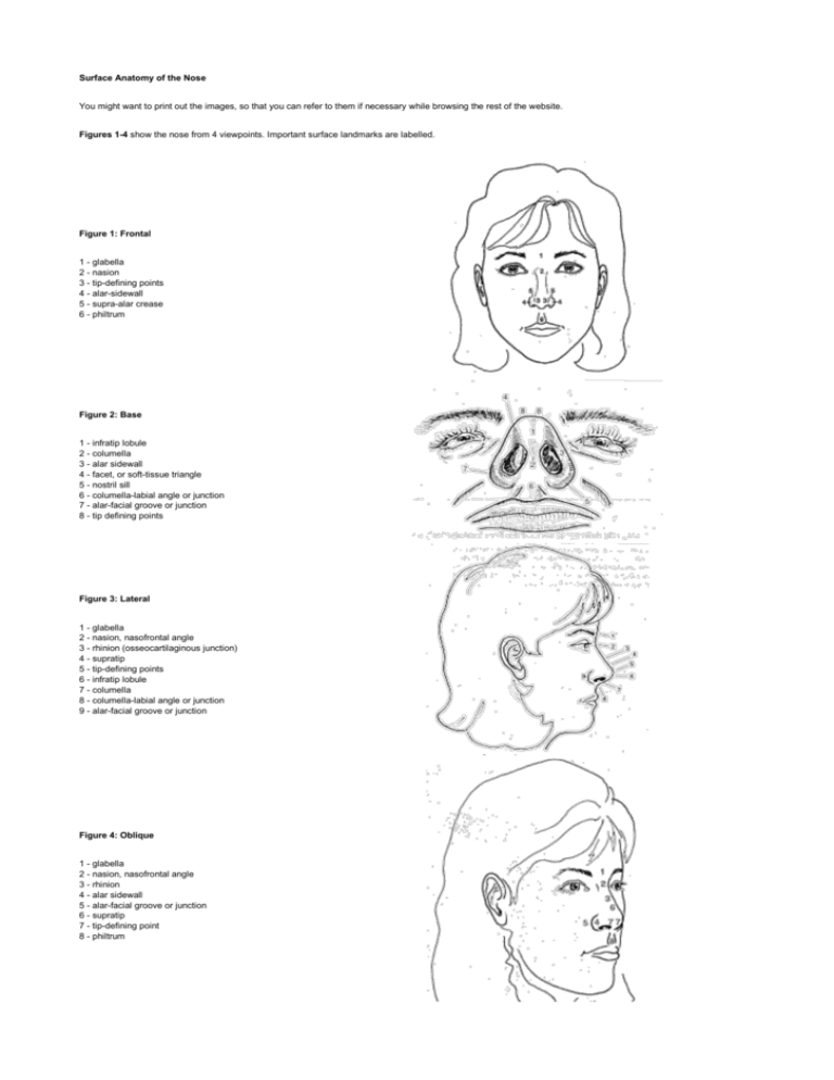
Surface Anatomy of the Nose You might want to print out the images, so that you can refer to them if necessary while browsing the rest of the website. Figures 1-4 show the nose from 4 viewpoints. Important surface landmarks are labelled. Figure 1: Frontal 1 - glabella 2 - nasion 3 - tip-defining points 4 - alar-sidewall 5 - supra-alar crease 6 - philtrum Figure 2: Base 1 - infratip lobule 2 - columella 3 - alar sidewall 4 - facet, or soft-tissue triangle 5 - nostril sill 6 - columella-labial angle or junction 7 - alar-facial groove or junction 8 - tip defining points Figure 3: Lateral 1 - glabella 2 - nasion, nasofrontal angle 3 - rhinion (osseocartilaginous junction) 4 - supratip 5 - tip-defining points 6 - infratip lobule 7 - columella 8 - columella-labial angle or junction 9 - alar-facial groove or junction Figure 4: Oblique 1 - glabella 2 - nasion, nasofrontal angle 3 - rhinion 4 - alar sidewall 5 - alar-facial groove or junction 6 - supratip 7 - tip-defining point 8 - philtrum Figures 5-7 show the internal anatomy, beneath the skin. Figure 5: Oblique 1 - nasal bone 2 - nasion (nasofrontal suture line) 3 - internasal suture line 4 - nasomaxillary suture line 5 - ascending process of maxilla 6 - rhinion (osseocartilaginous junction) 7 - upper lateral cartilage 8 - caudal edge of upper lateral cartilage 9 - anterior septal angle 10 - lower lateral cartilage - lateral crus 11 - medial crural footplate 12 - intermediate crus 13 - sesamoid cartilage 14 - pyriform aperture Figure 6: Lateral 1 - nasal bone 2 - nasion (nasofrontal suture line) 3 - internasal suture line 4 - nasomaxillary suture line 5 - ascending process of maxilla 6 - rhinion (osseocartilaginous junction) 7 - upper lateral cartilage 8 - caudal edge of upper lateral cartilage 9 - anterior septal angle 10 - lower lateral cartilage lateral crus 11 - medial crural footplate 12 - intermediate crus 13 - sesamoid cartilage 14 - pyriform aperture Figure 7: Base 1 - tip-defining point 2 - intermediate crus 3 - medial crus 4 - medial crural footplate 5 - caudal septum 6 - lateral crus 7 - naris 8 - nostril floor 9 - nostril sill 10 - alar lobule 11 - alar-facial groove or junction 12 - nasal spine The septum is the midline structure inside your nose that divides your nose into left and right. The septum is an important structure in septorhinoplasty. Its anatomy is shown here. Figure 8: Septum 1 - quadrangular caratilage 2 - nasal spine 3 - posterior septal angle 4 - middle septal angle 5 - anterior septal angle 6 - vomer 7 - perpendicular plate of ethmoid bone 8 - maxillary crest, maxillary component 9 - maxillary crest palatine component Figures 9-10 are not as important for the web viewer, but they highlight the important fact that the skin over the nose has muscles and blood vessels. This may seem obvious, but it is important because if the surgeon does not fully recognize the importance of this fact, they may operate in the incorrect tissue planes, which can result in violation of the muscle and blood vessels and subsequent abnormal scarring. Figure 9: Musculature A: Elevator muscles 1. Procerus 2. Levator labii alaequae nasi 3. Anomalous nasi B: Depressor muscles 4. Alar nasalis 5. Depressor septi nasi C: Compressor muscles 6. Transverse nasalis 7. Compressor narium minor D: Minor dilator muscles 8. Dilator naris anterior E: Other 9. Orbicularis oris 10. Corrugator Figure 10: Vasculature 1 - dorsal nasal artery 2 - lateral nasal artery 3 - angular vessels 4 - columellar artery

