Neuroscience 5 – Organisation of the CNS
advertisement
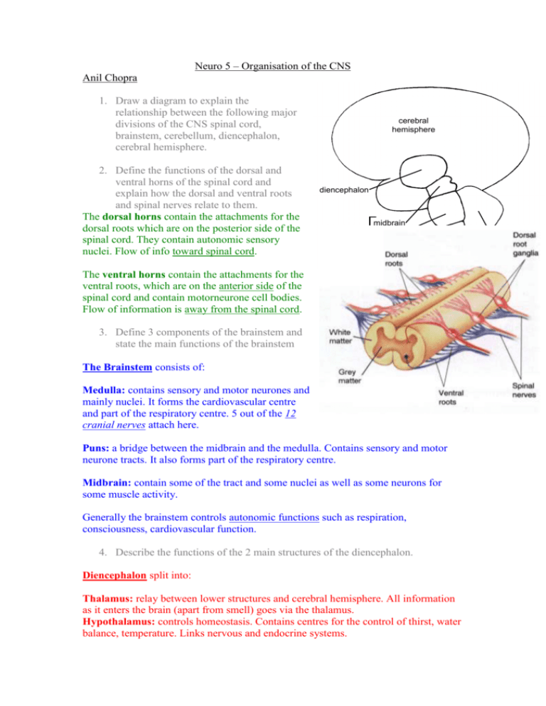
Neuro 5 – Organisation of the CNS Anil Chopra 1. Draw a diagram to explain the relationship between the following major divisions of the CNS spinal cord, brainstem, cerebellum, diencephalon, cerebral hemisphere. 2. Define the functions of the dorsal and ventral horns of the spinal cord and explain how the dorsal and ventral roots and spinal nerves relate to them. The dorsal horns contain the attachments for the dorsal roots which are on the posterior side of the spinal cord. They contain autonomic sensory nuclei. Flow of info toward spinal cord. cerebral hemisphere diencephalon midbrain brainstem pons medulla The ventral horns contain the attachments for the ventral roots, which are on the anterior side of the spinal cord and contain motorneurone cell bodies. Flow of information is away from the spinal cord. 3. Define 3 components of the brainstem and state the main functions of the brainstem The Brainstem consists of: Medulla: contains sensory and motor neurones and mainly nuclei. It forms the cardiovascular centre and part of the respiratory centre. 5 out of the 12 cranial nerves attach here. Puns: a bridge between the midbrain and the medulla. Contains sensory and motor neurone tracts. It also forms part of the respiratory centre. Midbrain: contain some of the tract and some nuclei as well as some neurons for some muscle activity. Generally the brainstem controls autonomic functions such as respiration, consciousness, cardiovascular function. 4. Describe the functions of the 2 main structures of the diencephalon. Diencephalon split into: Thalamus: relay between lower structures and cerebral hemisphere. All information as it enters the brain (apart from smell) goes via the thalamus. Hypothalamus: controls homeostasis. Contains centres for the control of thirst, water balance, temperature. Links nervous and endocrine systems. cerebellum spinal cord 5. State the functions of the basal ganglia and cerebellum. Basal ganglia: they receive information from the cerebral cortex and feed information back to the motor parts of the cortex. They are involved with the initiation and termination of some movement and are also involved with memory, learning attention and some emotions. Cerebellum: forms part of the hindbrain. It is involved with the maintenance of posture, muscular tone and coordinating voluntary movement. 6. Draw a diagram of the cerebral hemisphere the cortical lobes and primary cortical areas. parietal frontal occipital temporal Cerebral hemispheres: split into… Cerebral cortex: has primary areas and association areas which are involved in all functions but especially with language, memory, emotion. Corpus Callosum: connects 2 hemispheres across midline. (& basal ganglia) 8. Describe the 3 layers of meninges and their role in protecting the brain. Dura Mater: tough membrane attaché to bone. Folds over margins to form venous sinus. Arachnoid membrane: thin membrane beneath Dura mater Pia mater: delicate transport membrane attached to surface of brain and spinal cord. Meninges: synthesis CSF (cerebrospinal fluid) which lies in the subarachnoid space. It acts as a shock absorber (as well as providing nutrient to brain tissue) 7. Recognise the main structures in an MRI or Diagram 1 2 3 4 5 6 7 8 9 10 11 12 13 14 15 16 17 Lateral ventricle [may be cut through twice in horizontal or coronal plane] Third ventricle [may look like a hole or a slit in coronal and horizontal plane, depending on angle of section] Fourth ventricle Aqueduct Corpus callosum [may be cut through twice in horizontal plane] Frontal lobe Occipital lobe Parietal lobe Temporal lobe Basal ganglia [may be more than one part] Thalamus Internal capsule [both anterior and posterior limbs seen in horizontal plane] Optic chiasma Midbrain Pons Medulla Cerebellum 9. Explain how the major divisions of the brain relate to the crainae fossa at the base of the skull. Anterior cranial fossa holds frontal lobe of cerebral hemisphere. Middle cranial fossa holds temporal lobe of cerebral hemisphere. Posterior cranial fossa holds cerebellum Above sphenoid bone is hypothalamus Passing through foramen magnum medulla. 10. Explain the relationships between the spinal segments, spinal nerves and vertebrae and state at what level a lumbar puncture can be performed safely. 7 cervical vertebrae 8 cervical nerves (above corresponding vertebra except V8) 12 thoracic vertebrae 12 thoracic nerves (below corresponding vertebra) SPINAL CORD STOPS AT L2 5 lumbar vertebrae 5 lumbar nerves (below corresponding vertebra) Lumbar puncture is taken between L4 and L5 5 sacral vertebrae 5 sacral nerves (below corresponding vertebra) 11. Identify the components of the ventricular system and relate them to the divisions of the CNS. lateral ventricle – relates to cerebral hemispheres Aqueduct – relates to midbrain third ventricle – realtes to diencephalon central canal fourth ventricle – relates to pons and medulla 12. Explain to compensation, circulation and functions of CSF – cerebrospinal fluid. CSF Composition: similar to blood and has - glucose - oxygen - dissolved ions Different to blood in that - CSF has few cells - CSF has much less protein - CSF has fewer calcium and potassium ions - CSF has more magnesium and chloride ions. CSF Circulation Choroid plexus Lateral ventricle third ventricle aqueduct Dural venous sinus Subarachnoid space fourth ventricle Arachnoid villi Central canal of spinal cord CSF Functions Carries O2 glucose and other nutrients essential for brain tissue function. Removes waste products of CNS tissue. Provides optimal chemical environment for accurate neuronal signalling. Changes in ionic compensation can greatly affect neuronal function Acts as a shock absorber. Buoys the brain – brain “floats” in CSF. 13. State the average total volume and flow rate of CSF. Total CSF volume = 150ml Flow rate of CSF = 500ml/day 14. Define hydrocephalus and outline how it may be treated. Hydrocephalus Essentially, hydrocephalus is a block in the flow of CSF which results in pressure being built up around the brain. 2 types: Communicating: All 4 ventricles are affected. Blockage outside ventricular system Due to meningitis, head injury, congenital, sub-arachnoid haemorrhage, or a congenital disorder. Non-communicating: Only certain ventricles are affected Blockage within the ventricular system Due to aqueduct stenosis (narrowing of aqueduct) ventricular or paraventricular tumours. Symptoms include headache, drowsiness, blackouts, high intracranial pressure and increased head circumference in children, loss of upward gaze. Treatment includes: Removal of block e.g. tumour removal Diverting CSF to other route i.e. shunt. Draining CSF Open (create) an alternative pathway – ventriculostomy. 15. Distinguish between an epidural (extradural) and subdural haemorrhage. Epidural haemorrhage – damage to a meningeal artery between the skill and the dura after head trauma. Onset of symptoms should be almost instant (headache, drowsiness, vomiting and seizure) Subdural Haemorrhage – damage to a meningeal vein between dura and arachnoid membrane. Onset of symptoms can take hours Differentiation can be confirmed with imaging.
