Lab - KevinGoffMarineEdWiki
advertisement
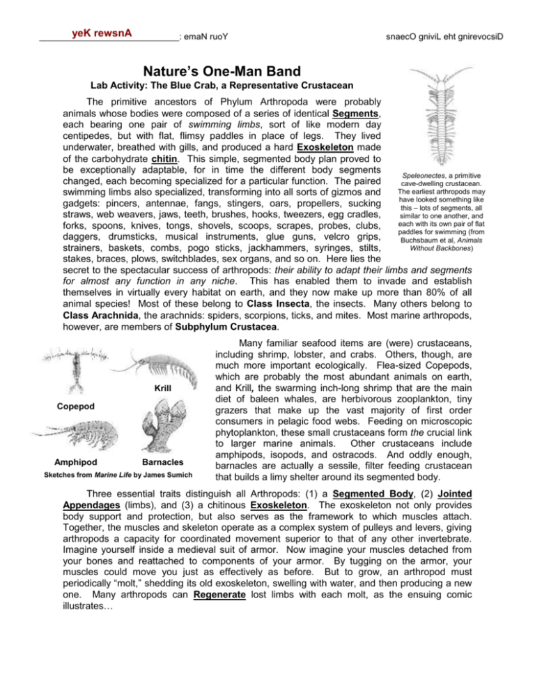
yeK rewsnA _____________________________: emaN ruoY snaecO gniviL eht gnirevocsiD Nature’s One-Man Band Lab Activity: The Blue Crab, a Representative Crustacean The primitive ancestors of Phylum Arthropoda were probably animals whose bodies were composed of a series of identical Segments, each bearing one pair of swimming limbs, sort of like modern day centipedes, but with flat, flimsy paddles in place of legs. They lived underwater, breathed with gills, and produced a hard Exoskeleton made of the carbohydrate chitin. This simple, segmented body plan proved to be exceptionally adaptable, for in time the different body segments Speleonectes, a primitive changed, each becoming specialized for a particular function. The paired cave-dwelling crustacean. The earliest arthropods may swimming limbs also specialized, transforming into all sorts of gizmos and have looked something like gadgets: pincers, antennae, fangs, stingers, oars, propellers, sucking this – lots of segments, all straws, web weavers, jaws, teeth, brushes, hooks, tweezers, egg cradles, similar to one another, and each with its own pair of flat forks, spoons, knives, tongs, shovels, scoops, scrapes, probes, clubs, paddles for swimming (from daggers, drumsticks, musical instruments, glue guns, velcro grips, Buchsbaum et al, Animals Without Backbones) strainers, baskets, combs, pogo sticks, jackhammers, syringes, stilts, stakes, braces, plows, switchblades, sex organs, and so on. Here lies the secret to the spectacular success of arthropods: their ability to adapt their limbs and segments for almost any function in any niche. This has enabled them to invade and establish themselves in virtually every habitat on earth, and they now make up more than 80% of all animal species! Most of these belong to Class Insecta, the insects. Many others belong to Class Arachnida, the arachnids: spiders, scorpions, ticks, and mites. Most marine arthropods, however, are members of Subphylum Crustacea. Krill Copepod Amphipod Barnacles Sketches from Marine Life by James Sumich Many familiar seafood items are (were) crustaceans, including shrimp, lobster, and crabs. Others, though, are much more important ecologically. Flea-sized Copepods, which are probably the most abundant animals on earth, and Krill, the swarming inch-long shrimp that are the main diet of baleen whales, are herbivorous zooplankton, tiny grazers that make up the vast majority of first order consumers in pelagic food webs. Feeding on microscopic phytoplankton, these small crustaceans form the crucial link to larger marine animals. Other crustaceans include amphipods, isopods, and ostracods. And oddly enough, barnacles are actually a sessile, filter feeding crustacean that builds a limy shelter around its segmented body. Three essential traits distinguish all Arthropods: (1) a Segmented Body, (2) Jointed Appendages (limbs), and (3) a chitinous Exoskeleton. The exoskeleton not only provides body support and protection, but also serves as the framework to which muscles attach. Together, the muscles and skeleton operate as a complex system of pulleys and levers, giving arthropods a capacity for coordinated movement superior to that of any other invertebrate. Imagine yourself inside a medieval suit of armor. Now imagine your muscles detached from your bones and reattached to components of your armor. By tugging on the armor, your muscles could move you just as effectively as before. But to grow, an arthropod must periodically “molt,” shedding its old exoskeleton, swelling with water, and then producing a new one. Many arthropods can Regenerate lost limbs with each molt, as the ensuing comic illustrates… Discovering the Living Oceans Complex Invertebrates Bloom County by Berke Breathed In this lab you’ll dissect the blue crab, Callinectes sapidus. The genus name means “Beautiful Swimmer” and the species name means “Tasty.” Blue crabs are indeed delicious, supporting the largest single-species fishery in the world. Most are steamed as “hard crabs,” but some are kept in captivity until they molt and then served as the delicacy “softshell” crab. Be aware that if you ever order a softshell crab sandwich at a restaurant, it will come with legs dangling out of the bun! Blue crabs belong to the Portunid family, the strongest, quickest, most agile swimmers of all arthropods. They spend most of their time, however, skulking on the soft seafloor of Atlantic estuaries from Nova Scotia to Argentina. They are a tough, opportunistic, omnivorous species, able to tolerate salinities from ocean water to nearly fresh, and adapted to eat everything from live prey to rotting carrion to vegetable matter. PROCEDURE 1. Get a crab and dissecting equipment. As you gather your utensils, ponder the fact that arthropods have invented all those same tools on their own! 2. Locate these external appendages: (a) Two Compound Eyes (pry them forward and notice that they sit atop stalks) (b) Two long Antennae (just inside the eyes) (c) Two shorter Antennules (tucked away between the eyes, near the center of the head; use a probe to extend them) (d) Two Chelae or Chelipeds (the claws) (e) Six Walking Legs (f) Two Swimming Legs (or “back fins”) Each of these appendages is a specialized tool, a gadget reshaped by evolution from an old swimming paddle (except for the eye, which has a different embryonic origin) to serve a new role in the crab’s niche. A basic principle of evolutionary biology is that “Form Follows Function.” Form refers to the shape, structure, and physical design of an organism’s body or body parts. Function refers to the role and reason for that form. To say that “Form Follows Function” means that a body part’s function determines its shape and structure. 2 Discovering the Living Oceans Complex Invertebrates For example, your skull is thick, hard, and bony (= form) in order to protect the brain (= function). Function comes first, and form follows from it! In the table below, sketch each limb, study and analyze its shape and its position on the animal, and describe it as a case of “Form Follows Function.” You are strongly encouraged to pluck off these limbs, especially the smaller ones, and study their sophisticated form with a magnifying glass or binocular scope! Sketches “Form Follows Function” Positioned atop a turret (with multiple lenses) for 360º vision. Compound Eye on Stalk Long and thin to flexibly reach out and feel/taste. Antenna Jointed and feathery to hoist up into the water for smell/taste or to detect vibrations in the water, much like a satellite dish. Antennule Jointed, sharp, and robust for a powerful pinch, and toothed for a firm grip. Chela Jointed and stilt-like for walking in soft sediment. Also laterally streamlined for sideways walking and swimming. (The hairs are sensory; they can taste w/ their feet) Walking Leg Flat, flexible, and robust for sculling and paddling. Swimming Legs (“back fins”) 3 Discovering the Living Oceans Complex Invertebrates The Far Side by Gary Larson 3. Compound Eyes are unique to Phylum Arthropoda. Remove one and observe it beneath a binocular scope, focusing clearly on the eye’s textured surface. Notice the hundreds of dimples. Each of these is a separate Lens. Now slice the eye in half. Notice that the eye is covered by a layer of exoskeleton, just like the rest of the body. When the crab molts, the outer eye must be shed, too! Peel away a piece of this outer layer and scrape away the black matter clinging to its inner surface. Place the exoskeleton in the well of a depression slide, add a drop of water and a cover slip, and study it beneath a compound microscope. What geometric shape are the lenses? Hexagonal We vertebrates have a “camera eye” with a single, flexible lens. The “compound eye” of arthropods, by contrast, has multiple lenses, each locked in place with a fixed focal length. What advantages might this type of eye give over the camera eye? What disadvantages? Advantages: panoramic vision; sensitive to slightest movements Disadvantages: unable to adjust depth/distance of focus 4. A nifty trick to impress your prom date at the seafood restaurant: Pluck a Chela (or pincer …and yes, it’s spelled “pincer” not “pincher”) off the crab, and then use scissors to cut out a section as diagrammed to the right. Remove the piece of exoskeleton to expose the underlying muscle. Resist the temptation to eat the Cut and Remove meat, as it’s either raw or marinated in embalming fluid. With a probe, tease away the muscle to uncover the thin, chitinous plate within. Now, wiggle the claw’s “thumb” a few times to loosen the joint. Finally, grasp the internal plate with forceps and push/pull it left and right. Explain the specific function of this plate (Hint: it’s a “tendon”). Why is it so wide? Muscle attachment. It’s wide for maximum musculature, hence strength. 5. Study the five pairs of mouthparts. Move them around with If available, feed a live blue probe or forceps to get a sense of how they work together crab and watch those crazy to collect, manipulate, dismantle, feel, and taste their food. mouthparts go, go, go! Blue crabs are omnivorous, opportunistic feeders. They prefer live prey – especially small clams and snails – yet often scavenge for dead, decaying animals, and will even eat plant matter. They are also quite cannibalistic, with smaller crabs making up as much as 20% of an adult’s diet! 4 Discovering the Living Oceans Complex Invertebrates The two outermost pairs of mouthparts are the 1st and 2nd Maxillipeds. The next two pairs (more delicate) are Maxillae, and the innermost pair (hard) are the Mandibles. Remember, all these used to be flat, flimsy little swimming paddles, but have since been fashioned through natural selection into utensils adapted for feeding! Based on their form, speculate on the current function of each: Maxillipeds: Handling food, manipulating it, shoveling, spooning, shredding… ____________________________________________________ Maxillae: Sorting food (edible from inedible), tasting, slicing... ____________________________________________________ Mandibles: Crushing, grinding, chewing… ____________________________________________________ 6. To the left and right of the mouth are openings into the Gill Chambers. See ‘em? _______. 7. The entire dorsal surface of the crab is covered by a single broad piece of exoskeleton, the Carapace. Projecting sharp spines and jagged edges, it makes the blue crab nearly impossible to swallow. On the underside, find the Genital Plate, or “apron.” Males (often nicknamed “Jimmies”) have a slender genital plate resembling the Washington Monument. Immature females (“Sally crabs”) have a triangular apron, like an Egyptian pyramid. Sexually mature females (“Sooks”) bear a broader, dome-shaped apron, like the U.S. Capitol Building, fit for carrying eggs. Is your specimen a Jimmy, Sally, or Sook? ___________________. Lift the genital plate to expose the Genital Openings in the abdominal groove beneath. Behind the apron of Jimmies are two brush-like appendages called Pleopods (also known as gonopods, or “sperm legs” …yet another pair of modified swimming limbs!). Sooks and Sallies have pleopods, too, but theirs are feathery. Crabs mate by facing each other, belly to belly, aprons open. The Jimmy ejects sperm from the tiny genital openings under his apron. He then inserts his two pleopods into the she-crab’s genital openings, using them to transfer sperm into her body cavity, where a mass of eggs waits to be fertilized. Crabs therefore reproduce through internal fertilization. A she-crab will mate only once in her life, and only after her final molt. When crabs molt, they are soft and vulnerable. Thus when a Jimmy meets a soon-to-molt, ready-to-mate she-crab, he will “cradle carry” her with his legs, swimming for days, even weeks, to find a sheltered area such as an eelgrass meadow where she can shed her exoskeleton in safety. When at last his “wife” (as crabbers call her) molts, the Jimmy fertilizes her eggs and then cradle-carries her for a couple more days while her new exoskeleton hardens. The following winter she buries herself in mud. In spring, she emerges and expels her eggs through her genital openings, and they cling to the feathery pleopods beneath her apron. At this stage crabbers call her a “sponge crab” because of the spongy orange egg mass bulging from her apron. Because blue crabs are not fully adapted to estuarine life and must begin life in saltwater, the sponge-bearing sook swims all the way to the ocean to release her eggs. The young crabs hatch out as Zoea larvae (say “zo-EE-uh”) that drift among the plankton. Several weeks later they metamorphose into the Megalops larval stage, before finally adopting their familiar benthic body form and lifestyle. When megalops swarm Atlantic beaches each summer, they nip wading tourists with their tiny claws, prompting complaints about “water fleas”! Sketches by Consuelo Hanks, swiped from William Warner’s Pulitzer Prize winning book, Beautiful Swimmers Zoeae 5 Discovering the Living Oceans Complex Invertebrates 8. If available, place a Zoea larva in the well of a depression slide and study it with a compound microscope on LOW power. Take your time, slowly dialing the fine adjustment knob up and down to alter your depth of focus. You should clearly see that the larva’s body is segmented, with jointed limbs. Next, examine a Megalops under a binocular scope. Again, take your time, alter the depth of focus, and move the specimen around to inspect the larva’s entire body. Notice that the Zoea is a furry, feathery fellow, with many tiny projections all over. Megalops, too, is quite hairy and spiny. The megalops is also rather flat and leaf-like. What might these structural adaptations accomplish for these drifting larvae? Help it stay adrift in the water column, catching currents like a kite while avoiding sinking. 9. Observe the contrast in coloration between the crab’s underside and its back. The ventral surface is light, while the dorsal surface (the carapace) is dark. Why? (Hint: these crabs are portunids, often swimming up in the water column.) The dark dorsal surface blends in with the seafloor, and while swimming, the light underside blends in with sunlight above (countershading) 10. Lay your crab belly-down in the dissecting tray. With scissors (blunt-tip down), cut around the rim of the carapace (the broad “shell” on top) just inside the spines. Gently lift off the severed piece while peeling away the underlying tissue. Snip and Remove 11. Use the diagram on the next page to identify internal structures. First examine the Gills (“dead man’s fingers”). How are they built for maximum gas exchange? Folded and feathery for amplified surface area. Find the Gill Bailer, the brush-like tool along the gills. Maneuver it to learn its range of movement. Strip away the gills on one side to find a similar tool underneath. These devices are yet another set of ancient swimming limbs, now modified into a new gadget …for what obvious function? 6 Discovering the Living Oceans Complex Invertebrates Cleaning the gills, and also circulating water across them (these are actually internal extensions of the maxillipeds) Notice that beneath the gills there is another wall of exoskeleton, which separates the gills from the internal organs like the stomach and heart. Why are the rest of the organs housed in a chamber separate from the gills? Gills are “external” organs that must be in contact with seawater, which is not good for the other organs, which are truly internal. Diagram from Invertebrate Zoology by Ruppert & Barnes 12. Around the base of the gills are the Gonads, which produce the gametes to be released through the genital openings under the apron. In males the gonads – or Testes – are a pair of coiled, zigzagging ribbons; in females, a mass of eggs, the Ovary. Extensions of the Jimmy’s testes may also be seen as whitish or pinkish structures between the heart and stomach. In this same area, a Sook has Sperm Storage Sacs, where her mate’s sperm may be stockpiled for years and repeatedly used to create new “sponges” of fertilized eggs …as many as 6 or 8 broods during her life, all from her one and only “roll in the eelgrass”! 13. Carefully strip away the thin membrane (mesentery) covering the internal body cavity. This is the Coelom (say “sea-loam”), a sealed off chamber where internal organs are free to move and operate. In the central dorsal area of the coelom you can locate the Heart (roughly between the points where the tips of all the gills converge). Notice that quite unlike our own, the heart resides in the lower back (holding a live softshell crab - one that’s just molted - you can feel the heart beating beneath its new, paper-thin carapace). The heart pumps oxygenated blood into a handful of arteries, which empty into open spaces within the body (“sinuses”). After delivering oxygen to the tissues, blood passes through channels in 7 Discovering the Living Oceans Complex Invertebrates the gills to pick up fresh oxygen, and finally returns via veins to the open space around the heart. Blood reenters the heart through small holes called Ostia, which you should be able to see. Which of the two basic types of circulatory system do arthropods possess? Open ____________________. 14. The stomach is the bag-like structure in the head region, just behind the mouth. Open it. Can you identify any contents? Inside is the Gastric Mill, hard toothy structures for grinding food (bet you wish you had teeth in your stomach, eh?). Behind the stomach and beneath the gonads are the brownish or yellowish Digestive Glands (or “hepatopancreas” …this is the soupy “mustard” inside a steamed crab). These secrete enzymes for the chemical breakdown of food. Feces (undigested food) follows a narrow intestine from the stomach to the anus, which you can find at the very tip of the genital plate (the apron). Did you? __________. 15. Carefully remove the stomach and look for the Brain, a small white mass in the head with white nerve cords exiting in various directions (it’s hard to find). A pair of major Nerve Cords run down the animal’s ventral surface. 16. On either side of the brain is a pair of Green Glands (they aren’t green). These help to regulate the concentration of certain salts in the blood, and they also filter out some nitrogen wastes (ammonia-like compounds), which they expel through openings at the base of the antennae (yes, crabs urinate from between their eyes). It is the gills, however, that excrete the majority of cellular wastes and that primarily control osmotic balance. In fact, unlike most crustaceans, blue crabs are good osmoregulators. Given that the blue crab is an estuarine animal, living where the salinity is ever shifting, and given that the soft crustacean body is encased in an inflexible exoskeleton, why is it crucial that the blue crab have an ability to osmoregulate? (Hint: think about the effect that changing salinity has on osmoconformers.) If the blue crab were an osmoconformer, the ever-changing salinity in an estuary would cause its soft body to shrink/swell against/away from the rigid exoskeleton. Shrinking and swelling is especially traumatic if your exoskeleton can’t shrink or swell with you. 17. Here’s another trick to impress your dinner date: In the area beneath the stomach (now removed), find a pair of tendons that connect the mandibles (“jaws”) to the mandibular muscles. These ligaments move on a pivot. Wiggle them to make the mandibles move. Notice that unlike our own, a crab’s jaws chomp side-to-side. 18. Gut your specimen and cut into the chambers in the ventral region of the body. Leg muscles are rooted in each chamber. Notice that the heftiest (and most delicious!) muscle is that of the “back fin” or Swimming Leg. Account for its large size: The muscle is large for powerful, sustained swimming, able to hoist the entire animal off the seafloor against the pull of gravity. 8 Discovering the Living Oceans Complex Invertebrates In terms of the evolutionary history of crabs, why are the limbs rooted in separate chambers? (Hint: think about the ancestral arthropod, about which you can re-read in the lab’s opening paragraph). This is a trace of their segmented past. Metamerism is the condition of having segments, or the evolutionary process of developing those segments in the first place. It was a trick by which evolution lengthened or expanded the bodies of some primitive animals, achieving size and complexity simply by “repeating” the body parts that were already there. After metamerism had occurred, different segments could evolve into specialized body parts, as you have clearly seen with the blue crab. Look at your crab’s underside and you can easily spot the vestiges of metamerism. Internally, you see it in the repetition of the gills. Tagmosis is sort of the opposite of metamerism. It is the process by which an animal’s body segments may later fuse together or vanish altogether – a reduction in segmentation. This simplifies the animal, trimming the many repeating segments into a handful of major body regions. You can see this, too, in the crab’s single body cavity and collective gill chambers. But what about us Vertebrates? We seem to be quite different animals from the segmented Arthropods and Annelids, as we lack any obvious external segmentation. Think below the skin, though. Do we bear any internal remnants of metamerism and tagmosis? Describe fully. Vertebrae, ribs, phalanges, and the bones of the arms and legs reveal our segmented heritage. Less obviously, so does our musculature. The single thorax shows later tagmosis. 9
