How the Brain Sees Smells - Leslie Vosshall's Lab
advertisement
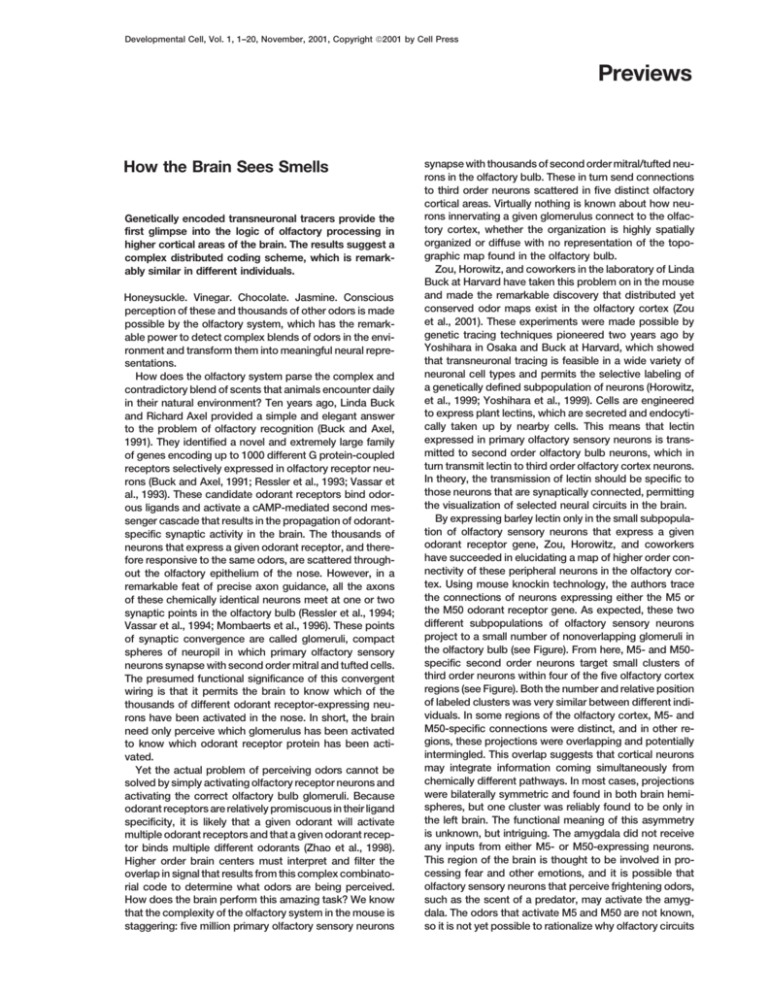
Developmental Cell, Vol. 1, 1–20, November, 2001, Copyright 2001 by Cell Press Previews How the Brain Sees Smells Genetically encoded transneuronal tracers provide the first glimpse into the logic of olfactory processing in higher cortical areas of the brain. The results suggest a complex distributed coding scheme, which is remarkably similar in different individuals. Honeysuckle. Vinegar. Chocolate. Jasmine. Conscious perception of these and thousands of other odors is made possible by the olfactory system, which has the remarkable power to detect complex blends of odors in the environment and transform them into meaningful neural representations. How does the olfactory system parse the complex and contradictory blend of scents that animals encounter daily in their natural environment? Ten years ago, Linda Buck and Richard Axel provided a simple and elegant answer to the problem of olfactory recognition (Buck and Axel, 1991). They identified a novel and extremely large family of genes encoding up to 1000 different G protein-coupled receptors selectively expressed in olfactory receptor neurons (Buck and Axel, 1991; Ressler et al., 1993; Vassar et al., 1993). These candidate odorant receptors bind odorous ligands and activate a cAMP-mediated second messenger cascade that results in the propagation of odorantspecific synaptic activity in the brain. The thousands of neurons that express a given odorant receptor, and therefore responsive to the same odors, are scattered throughout the olfactory epithelium of the nose. However, in a remarkable feat of precise axon guidance, all the axons of these chemically identical neurons meet at one or two synaptic points in the olfactory bulb (Ressler et al., 1994; Vassar et al., 1994; Mombaerts et al., 1996). These points of synaptic convergence are called glomeruli, compact spheres of neuropil in which primary olfactory sensory neurons synapse with second order mitral and tufted cells. The presumed functional significance of this convergent wiring is that it permits the brain to know which of the thousands of different odorant receptor-expressing neurons have been activated in the nose. In short, the brain need only perceive which glomerulus has been activated to know which odorant receptor protein has been activated. Yet the actual problem of perceiving odors cannot be solved by simply activating olfactory receptor neurons and activating the correct olfactory bulb glomeruli. Because odorant receptors are relatively promiscuous in their ligand specificity, it is likely that a given odorant will activate multiple odorant receptors and that a given odorant receptor binds multiple different odorants (Zhao et al., 1998). Higher order brain centers must interpret and filter the overlap in signal that results from this complex combinatorial code to determine what odors are being perceived. How does the brain perform this amazing task? We know that the complexity of the olfactory system in the mouse is staggering: five million primary olfactory sensory neurons synapse with thousands of second order mitral/tufted neurons in the olfactory bulb. These in turn send connections to third order neurons scattered in five distinct olfactory cortical areas. Virtually nothing is known about how neurons innervating a given glomerulus connect to the olfactory cortex, whether the organization is highly spatially organized or diffuse with no representation of the topographic map found in the olfactory bulb. Zou, Horowitz, and coworkers in the laboratory of Linda Buck at Harvard have taken this problem on in the mouse and made the remarkable discovery that distributed yet conserved odor maps exist in the olfactory cortex (Zou et al., 2001). These experiments were made possible by genetic tracing techniques pioneered two years ago by Yoshihara in Osaka and Buck at Harvard, which showed that transneuronal tracing is feasible in a wide variety of neuronal cell types and permits the selective labeling of a genetically defined subpopulation of neurons (Horowitz, et al., 1999; Yoshihara et al., 1999). Cells are engineered to express plant lectins, which are secreted and endocytically taken up by nearby cells. This means that lectin expressed in primary olfactory sensory neurons is transmitted to second order olfactory bulb neurons, which in turn transmit lectin to third order olfactory cortex neurons. In theory, the transmission of lectin should be specific to those neurons that are synaptically connected, permitting the visualization of selected neural circuits in the brain. By expressing barley lectin only in the small subpopulation of olfactory sensory neurons that express a given odorant receptor gene, Zou, Horowitz, and coworkers have succeeded in elucidating a map of higher order connectivity of these peripheral neurons in the olfactory cortex. Using mouse knockin technology, the authors trace the connections of neurons expressing either the M5 or the M50 odorant receptor gene. As expected, these two different subpopulations of olfactory sensory neurons project to a small number of nonoverlapping glomeruli in the olfactory bulb (see Figure). From here, M5- and M50specific second order neurons target small clusters of third order neurons within four of the five olfactory cortex regions (see Figure). Both the number and relative position of labeled clusters was very similar between different individuals. In some regions of the olfactory cortex, M5- and M50-specific connections were distinct, and in other regions, these projections were overlapping and potentially intermingled. This overlap suggests that cortical neurons may integrate information coming simultaneously from chemically different pathways. In most cases, projections were bilaterally symmetric and found in both brain hemispheres, but one cluster was reliably found to be only in the left brain. The functional meaning of this asymmetry is unknown, but intriguing. The amygdala did not receive any inputs from either M5- or M50-expressing neurons. This region of the brain is thought to be involved in processing fear and other emotions, and it is possible that olfactory sensory neurons that perceive frightening odors, such as the scent of a predator, may activate the amygdala. The odors that activate M5 and M50 are not known, so it is not yet possible to rationalize why olfactory circuits Developmental Cell 2 Perception of Olfactory Inputs in the Nervous System Neurons expressing different odorant receptor genes are intermingled in the olfactory epithelium (left), converge to specific glomeruli in the olfactory bulb (center), and form complex and intermingling connections in different regions of the olfactory cortex (right). The spatial maps in both olfactory bulb and olfactory cortex are conserved in different individuals. specific to these odorant receptors would bypass the amygdala. This paper demonstrates that higher order olfactory projections are strikingly similar in different individuals and that specific odors may be represented by spatial maps in the olfactory cortex. This suggests that while different people may have different preferences for odors, the underlying neural network that perceives the odors is genetically hard wired. Leslie B. Vosshall Laboratory of Neurogenetics and Behavior The Rockefeller University New York, New York 10021 Selected Reading Buck, L., and Axel, R. (1991). Cell 65, 175–187. Horowitz, L.F., Montmayeur, J.-P., Echelard, Y., and Buck, L.B. (1999). Proc. Natl. Acad. Sci. USA 96, 3194–3199. Mombaerts, P., Wang, F., Dulac, C., Chao, S.K., Nemes, A., Mendelsohn, M., Edmondson, J., and Axel, R. (1996). Cell 87, 675–686. Ressler, K.J., Sullivan, S.L., and Buck, L.B. (1993). Cell 73, 597–609. Ressler, K.J., Sullivan, S.L., and Buck, L.B. (1994). Cell 79, 1245– 1255. Vassar, R., Ngai, J., and Axel, R. (1993). Cell 74, 309–318. Vassar, R., Chao, S.K., Sitcheran, R., Nunez, J.M., Vosshall, L.B., and Axel, R. (1994). Cell 79, 981–991. Yoshihara, Y., Mizuno, T., Nakahira, M., Kawasaki, M., Watanabe, Y., Kagamiyama, H., Jishage, K., Ueda, O., Suzuki, H., Tabuchi, K., et al. (1999). Neuron 22, 33–41. Zhao, H., Ivic, L., Otaki, J.M., Hashimoto, M., Mikoshiba, K., and Firestein, S. (1998). Science 279, 237–242. Zou, Z., Horowitz, L.F., Montmayeur, J.-P., Snapper, S., and Buck, L.B. (2001). Nature, in press.


