Microbial death
advertisement
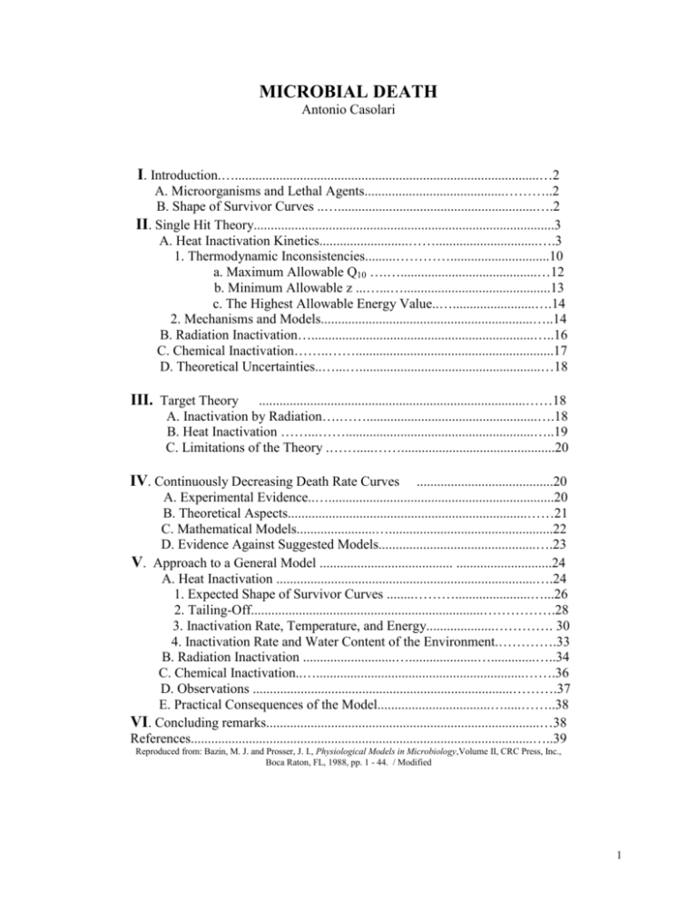
MICROBIAL DEATH Antonio Casolari I. Introduction.….........................................................................................…2 A. Microorganisms and Lethal Agents.........................................………..2 B. Shape of Survivor Curves ..…..........................................................….2 II. Single Hit Theory........................................................................................3 A. Heat Inactivation Kinetics..........................……..............................….3 1. Thermodynamic Inconsistencies.........………….............................10 a. Maximum Allowable Q10 ….…........................................…12 b. Minimum Allowable z ...…...…...........................................13 c. The Highest Allowable Energy Value..…........................….14 2. Mechanisms and Models..............................................................…..14 B. Radiation Inactivation….................................................................…..16 C. Chemical Inactivation……..……..........................................................17 D. Theoretical Uncertainties..…...….....................................................…18 III. Target Theory ..............................................................................……18 A. Inactivation by Radiation….……..................................................….18 B. Heat Inactivation ……...…….......................................................…..19 C. Limitations of the Theory .…….....…….............................................20 IV. Continuously Decreasing Death Rate Curves ........................................20 A. Experimental Evidence..…..................................................................20 B. Theoretical Aspects......................................................................……21 C. Mathematical Models.......................…................................................22 D. Evidence Against Suggested Models..............................................….23 V. Approach to a General Model ....................................... ............................24 A. Heat Inactivation ............................................................................….24 1. Expected Shape of Survivor Curves ........………......................…...26 2. Tailing-Off....................................................................…………….28 3. Inactivation Rate, Temperature, and Energy....................…………. 30 4. Inactivation Rate and Water Content of the Environment.………….33 B. Radiation Inactivation ...........................…....................….............…..34 C. Chemical Inactivation..….............................................................…….36 D. Observations ............................................................................……….37 E. Practical Consequences of the Model.................................….....……..38 VI. Concluding remarks................................................................................…38 References....................................................................................................…..39 Reproduced from: Bazin, M. J. and Prosser, J. I., Physiological Models in Microbiology,Volume II, CRC Press, Inc., Boca Raton, FL, 1988, pp. 1 - 44. / Modified 1 I. INTRODUCTION Microbial inactivation studies seem to be dominated by the exponential-single hit and multitarget theories, the former being applied mostly to heat inactivation kinetics and the latter to microbial inactivation by radiations. However, both theories are unable to satisfactorily explain some relevant experimental results, although they are valid enough to be of some predictive value in sterilization technology. Several satellite theories of lesser relevance do not add significantly to the understanding of inactivation phenomena. In this chapter an attempt is made to avoid interpreting experimental data in too simplistic a fashion, and a general theory is presented which provides a unifying description of microbial inactivation kinetics. A. Microorganisms and Lethal Agents Microorganisms can be regarded as elementary biological particles able to undertake functional relationships within aqueous surroundings, whether provided with levels of autonomous functional organization (bacteria, yeasts, molds, etc.) or not (viruses). Characteristically, the primary function of microbial interaction with the environment is the production of progeny. Hence, the single practical criterion of death of microorganisms is the failure to reproduce in suitable environmental conditions.1 Physical and chemical agents affecting microbial activities to such an extent as to deprive microbial particles of the expected reproductive capacity can be regarded as lethal agents. Heat, ionizing, and UV radiation are the most relevant physical lethal agents. Ultrasonic frequencies, pressure, surface tension, etc. are mostly employed as cell disrupting agents in studying subcellular components. Chemical lethal agents compose a wide range of compounds employed in the microbial inactivation process called disinfection. The most important are oxygen, hydrogen peroxide, halogens, acids, alkalis, phenol, ethylene oxide, formaldehyde, and glutaraldehyde. B. Shape of Survivor Curves Survivor curves are usually described by plotting the logarithm of the number of microorganisms surviving against the size of treatment (time, dose of radiation, concentration of the chemical lethal agent). 9 8 7 Log Cs 6 A 5 4 B 3 2 1 C D 0 T re a t m e n t s ize , S Figure 1. Shape of survivor curves of microorganisms treated with lethal agents: convex curve [A], sigmoid [B], concave or continuously decreasing rate curve [C & D]. Cs is the concentration of survivors after the lethal treatment of size S. 2 The semilogarithmic plot of survivor curves may have various shapes such as convex, sigmoid, concave, or linear (Figure 1). Different shapes can be obtained: (1) under identical experimental conditions with different microorganisms or with the same organism in a different physiological condition (i.e., vegetative cell or spore, the former being in the lag or log phase, the latter being activated or dormant, etc.); 2) using the same population of organisms while changing the destructive potential of the applied lethal agent (viz., changing the treatment temperature or the concentration of the chemical lethal agent); (3) using the same population of organisms while changing the environment in which the microorganisms are suspended or the culture medium employed to detect surviving fractions, etc. Radiation inactivation studies show the typical convex survivor curve (often erroneously called sigmoid) characterized by a more or less extended lag in inactivation at lower doses (shoulder), followed by a nearly exponential decay phase (Figure 1(A)). Such a shape has also been found in heat inactivation experiments, although less frequently, and in microbial inactivation by chemical compounds. Often disregarded, though not uncommon, is the true sigmoid shape characterized by a more or less pronounced tail occurring after initial shoulder and exponential decay phases (Figure 1(B)). Concave survivor curves characterized by a continuously decreasing death rate (CDDR) with increasing treatment time or size, and often described as bi- or multiphasic, can be produced by many physical and chemical lethal agents. The complexity of the situation was recognized about 70 years ago.2-4 Nevertheless, most of the early authors attempted to describe semilog survivor curves by a straight line (exponential inactivation) regardless of shape, claiming that the direct utilization of the data would have been otherwise quite difficult. The most relevant practical application of the exponential simplification has been in heat sterilization technology. Reports of Bigelow and Esty5 and Esty and Meyer6 on the destruction rate of bacteria subjected to moist heat aided greatly in strengthening the belief in exponential inactivation, although counter to experimental evidence from their own results. Fundamental books on heat sterilization technology7,8 perpetuated the belief in this exponential relationship, particularly the book by Stumbo,8 who tried to explain any deviation from exponential decay with a series of conjectures, rather than with experimental evidence. Meanwhile, Pflug and Schmidt9 claimed that "the majority of survivor curves are not straight lines on semilog plot", but did not propose an acceptable alternative theory. II. SINGLE HIT THEORY According to the single hit theory, the death of microorganisms results from the inactivation of a single molecule or site per cell; the death rate is expected to be proportional to the number of organisms remaining alive and follows first-order kinetics. A. Heat Inactivation Kinetics Plotting logarithm of surviving cell concentration, Nt (Nt /g, Nt /mt, Nt /cm2) against the time, t, of treatment at temperature T, a linear relationship is expected to occur: Log10 Nt = Log10 No - k’ * t (1) where No is the concentration of the untreated population and k' is the inactivation rate constant. This type of plot is regarded as very convenient if the resistance of different microorganisms is to be compared. In fact, a parameter called the decimal reduction time is easily obtained from the regression coefficient k' and is denoted by D90 , D10 or DT: 3 DT = 1 / k' (2) The use of D90 or D10 refers to the fact that after a treatment time t = 1 / k', 90% of the microbial population is destroyed or, alternatively, 10% of the population survives. In heat inactivation studies DT is preferred, where T is the treatment temperature. In exponential form, Equation 1 may be written: Nt = No * 10 – k’ * t (3) so that after a treatment time t = t1 Equation 3 becomes: N1= No * 10 – k’ * t1 (4) and after treatment time t = t2: N2 = No * 10 – k’ * t2 (5) N2 / N1 =10 – k’ * (t2 – t1) (6) Solving for k': which in logarithmic form yields: Log10 (N2 / N1) = - k' * (t2 - t1) (7) - k' = (Log10 N2 – Log10 N1 ) / (t2 - t1) (8) k' = (Log10 N1 – Log10 N2 ) / (t2 - t1) (9) so that: and From Equation 2: DT = 1 / k' = (t2 - t1) / (Log10 N1 – Log10 N2) (10) and, as expected, if the treatment time is increased from t1 to t2 , 90% of the population is destroyed, (Log10 N1 – Log10 N2) equals 1 and the decimal reduction time at temperature T is: DT = t2 - t1 (11) Each type of microorganism (virus, bacterial vegetative cell or spore, yeast vegetative cell or spore, fungal cell or spore), as well as each species and strain of the same group of organisms, has its own resistance at a particular temperature T under defined environmental conditions, that is, its own D,. On changing the environment, the microbial resistance changes accordingly. The most relevant physicochemical factors affecting heat resistance are the water content in the environment, pH, and temperature. Changing the solute concentration in the medium also changes the osmotic pressure or, referring to the usual parameter employed in food microbiology, the water activity a (aw = p/po where p and po are the water pressures of the medium and of pure water, 4 respectively, in isothermal and isobaric conditions). The water activity is described by Raoult's law, so that: aw = f [nw / (nw + ns)] (12) where nw and ns are the concentrations of water and solute, respectively. It follows that by increasing the solute concentration, the aw of the environment decreases. As aw decreases, the thermal resistance of microorganisms increases. The DT value in dry conditions may be more than 103 times higher than that in moist (high aw) conditions.9 An exact relationship between DT and aw has not been developed. Thermal resistance is usually higher at neutral pH and it decreases when the pH is increased or decreased. A defined relationship between DT and pH is not known, though a tenfold change in DT is often observed for each 2 pH units. With respect to chemical reactions, a defined relationship between DT and temperature can be established. Plotting Log D against temperature, a linear relationship is usually obtained: Log10 DT = Log10 U - b * T (13) where U is a proportionality constant and b is the rate at which D changes with temperature. Equation 13 may be written: DT = U * 10 – b * T (14) The above equations yield the parameter z: z=1/b which is related to the temperature coefficient Q10 by: z = 10 / Log10 Q10 where Q10 = K(T+10) / KT (15) (16) (17) z is the number of degrees required to achieve a tenfold change in DT . It follows that: 0.1 * DT- z = DT = 10* DT + z (18) If we let D1 and D2 be the DT values at the temperature T1 < T2 respectively, it follows that: and so that, solving for b: D1 = U * 10 – b * T1 (19) D2 = U * 10 – b * T2 (20) D2 / D1 = 10 – b * (T2 – T1) (21) Log10 (D2 / D1) = - b * (T2 - T1) (22) b = (Log10 D1 – Log10 D2) / (T2 - T1) (23) and in logarithmic form: and then: 5 and z = 1 / b = (T2 - T1) / (Log10 D1 – Log10 D2) (24) As expected, when D1 = 10* D2 , the z value will be equal to T2 - T1. Knowledge of the DT and z values for microorganisms in given environmental conditions is very useful in practice, when heat sterilization times at an unknown temperature Tu must be determined when DTu is not known. In such a case, Tu - T = n*z (25) DTu = DT * 10 – (Tu – T) / z (26) DTu = DT * 10 – n (27) D(T + n*z) = DT / 10 n (28) t (T + n*z) = t T / 10 n (29) and so: where ' t (T + n*z)' is the treatment time at the temperature T + n*z equivalent to the time t T at the reference temperature T. Sterilization cycles are usually based on a minimum time required to obtain 12 decimal reductions of a particular microorganism (Clostridium botulinum spores in food sterilization technology, for instance). Usually Equation 29 is employed, although using the symbolism: (T + n*z) = T / 10 n (30) where is the number of minutes required to obtain the expected number, i, of decimal reductions ( = i * D). In heat sterilization technology, at the reference temperature T =121.1°C = 250°F, the 121.1°C = 250°F value is Fo and z = 10°C = 18°F, so that Equation (30) becomes: 121.1°C / (T + n * 10) = 10 n (31) or in a better known form, putting T = T(T+n*10) F / = 10 (T - 121.1°C) / 10 (32) Equation 32 is currently employed to evaluate the efficiency of sterilization cycles.7,8 By analogy with chemical kinetics, the exponential heat inactivation curve can be described by the relationship: - dNt /dt = k * Nt (33) yielding, by integration: Log ( Nt / No ) = - k* t (34) Nt / No = exp ( - k*t) (35) and: 6 It follows that DT as defined by Equation 10 is DT = 2. 303 / k (36) The analogy can be extended to the relationship between k and temperature. According to the chemical kinetics: k = A * exp( - Ea /R*T) (37) where A is a frequency factor, Ea is the Arrhenius activation energy, R is the gas constant (1.987 cal/mol), and T is the absolute temperature (degrees Kelvin). From Equation 37: k1 = A * exp( - Ea / R* T1) (38) and k2 = A * exp( - Ea / R* T2) (39) so that: k1 / k2 = exp [ - Ea (1/T1 - 1/T2 ) / R (40) From Equation 40, Log (k1 / k2) = ( - Ea / R)(T2 - T1 ) / (T2 * T1) (41) and since k = 2. 303 / DT, from Equation 36, Log(D2 / D1) = ( - Ea / R)(T2 - T1 ) / (T2 * T1) (42) And Log10 (D2 / D1) = ( - Ea / 2.303*R)(T2 - T1 ) / (T2 * T1) (43) Taking into account Equation 24, z = 2.303*R*T2*T1 / Ea (44) It follows that the z value cannot be regarded as being constant, but varies with temperature. It follows that a z value of 5°C obtained between 60 and 65°C increases to 5.23 between 65 and 75°C, and to 5.53 between 75 and 85°C. Analogously, a z value of 10°C obtained in the temperature range 100 to 110°C will reach the value of 11.09 in the range 120 to 130°C. According to Equation 44, the Arrhenius relationship requires that z increases as the temperature increases. The constancy of z values obtained by plotting Log10 DT against temperature is difficult to ascertain in practice, as the evaluation of D is not sufficiently accurate. In some instances, a trend toward an increase of z with increasing temperature has been shown.10-14 Following chemical kinetics, the Arrhenius activation energy Ea may be obtained using Equation 37, plotting Log k against T-1. Alternatively, Ea can be obtained from: Ea = a * Log Q10 (45) or Ea = c / z (46) where a = 0.1*R* T1* T2 and c = 2. 303*R* T1* T2 7 since, according to Equation 43: Ea = 2.303*Log10 (D1 / D2) *R*T2*T1 ) / (T2 - T1) (47) Nevertheless, z is not constant, according to Equation 44, so Ea values obtained from Equation 46 differ from those from the Arrhenius equation (Equation 37) by about 1 kcal / mol (4.18 kJ/mol). Figure 2 shows the expected value of Ea as a function of Q10 and ‘z’ in temperature ranges resulting in the thermal destruction of microorganisms. The standard energy of activation, G, the standard enthalpy of activation, H, and the standard entropy of activation, S, are sometimes reported for the thermal death of microorganisms. According to Eyring's relationship: k = k ' (K*T / h)* kc (48) where k is the reaction rate constant observed at the absolute temperature T, k' is the transmission coefficient (usually considered = 1), K is Boltzmann's constant (1.38*10-16erg/degree), h is Planck's constant (6.624 *10-27 erg/sec), and kc is the equilibrium constant; kc may be expressed in terms of the standard energy of activation: kc = exp ( - G/R*T) (49) and since: G = H – T*S Equation 48 becomes: k = (K*T / h) * exp (S / R) * exp( - H / R*T) (50) (51) FIGURE 2. Relationship between Q10, corresponding ‘z’ values, and Arrhenius activation energy E a, predicted by thermodynamic treatment of first-order inactivation kinetics of microorganisms subjected to low temperatures (50 to 60°C) (heat sensitive organisms, HSO, with an average Q10 = 100), or to high temperatures (100°C; heat resistant bacterial spores, HRBS, with an average Q10 = 10). Equation 51 is very similar to the Arrhenius relationship, taking into account that: exp( - H/R*T) = exp( - Ea /R*T) (52) 8 when Ea >> R*T, since: H = Ea – R*T and from Equations 37 and 51: A = (K*T / h)* exp(S / R) (53) (54) Because, in microbial heat inactivation kinetics R*T is more than 100 times lower than Ea Equation 51 can be written: k = (K T / h)*exp(S / R)*exp(- Ea / R*T) (55) S = R*(Log k – Log (K*T / h) + Ea /R*T) (56) after which: or, taking into account that K = R / N, where N is Avogadro's number and R = 8.32*107 erg/mol/degree: S = R*(Log k – Log (R*T / N*h) + Ea /R*T) (57) FIGURE 3. Relationship between Arrhenius activation energy Ea (i.e., the activation enthalpy minus RT) and activation entropy (S), predicted by thermodynamic treatment of first-order inactivation kinetics of microorganisms subjected to 50°C (heat sensitive organisms, HSO) or to higher temperatures (heat resistant bacterial spores, HRBS). As shown in Figure 3, there is a linear relationship between E, or H and S at any given temperature. It follows that Equation 57 (or Equation 56) can be reduced to: S = (Ea / T) + B (58) where B equals R*(Log k + Log (R*T / N*h)) or R*(Log k + Log (K*T / h)). Equation 58 is often called the "isokinetic relationship" or the "compensation law", since an increase in the activation energy or enthalpy is exactly compensated by an increase in entropy.15-17 Nevertheless, when activation energy and activation entropy values are obtained by a series of computations based on Equations 37 to 58, the compensation law is not such a mysterious 9 relationship as it is sometimes regarded, but follows directly from the premises (Equation 48).18,19 At this point, the analogy between microbial heat inactivation kinetics and chemical reaction kinetics should be examined closely. 1. Thermodynamic Inconsistencies Figure 4 shows the frequency distribution of 231 z values collected from the literature. As can be seen, the spread is very large. After grouping collected data into 5°C classes the most frequent z value obtained for viruses and microbial vegetative cells is 5°C (Q10 = 100) and for bacterial spores is 10°C (Q10 = 10). It must be remembered that viruses and vegetative microbial cells are usually 102 to 108 times less heat resistant than bacterial spores. Vegetative cells are destroyed at temperatures ranging from 50 to 60°C at a rate k = 3.838*10 - 3 (DT 10 min). Bacterial spores are destroyed at a similar rate at temperatures ranging from 100 to 120°C. FIGURE 4. Frequency distribution curves of 231 ‘z’ values collected from the literature, together with corresponding Q10 values, for viruses, vegetative bacteria, yeasts, and molds (o) and bacterial spores (). As shown in Figure 2, the activation energy obtained for less resistant vegetative cells (mean z = 5°C = 9°F) is much higher than the Ea value of more resistant bacterial spores (mean z = 10°C = 18°F), the former being about 90 kcal/mol (380 kJ/mol) and the latter about 60 kcal/ mol (250 kJ/mol). Disregarding the parameter A in Equation 37, reaction rates of analogous reactions are expected to be inversely related to the activation energy value required for the occurrence of single reactions. Contrariwise, the heat inactivation of bacterial spores has a lower activation energy, while occurring at a rate very much slower than that of heat-sensitive viruses or vegetative microbial cells. Consequently, it can be argued, the A value in the Arrhenius equation cannot be disregarded and Eyring's relationship must be taken into account. As previously shown, the so-called compensation law requires that high Ea (or H) values necessarily follow high S values. In fact, as shown in Figure 3, S for vegetative cells is about 210 cal/mol/degree (880 J/mol/degree) and S for bacterial spores is about 90 cal/ mol/degree (380 J/mol/degree). Accordingly, higher activation energies required to inactivate vegetative cells would 10 be justified on the basis of the greater activation entropy involved in the process. Disregarding uncertainties associated with the rigid application of the compensation law, it seems hard to envisage a greater S for the heat inactivation of vegetative cells rather than of spores, since it is reasonable to assume that the entropy level of the former is higher than that of the more orderly assembled molecular structures of bacterial spores; however, this may not be the case. Nevertheless, the reservations regarding what results merely from a thermodynamic treatment of exponential heat inactivation kinetics are further strengthened by the very high values of the ensuing Ea or H. Using the Maxwell-Boltzmann's law for the distribution of velocities among molecules: nE /no = 2 - 1/2 (Ea / R*T)1/2 exp( - Ea / R*T) (59) where nE and no are the number of molecules having energy Ea and the total number of molecules, respectively; or in the simplified form (i.e., on the basis of two axes): nE /no = exp( - Ea / R*T) (60) For values of 2 - 1/2 (Ea / R*T)1/2 ranging between about 5 and 15 at temperatures 273.15°K and Ea values lower than about 100 kcal/mol, it can be computed that the probability of finding Ea values as high as those obtained for vegetative cells exposed to lethal temperatures is so low as to be unreasonable. The probability P(E) = nE/no of occurrence of molecules having Ea = 90 kcal/mol at 60°C is expected to be: n90,000 / 6*1023 = exp ( - 90,000 / ((60 + 273.15)* 1.987)) = 8.99*10 - 60 which means that a single molecule carrying more than 90 kcal could occur, at 60°C, in about 10 36 mol of a substance (i.e., a mass of hydrogen about 103 times that of the sun). A mole of cells weighs about 1011 g; therefore, we would find a single molecule carrying the above energy in 1047 g of cells, a weight about 103 times that predicted for our galaxy. Summing the number of intermonomer chemical bonds of about 106 protein molecules (average mol wt, 2*104) and of DNA (mol wt about 109) occurring in a microbial cell, we obtain about 1010 chemical bonds per cell. This implies that we would expect to find a single one of the above 10 10 bonds with about 100 kcal in a mass of cells approximately 104 times that of the sun. The above results seem to suggest that E, values as high as those obtained by the thermodynamic treatment usually applied to microbial heat inactivation kinetics can be regarded to be of doubtful meaning. a. Maximum Allowable Q10 Q10 values obtained in microbial heat inactivation studies are usually much higher than those expected to occur in chemical kinetics. The maximum Q10 (MQ10), the value above which thermodynamic treatment of reaction rate loses any meaning, can be evaluated. According to Equations 45 and 60, and letting P(E) be the probability of occurrence of molecules carrying more than Ea energy, it follows that: P(E) = exp ( - a * Log Q10 / R*T) (61) The minimum value of the probability (mP(E)) required for a reaction to occur at the temperature T, = T, + 10§ can be regarded to be 1, that is, at least one n, molecule can be expected to be present in the total amount of available reactants, or one n, per total number of available molecules; so that: 11 mP(E) = 1 / i*N (62) where i is the number of moles of available reagents and N is the Avogadro's number. If the above probability is higher at T2 than at T1 Equation 61 can be written: mP(E) = exp (- a*Log Q10 / R*T2) (63) so that combining Equations 62 and 63: (i*N)- 1 = exp ( - a * Log Q10 / R*T2) (64) As a = 0.1*R*T1* T2 , as previously shown, and rearranging: MQ10 = (i*N)10 / T1 (65) it follows that, letting the highest conceivable value of i = 103, at T1 = 50 or 100°C the maximum allowable Q10 would be 6.74 or 5.22, respectively. A value of Q10 = 2 to 3, as is usually found in chemical kinetics, is expected to be obtained at about 100°C = 212°F in an amount of solution containing about 10-12 to 10- 6 mol of reagents, that is, about 1011 or 1018 molecules, respectively (i = (M Q10)T1 /10 *N-1 ). Table 1 shows the M Q10 values expected from a thermodynamic treatment of microbial heat inactivation, according to Equation 65, for two amounts of cells (1 g and 1 kg), exceedingly higher than those employed in practice. By comparing values reported in Table 1 with those in Figure 4, it can be seen that all Q10 values obtained by treating microbial heat inactivation as a first-order reaction, can be regarded as unreasonably high. 12 b. Minimum Allowable z According to Equation 17, if Q10 increases, z decreases. The minimum allowable z value, zm , under which P(E) must be regarded as being unreasonably low, can also be obtained. From Equations 17 and 65: (i * N)10/T1 = 10 10 / Zm (66) Taking logarithms and rearranging: zm = T1 / Log10 (i * N) (67) Microbial heat inactivation experiments are usually done employing less than 1012 particles per liter of medium. Therefore, while taking into account the maximum allowable concentration of cells that could be employed in practice, the minimum allowable z values following Equation 67 are two to six times higher than those found in the literature (Table 1). We could assume ah absurdo that a microorganism is inactivated if a single chemical bond in the cell is broken, so that the quantity ‘i*N’ in Equation 67 could be regarded to be about 1025; the resulting z, will be 13.33, 14.13, 14.93, or 15.73 at T1 = 60, 80, 100, or 120°C, respectively. As a consequence, all z values reported in the literature (Figure 4) can be regarded as unreasonably low. c. The Highest Allowable Energy Value It would he interesting to know the highest reasonable E,. value. To this end, Equation 63 can be written: mP(E) = exp( - MEa / R*T2) (68) where MEa is the maximum value of the energy of activation at the temperature T2 = T1+10°C, and thus the Ea value above which P(E) is too low to be acceptable. From Equation 68: MEa = R*T2 *( - Log mP(E)) (69) and then: MEa = R*T2 * (- Log (1/(i*N))) (70) = R*T2 * (Log i*N) It follows that at T2 = 50 or 100°C the maximum allowable Ea value is expected to be 39,591 or 45,716 cal/mol (165,708 or 191,348 J/mol). Table 2 shows expected values of MEa according to Equation 70 for first-order kinetics of microbial heat inactivation. As can be seen, all Ea values reported in the literature are unreasonably high. Perhaps, as a provisional conclusion, we may say that the thermodynamic treatment of reaction kinetics on an exponential basis is not readily applicable to the heat inactivation of microorganisms. It can be rather useful to retrace our steps in order to answer, first, the most relevant question: could microbial heat inactivation kinetics be regarded as an exponential phenomenon? Several observations seem to suggest that this is not always the case. The question will be examined later. 13 2. Mechanisms and Models The greatest insight into heat inactivation mechanisms was obtained by Ball and Olson,7 as presented in the book Sterilization in Food Technology, still regarded as the bible of the field. Through an analysis of the concepts of heat and temperature on a macroscopic and microscopic scale, they pointed out that "whenever Brownian movement is observed, our macroscopic concepts of temperature and heat transfer break down and must be replaced by energy considerations involving molecules in the discrete, and not in the statistical sense" (see also Tischer and Hurwicz20). Soon after "... it is not something within the cell (such as temperature) which is the cause of death. The cause must be outside the cell. It must be in the medium." Accordingly, Ball and Olson suggested a mechanism of microbial death brought about by "... one or more molecules in the surrounding medium", having "the greater mean velocity ... according to the velocity distribution curve". Nevertheless, they did not develop a mathematical model for microbial death. Charm21 presented a model referring to the discrete nature of molecules involved in heat inactivation mechanisms, although diverging somewhat from the work of Ball and Olson7. Charm's model leads to exponential inactivation, but some uncertainties cast doubts on its reliability. Charm21 regards the microbial cell as "... composed of a number of sensitive volumes surrounded by a number N of water molecules; when the water molecule, in contact with a sensitive volume, is able to impart sufficient energy to the sensitive volume inside the cell, it is thought to cause inactivation of the cell". The model yields the equation: log(Nt/No ) = - S * t * exp( - E/RT) (71) where S (min-1) is the frequency with which water molecules exchange the energy, E, with the sensitive volume. Some inconsistencies arising from the model can be pointed out: (1) Charm defined E as energy / cell, while energy/mol was considered, since R = 1.987 cal/mol/degree was seemingly taken 14 throughout for computation purposes; (2) reported E values seem to be too high (for C. botulinum spores heated in distilled water, for instance, E = 73,500 Btu/spore, equivalent to about 1.85 *10 7 cal or 7.35 * 107 J); (3) the reported values of the frequency factor S are much too high to be reasonable (S 1020), being equal to or greater than the frequency of X-rays produced by a potential difference of about 1.7 * 105 V. In addition, Charm's model contains the same inconsistencies pointed out previously regarding the thermodynamic treatment of exponential kinetics (i.e., very high energy values, energy values greater for less than for more resistant organisms, etc.) and substantially, it leaves inactivation kinetics still unresolved. B. Radiation Inactivation At present, studies of microbial inactivation by UV radiation are usually carried out using low pressure mercury lamps which emit about 95% of their light at a wavelength very close to 253 nm. The plot of the logarithm of surviving organisms against UV dose yields, very frequently, nonlinear curves, with a pronounced tendency to tail off.22-25 A quantum of UV radiation has an energy level of about 5 eV, insufficient to eject electrons from atoms or molecules, and able to cause only excitation processes. High energy radiations (y and X-rays, n, p, and neutron particles) have, characteristically, the property of causing ionization in the absorbing material; many atoms and molecules are excited and converted to free radicals.26 The density of ionization and excitation events produced along the path of radiation depend upon the photon energy and the type of absorbing material, and is usually expressed as a linear energy transfer (keV/p,m). Linear energy transfer (LET) increases with the square of the charge carried by the particle and decreases as its speed increases.26 The relative biological effectiveness usually increases as the LET of the employed radiation increases, but this is not always the case.27, 28 A very limited number of experimental survival curves of irradiated microorganisms can be safely described by the law of exponential decay: Nd / No = exp( - a * d) (72) where Nd is the number of microorganisms surviving the dose d of radiation and a is the rate constant of inactivation. The coefficient a in Equation 72 is expected to represent the probability of inactivation of microorganisms per unit dose (rad or gray) of radiation. Inactivation is expected to result from a single hit, that is, through excitations and ionizations occurring along the track of photons crossing the cell. Hence, the concept of "direct action" arises, according to which the target is a molecule or a narrow group of molecules within the cell, in which the primary event (i.e., an ionization) has to occur, or through which or near which a unit of ionizing radiation must pass.29 On the other hand, the "indirect action" theory assumes that the whole solution, in which microorganisms are suspended, is the true target, lethal events being produced by reactions of chemical active agents induced by radiation in water, outside, and/or inside the cell. In fact, a number of reactions do occur in irradiated water: H2O H2O+ + eH2O+ OH. + H+ e- + H2O OH- + H+ 15 2H. H2 2OH. H2O2 H. + OH. H2O H. + O2 .HO2 2 .HO2 H 2 O2 + O2 The most active and long-living radicals are regarded to be OH. . For the single-hit theory to be tenable, direct interaction between the radiation and a single sensitive target inside the cell must occur.30 However, many environmental factors can modify the sensitivity of microorganisms to ionizing radiations such as oxygen, moisture, the presence of sulfydryl compounds and/or - SH reagents in the medium, previnus treatments of the microorganisms, physiological age of the cells, etc.31-36 Actually, exponential survival curves of irradiated microorganisms usually result from experiments carried out either using very sensitive organisms or are not prolonged enough to determine the shape of survivor curves at very low surviving fractions. Nevertheless, it is well known that survivor curves of irradiated micro organisms are not usually exponential, but are typically sigmoid. C. Chemical Inactivation The concept of "effective concentration" requires that a lethal chemical compound must possess the ability to reach, and to accumulate at, the site(s) of action, whether on the surface of, or within, the microbial cell. Thus, a dose-effect curve is expected to be characteristically sigmoid. At very low concentrations of the lethal compound the microbial inactivation rate will be very low and approaches zero; at higher doses, the death rate is expected to increase as the dose itself is increased; at the highest doses, the rate of the process would not increase further, since it would be sorption-limited. In spite of these expectations, Madsen and Nyman' and Chick' established a mathematical model for chemical disinfection on the basis of an analogy between microbial inactivation process and first-order reaction kinetics. This model has formed the basis of most subsequent investigations. According to the model, the relationship between the surviving organisms and the contact time, t, with a given concentration of a lethal chemical compound is expected to be Nt / No = exp( - kt * t) (73) where kt is the rate constant. The inactivation rate constant kt changes as a function of the concentration of the disinfectant. The relationship between the rate constant kt and the concentration C of the lethal agent is expected to be: kt = B * exp(kc * C) (74) where kc is the rate of change of inactivation rate per unit change in the concentration C of the lethal compound . Usually Equation 75 is used instead of Equation 74: k’t = B' * 10kc’ * C (75) 16 and 1/kc' is the concentration required to achieve a tenfold change in the inactivation rate. However, subjecting microbial populations to increasing concentrations of lethal compounds for a fixed contact time seldom produces survival curves described by a function of the type: Nd / No = exp (- kd * D) (76) where D is the concentration of the lethal compound. Usually Equation 77 is used instead of Equation 76: Nd / No = 10 – kd’ * D (77) and 1/kd' equals the change in concentration required for the survival probability Nd /No to change ten times. Reaction rate constants kt , kc and kd are expected to increase as temperature increases, following the Arrhenius law: kt, c, d = F * exp(- Ea /R*T) (78) where F is a frequency factor whose value is linked to the rate constant considered (kt , kc or kd). Actually, a relationship of the above type is expected to occur in disinfection processes at temperatures lower than about 45 or 100°C for vegetative cells (heat sensitive) or bacterial spores (heat resistant), respectively. In a range of temperatures sufficiently high for heat inactivation, a relationship more complex than Equation 78 is expected to be more appropriate. A strikingly limited number of Arrhenius plots is reported in the literature, notwithstanding the relevance of temperature coefficients of chemical lethal compounds to the practice of disinfection, especially with respect to the choice of suitable agents of disinfection. Q10 values for microbial inactivation by chemical compounds reported in the literature range between about 1.6 and 3.3 and have been obtained by treating bacterial spores with hydrogen peroxide, formaldehyde, glutaraldehyde, -propiolactone, ethylene oxide, chlorine, and iodine, at temperatures ranging from - 10 to 95°C.37-42 Therefore, Arrhenius activation energies are expected between about 10 and 25 kcal/mol. Recently, Gelinas et a1.43 reported Ea values ranging from 0 to about 37 kcal/mol, so that Q10 fell between about 1 and 7.7, although, the method used by the authors to assay the sensitivity of vegetative bacterial cells cannot be regarded as being reliable. Higher Ea values are reported for Escherichia coli treated with phenol at temperatures from 30 to 42°C: 52 kcal/mol;44 a Q10 = 10 was reported using peracetic acid.45 Nevertheless, a disinfection model is difficult to develop on the basis of these sorts of results, since several observations suggest that survivor curves may have different shapes (mostly convex, sigmoid, and concave, with a more or less pronounced tail), according to the experimental conditions employed.37, 40-42, 45-49 A comprehensive theory of the disinfection process is, unfortunately, still lacking. D. Theoretical Uncertainties Characteristically, the single-hit theory is applied to heat inactivation kinetics. Heat sterilization technology is based on the tenet of exponential inactivation. If this order of death should be found to be invalid, the efficiency of the sterilization process becomes questionable. As pointed out by several authors, first-order kinetics are expected to be produced by unimolecular reactions, without regard to the underlying reasons (pseudo-first-order reaction, for instance). As far as microbial inactivation is concerned, a unimolecular reaction would comply with the single-hit or single-site theory, after which a single damage produced in the cell would 17 unequivocally lead to the death of the cell, whether affecting an enzyme, the DNA, or a different molecule. The concept is quite limiting, since microorganisms subjected to a lethal agent are not unequivocally dead or alive, but they either die or recover, depending on the environmental conditions applied after the treatment. The phenomenon is well known to microbiologists and is called "sublethal injury or damage". Usually, microorganisms treated with lethal agents become more exacting about their environmental conditions than untreated organisms.12, 24, 34, 50-57 As a consequence, survivor curves obtained using a population subjected to a lethal treatment might differ depending on the experimental conditions applied after the treatment. As a rule, the extent of the damage becomes increasingly difficult to demonstrate as the intensity of the applied lethal condition increases. The sublethal injury phenomenon can hardly be reconciled with the single-hit theory. On the contrary, it seems to suggest that microbial death can be rather more satisfactorily envisioned as an end point of a gradual, damaging process. III. TARGET THEORY A. Inactivation by Radiation The effects of ionizing radiation upon microorganisms have led to the development of some very interesting models, usually described under the name of "target theory". The concept of microorganisms, or something(s) inside microorganisms, as targets being hit by photons or particles, reflects quite closely what is believed to occur in radiological phenomena. The basic assumption of the "multiple hit" theory was that a single target must be hit n times before the organism is destroyed.58-60 Let a be the sensitive volume of the cell and h the average number of hits per unit volume of the microbial suspension; then, ‘ah’ is the average number of hits within the sensitive volume a. If the hits occur independently and at random, the probability that f hits fall within the volume a is given by the Poisson distribution: P(f) = exp ( - a*h) (a*h)f / f! (79) If n is the number of hits required to inactivate a microorganism, then all cells receiving less than n hits will survive. The probability Nh /No that after the dose h only a fraction f < n of hits had occurred, i.e., the probability of survival, is given by: n-1 Nh /No = exp (- a*h) (a*h)f / f! (80) o The second multiple-hit hypothesis of the target theory follows directly from the single-hit theory. If a population of organisms contains n sensitive targets that are inactivated exponentially, the viability of the organism is ensured if fewer than n targets are hit. The probability that all the targets in such a group of n becomes inactivated is given by: P(n) = (1 – exp (- k*d))n (81) since exp(- k*d) is the probability that targets are not hit. This assumes that the rate ‘k’ of occurrence of hits is the same for all the targets. Therefore, the assumption is not simply that n hits per organism are required, but that each of n particles or targets within the cell must be hit at least once.61 18 Both Equations 80 and 81 describe convex curves, characterized by an initial shoulder followed by a nearly exponential behavior at higher doses. The fitting procedures of experimental dose-effect curves by one or other of either equations are not critical. Discrimination between the two models would require a level of experimental accuracy that is not attainable in practice. It has been suggested that the n value should be referred to as the "extrapolation number", whether the multihit or the multi-target theory is considered.62 The multi-target theory suggests a likely explanation for the observed variations in recovery rate in different environmental conditions. In fact, it envisages microbial death as an end point of a process of gradually increasing damage as the applied dose increases, since the cell dies only when all the n vital sites are hit. Under suitable environmental conditions the cell can recover. B. Heat Inactivation As indicated above, convex survival curves are seldom reported in heat inactivation studies. Moats57 succeeded in fitting convex survival curves based on a multi-target model he developed for heat-treated bacteria. However, according to his model the survival curve is expected to be characterized, as in the multi-target theory developed by Atwood and Norman,61 by an initial lag (shoulder) followed by essentially exponential behavior over a wide range of intensities. The fundamental equation developed by Moats57 was Xp-1 P(S) = (N/X) exp( - k*t *(N - X)) (1- exp ( - kt))X (82) X=0 where P(S) is the survival probability, X is the number of critical targets inactivated at any time t, X, is the number of critical sites that must be inactivated to cause death, N is the total number of targets, and k is the rate constant for inactivation of individual targets. The values of k, N, and X, can be obtained from experimental data. Solving simultaneously for k: d / s = N(exp(- k*t) - 2 * exp(- k*t + k*t50) + exp(- 2*k*t")) / (exp(- k*t) - exp(- 2*k*t) ) (83) where d is deviation from the mean (i.e., X – XL), s = (Npq)1/2 where p = X/N and q = (N - X)/N, and t50 is the time at which 50% of the population is killed.54 According to the model, cells of Salmonella anatum heated at 55°C can survive if plated on trypticase soy agar (a relatively rich medium) having about 7.8 critical sites inactivated, while if plated on basal medium (a less rich medium), only about 3.3 sites need to be inactivated to cause death. Estimation of N and the procedure for calculating the rate constants is statistically difficult.63 In the example quoted above, N ranges between 38 and 173 (see Reference 57). As pointed out by Moats himself, the model is unable to explain all survival curves and especially those with a tail.56, 57 Alderton and Snell64 developed an empirical expression which gave a reasonable fit to heat-treated bacterial spore survivor curves, showing a shoulder followed hy an exponential decay: Log10 (No / Nt)a = k’ * t + C (84) where “a” is a constant characterizing the degree of resistance of the microorganism and C is a constant whose value increases with both treatment temperature and the sensitivity of the 19 microorganism. Equation 84 allows the linearization of survivor curve obtained following treatment with heat, radiation, and disinfectants.63, 65 Alderton and Snell64 did not supply an explanation of the parameters “a” and C, nor of the inactivation mechanism. C. Limitations of the Theory The target theory was developed to explain the shoulder in survivor curves of irradiated microorganisms. The single-hit survivor curve is a special case of the tbeory. Nevertheless, the target theory does not explain other types of survival curves occurring in many radiation inactivation experiments, such as true sigmoid curves, continuously decreasing death rate curves, and curves with long tails. The merit of the theory is that it suggests a multiplicity of events leading to death as a possible general mechanism of microbial inactivation. IV. CONTINUOUSLY DECREASING DEATH RATE CURVES A. Experimental Evidence Moats et al.56 stated that "... examples of non-exponential survivor curves found in the literature are too numerous to list". Actually, as shown in Table 3, the list of only more prominent sigmoid or concave survivor curves reported in the literature is long. Nevertheless, some authors8, 50, 66 seem to ignore factual evidence, proposing hypothetical biological or experimental reasons for deviation from exponential behavior, although their proposals are not convincing and they provide little in the way of experimental evidence. The early experimental evidence of Bigelow and Esty5 showing many data incompatible with the exponential tenet and by Esty and Meyer's6 data showing a Clostridium botulinum spores heat destruction curve with a tail lasting about 40 min seem to be disregarded.The experimental evidence of deviations from exponential kinetics by many lethal agents is too widespread to be ignored. B. Theoretical Aspects A concave or biphasic survival curve suggests a phenomenon brought about by population heterogeneity. Chick's4 proposal that heterogeneity in heat resistance could be responsible for a concave survivor curve seemed to be the only explanation for more than 70 years, although based on very little experimental evidence. Microbial heterogeneity can explain biphasic survivor curves.7, 133-136 A mixture of two populations of organisms having different resistances to a lethal agent yields a biphasic survival curve. For example, mixing 107 spores of C. botulinum with 103 spores of PA 3679 and then subjecting the suspension to a temperature of 110°C, results in two survivor curves which intersect after about 15 min of treatment. Biphasic survivor curves could also occur when cells of a single species are composed of two populations with respect to their resistance to the lethal agent employed. Based on the same reasoning, multiphasic survivor curves, or continuously decreasing death rate curves (CDDRC), would be expected when heterogeneous populations are treated.137 The first problem raised by several authors was whether the distribution of resistance among individuals in a population was permanent ("innate heterogeneity'' theory)7, 8, 138-140 or if it was acquired during the treatment ("adaptation model").141-143 The second problem was which type of probability distribution could explain different shapes of the survivor curves. 20 C. Mathematical Models Han et al.135 developed a model for both the innate heterogeneity hypothesis and the adaptation hypothesis. The following equation was derived for the former: Log10 (Nt / No) = - K*t + (s2 / 2)* t2 (85) where K is the most probable value of the destruction rate, “s” is the standard deviation, and “t” is the treatment time. The heat adaptation approach leads to the following equation: Log10 (Nt / No) = - Ko * X ((1 - a) * t – a*b*(exp(- t / b) - 1)) (86) where Ko is the initial rate of destruction, “a” is a constant representing the maximum amount of resistance attainable for a unit amount of destructive power, “b” is a constant representing the rate of development of resistance, and “t” is the time. By applying the two models to some bacterial 21 inactivation curves, the authors concluded that curvilinearity in the survival curves resulted from the development of resistance during the treatment, rather than from innate heterogeneity. Sharpe and Bektash136 modified the models developed by Han et al.135 by utilizing other types of probability distributions of resistance in the population, including the normal, , shifted , and a modified Poisson distribution, and suggested that a combination of the innate heterogeneity and adaptation models might be appropriate. They proposed that the distribution of the initial rate of destruction (Ko) could be represented by any distribution having a probability P(K < 0) = 0, that the life of a cell follows an exponential distribution with mean 1/K, and that the state of the cells (i.e., living or dead) is independent. These considerations lead to a probability of survival S(t) at time t of the following form: S(t) = Log (Nt / No) = Log Lt ( t ) (87) where Lt ( t ) is the Laplace transform of the density function of K at time zero, that is for a normal distribution, Lt ( t ) = exp( - Ko * t + (s2 * t2) /2) (88) for a distribution, Lt ( t ) = (1 + t / )- r (89) for a shifted distribution, Lt ( t ) = exp( - a*t) (1 + t / )- r (90) where “a” is the minimum value of the rate of destruction and for a modified Poisson distribution, Lt ( t ) = exp(- Ko * (1 - a)* t + Ko * a*b*(exp(t/b) - 1) (91) Sharpe and Bektash136 concluded that it is not possible to distinguish between the two possibilities (innate heterogeneity or development of resistance) on the basis of the survival data alone; the heat adaptation model of Han et al.,135 for instance, has been shown to be equivalent to an innate heterogeneity model with a modified Poisson distribution. Nevertheless, from the mathematical analysis carried out by the authors, it can be argued that all concave survivor curves can be reasonably explained by the innate heterogeneity theory. Two objections can be made to the purely phenomenological models described above: first, the adaptation model cannot be established if the acquisition of resistance during the treatment is not permanent; second, the distribution of resistance in an untreated population can be demonstrated experimentally. Furthermore, none of the models examined is able to explain the tailing phenomenon. As shown later, the experimental evidence is contrary to both models. Brannen144 developed a model based on the assumption that survival depends on a number of subsystems, whose functionality is affected by heat. The model yields the four classical types of survivor curves, although "testing of the model is extremely difficult" 144 and the relationship between heat resistance and water content of the environment is not explained. D. Evidence Against Suggested Models As pointed out by several authors, the occurrence of small numbers of very resistant individuals could be regarded as a normal feature of a population of microorganisms. To verify the assumption that CDDR curves and tailing result from the type of the distribution of resistance in the population, at least two types of experiments must be done: (1) particles surviving more drastic treatments must be assayed in order to show if their resistance is greater than that of the majority of individuals in the population and (2) the resistance of decreasingly smaller fractions of the population must be 22 assayed to determine whether the CDDR curves become progressively exponential as cell counts decrease. The first type of experiment has been performed by few authors, and all failed to show survivors of greater resistance (to heat, radiation, or disinfectant) than in the parent population.14, 56, 73, 138, 145 The second type of experiment has been performed by more authors. Bigelow and Esty5 reported heat resistance data obtained using thermophilic bacterial spores treated at temperatures ranging from 100 to 140°C. They used mother suspensions containing more than 105 spores per sample and diluted these down to 3 spores per sample. Surprisingly, since it was unexpected both on the basis of a hypothetical distribution of the resistance among individuals in the spore population and on the basis of expected exponential inactivation, it was found that the time required to destroy 90% of the spores increased as particle concentrations (No) decreased. More than 80% of the assays carried out by Bigelow and Esty5 showed such an effect. The decreasing death rate found as spore concentrations decreased was increasingly evident (and obviously statistically more reliable) as treatment temperatures were lowered. This phenomenon escaped the attention of many researchers. Many authors followed the suggestion of Stumbo et al.146 and averaged the DE values they obtained, disregarding the actual meaning of the decrease in death rate with increasing treatment time. Reed et al.147 found a similar phenomenon in heat destruction rate studies on PA 3679 spores. Pflug and Esselen148 found increased resistance of PA 3679 spores at all 14 temperatures tested (ranging from 112.8 to 148.9°C), as treatment time increased. Kempe et al.149 found a linear relationship between the logarithm of Clostridium botulinum 62A spores counts and a dose of -radiation. At spore concentrations ranging from 4*104 to 4 *102 the decimal reduction dose ranged from 0.6 to 0.8 Mrad; the D90 increased to 1.5 Mrad at lower concentrations and reached as high as 12.0 Mrad at a spores concentration of four in ten samples. Amaha12 found a linear relationship between Log10 (time) and Log10 (No) using spores of C. sporogenes, Bacillus megaterium, and B. natto treated at temperatures ranging from 105 to 120°C. Using PA 3679 spores Casolari 82 found an increase in DT value as spore concentrations decreased from 9 *106 to 1.2 * 100, employing ten different media for the recovery of treated spores. Using five C. botulinum strains (types A, B, and E) and six PA 3679 strains, the extent of inhibition of growth of the vegetative cells brought about by nitrite-dependent-compounds (inhibitory substances present in heat treated solutions containing nitrite) was found to be linearly correlated with the logarithm of the initial cell concentration, ranging from 4 to 106 per sample.150 Analogous results, although unrecognized or disregarded, were obtained by Greenberg,151 Crowther et al.,152 and Roberts and Ingram153 among others, using heat treated substrates containing nitrite. Greater heat resistance by low concentrations of yeasts was shown by Williams (see Morris85) and Casolari and Castelvetri,86 Campanini et al.75 found the same phenomenon using S. faecalis. Spores of B. polymyxa (4 No 104 per experimental unit) and vegetative cells of Staphylococcus aureus (4 No 200/ml) and E. coli (0.1 No 102 cells per 10 ml) were treated with -radiation from a 60Co source using three dose rates (from 1 to 22 krad / min).154 The results showed clearly that the initial concentration of particles affected survival probability at all the dose rates tested.155 When S. aureus was irradiated in solutions containing cysteine at 10-3 M, the D90 doubled at No = 4.6 cells per 10 ml [D90 (200 cells) = 31 krad, D90(4.6 cells) = 73 krad] and was three times higher at a cell concentration of 0.93/10 ml (D90 = 214 krad).154, 155 According to the above observations, CDDR curves neither result from heterogeneity in the resistance of individuals in a population, nor from the acquisition of resistance during treatment. Therefore, a different hypothesis must be formulated. 23 V. APPROACH TO A GENERAL MODEL Some years ago a model was devised155 which related in some way concepts suggested by Ball and Olson7 about the likely mechanism of microbial heat inactivation. The model was based originally on what might be envisioned to occur in the process of heat inactivation, although, as will be shown later, it applies to radiation and chemical inactivation processes as well. A. Heat Inactivation The basic reasoning was that as shown experimentally, a single factor is of paramount importance in the heat inactivation process. This is the water content of the environment. A suspension of microbial cells in aqueous medium can be regarded as a biphasic system consisting of about 3 * 1022 water molecules and less than 109 microbial particles per milliliter.Energy supplied to a system will be taken up by the more concentrated components of the system and then transferred by collision to less concentrated ones. Accordingly, kinetic energy supplied to a microbial suspension is expected to be taken up by water molecules and then transferred to microbial cells. Brownian movement of particles results from this collision process. If the energy transferred between the particles is sufficiently high, the physicochemical structure of the microbial particle is damaged. If the damage is great enough the particles lose their ability to functionally relate with their environment and become unable to multiply, viz., they die. A more detailed hypothesis about the death mechanisms has been reported elsewhere. 155 Let the survival probability P(S) = Ct / Co where Co and Ct represent the concentration of living organisms initially and after a treatment time t, respectively. Based on the experimental evidence outlined above, the probability with which particles elude collisions (q) with water molecules carrying lethal energy Ed can be regarded as inversely related to the living particle concentration in the suspension at the time t: q = 1/Ct (92) The probability P(T) with which particles elude lethal collisions at temperature T is expected to depend on the frequency Pc of collision at the temperature T, so that: Po(T) = qPc (93) and during t min at the temperature T: Po(T) = qt * Pc (94) The collision frequency Pc depends on both the probability of there being a given number of water molecules with more than Ed energy, that is P(nE), and on the probability P(h) that available n, molecules strike microbial particles, so that: Pc = P(nE) * P(h) (95) Taking into account the relative size of microbial particles (about 1011 times greater than a water molecule), the probability P(h) almost equals the probability of having n, molecules per unit volume, so that Equation 95 can be rewritten: Pc = (P(nE))2 (96) 24 The P(nE) value comes from the Maxwellian distribution of energy from which, in the simplified form (Equation 60), the number of molecules carrying more than Ed energy present in 1 ml of water will be P(nE) = (6.02295 * 1023 / 18) * exp( - Ed / R*T) (97) so that: Pc = M = (6.02295 * 1023 / 18)2 * exp( - 2*Ed / R*T) (98) that is M = exp(103.7293 – 2*Ed / R*T) (99) It follows that the survival probability after time t at temperature T is described by the equation: P(S) = Ct /Co = q M*t = (1/Ct) M * t = Ct- M * t Dividing by Ct: (100) Co = Ct (1 + M * t) (101) Ct = Co 1 / (1 + M*t) (102) or that is: Ct = Co (1+t) * exp[103.7293 – 2*Ed / (R*T)] (103) To fit experimental data using Equation 103 we must know values for Co, Ct and ‘t’. With these values for a single temperature, the value of M can be obtained from Equation 102: M = ((Log Co / Log Ct) - 1) / t (104) and hence the Ed value: Ed = 0.5*R*T*(103.7293 - Log M) (105) The Ed value is the most important single parameter characterizing the heat resistance of microbial cells in defined environmental conditions. It can be expected that for a given microorganism, Ed will depend on the environmental conditions pertaining after heat treatment.Knowing the Ed for a given microorganism, the inactivation curves at any temperature and at any environmental water content can be obtained by simple computation. l. Expected Shape of Survivor Curves Survivor curves obtained by plotting Log10 Ct against time t (min) are expected to be, according to the model, fundamentally concave. Nevertheless: 1. At a given temperature T, survivor curves of microorganisms having high Ed are expected to be nearly exponential (i.e., statistically indistinguishable from an exponential decay curve); those of organisms having intermediate values of Ed are expected to be concave (i.e., of the CDDR type); and survivor curves of microorganisms having low Ed are expected to be nearly exponential initially, followed by a phase of CDDR type and finally tailing (Figure 5). 25 FIGURE 5. Predicted shape of survivor curves of microorganisms heated at the temperature T, as a function of the lethal energy E, (kcal/mol), according to the general model. Correlation coefficients of linear regression obtained from ten pairs of data were r = - 0.9994, - 0.9902, or - 0.9485, for Ea = 36, 35.4, or 35 kcal/mol, respectively. Ct is the concentration of organisms surviving t (min). 2 . A population of organisms having a given Ed is expected to yield nearly exponential inactivation curves at low temperature, concave (CDDR type) survivor curves at intermediate Figure 6. Predicted shape of survivor curves of a heat sensitive organism [Ed = 35 kcal /mol] heated at different temperatures [°C], according to the general model. Correlation coefficients of linear regression obtained from ten pairs of data were r = -0.9985 or 0.9856 at 53° or 57°C, respectively. 26 temperatures, and curves nearly exponential at first (short treatment times), followed by a CDDR phase and then tailing (Figure 6). FIGURE 7. Predicted shape of survivor curves of a heat sensitive microorganism (Ea = 35 kcal/mol) as a function of the concentration of untreated organisms (Co) and of the temperature, according to the general model. The correlation coefficients of linear regression from ten pairs of data obtained at 55°C are all greater than - 0.99. 3 . Depending on the concentration of cells, survival curves at a given temperature T are more concave (high concentration) or less concave (low particle concentration) (Figure 7). Equation 103 agrees quite well with experimental data, as already shown.155 Pflug and Esselen148 reported a total of 95 experimentally determined decimal reduction times obtained by treating PA 3679 spores at 14 temperatures; the relationship obtained by plotting Log10 DT against temperature yields Log10 DT = 12.986 - 0.107*T (r = - 0.9992). Figure 8 shows the ratio between experimental DT values and the DT expected by interpolation, using the above equation, together with those expected from the model. The computation of D T values as performed by authors was possible only if a fraction of heat treated samples was sterile, since in order to obtain the number of survivors, they used the first term of the Poisson distribution, known as the Halvorson and Ziegler formula: __ Nt = Log (A / H) (106) where Nt is the average number of survivors per sample, A is the total number of treated samples, and H is the fraction of sterile samples. To use the model, the first term of the Poisson distribution was used in order to obtain a Ct value and to compute an M value from Equation 104. A parameter analogous to the decimal reduction time, subsequently called P(10,T), was obtained from Equation 100: P( 10, T) = 1 / M* Log10 Ct (107) 27 To use the above equation, a Ct value corresponding to 37% sterility of treated samples was chosen for all temperatures; Pflug and Esselen148 used Nt values derived from sterility fractions ranging from 4 to 96%. As can be seen from Figure 8, the experimental data are quite scattered around the interpolated values. Nevertheless, both experimental DT values and those predicted by the model show a defined trend; that is, they are not equally distributed about values expected from the regression obtained using all data, although they show a defined concavity around interpolated values obtained by assuming that z is constant. Such behavior is pertinent to the controversy regarding the linear dependence of inactivation rate against temperature.156, 157 The question arises as to whether the Arrhenius activation energy or the z value can be regarded as being constant. The moderate concavity obtained by plotting microbial heat destruction rates against T - 1 (that is Log10 DT vs. 1/T) is statistically indistinguishable from the plot of Log 10 DT against T, taking into account the low level of experimental accuracy attainable in practice. Hence, the controversy is fed.156, 157 FIGURE 8. Relationship between temperature and (1) the ratio of single experimental D, values (D,(e)) reported by Pflug and Esselen'" to those obtainable from the regression equation (D,(i)) calculated using the 95 experimental data (r = - 0.9999), (2) the ratio of P(10,T) (the parameter analogous to D T from the general model) to DT(i). Full circles = DT(e) / DT(i); empty circles = P(10, T) / DT(i). In using P(10,T) = I/M Log10 Ct five experimental survival fractions were chosen at random and M values calculated using Equation 104. The fact that DT-equivalent data arising from the model (i.e., P(10,T)) do show concave behavior is not surprising, since the model requires that Ed must be constant; the unexpected evidence coming from the experimental data reported by Pflug and Esselen,148 on the contrary, suggests that Log DT is linearly correlated with 1/T and not with T (i.e., it is lethal energy which is constant, not z). It follows that a plot of Log10 DT against T is not appropriate. 28 2. Tailing-Off According to Equation 107, the inactivation rate decreases as C, decreases. At a low C t level following the M level in the environment (i.e., temperature, free water, etc.), the inactivation rate is expected to be very low and it approaches zero as Ct approaches unity. Tailing can be regarded as a phenomenon produced by the increasingly low probability of collision between water molecules having more than Ed energy and microbial particles. If microbial particles are about 10 µm apart on the average (as in suspensions containing 109 particles per milliliter), water molecules carrying lethal energy can be expected to have a greater probability of striking the particles than when the particles are more than 1000 µm apart (as in suspensions containing less than 103 particles per milliliter). Actually, a water molecule having high energy behaves like a long-lived bullet, able to overcome a large number of collisions with other water and/or solute molecules, while traveling through the medium, keeping enough energy to kill living particles it meets along its path. Nevertheless, in environmental conditions which result in more tailing-off, the concentration of M molecules is very low. In heat inactivation curves, the tail appears after shorter treatments as temperature increases. The probability of finding the tailing-off of survivor curves decreases as temperature increases, since microbial particles are inevitably struck with increasing frequency and also by molecules having less than Ed energy while having a value high enough to damage the particles. The probability of occurrence of molecules having enough energy to damage the particles is expected to increase with temperature, according to the Maxwell-Boltzman distribution of energy. It follows that a fraction of particles could become incapable of growing, although surrounded by suitable environmental conditions, being extensively damaged not by molecules carrying more than Ed energy, but by those carrying less than Ed energy. Such an event is not accounted for by the model developed, while it can be expected to occur following the premise of random collisions. However, these considerations concern almost exclusively the last living particle, since the model predicts that inactivation curves tend to become exponential as temperature increases owing to the ensuing increase of M value. According to the model, the death of the last particle cannot be expected to occur. Tailing occurs at decreasingly low particle concentrations, as temperature increases. There are intermediate temperatures at which tailing occurs with a greater probability, as well as extreme ones (low or high temperatures) at which inactivation curves resemble a straight line. Very low survivor values are often disregarded on the basis that they are not statistically reliable. 158 – On the other hand, they are accepted for exponential inactivation curves at values as low as 10 12 . 3. Inactivation Rate, Temperature, and Energy As noted previously, a relevant parameter employed to define heat resistance in microorganisms is z (Equation 15), related to the temperature coefficient, Q10, by the relationship expressed in Equation 16. According to Equations 99 and 107, the inactivation rate increases as the M value increases, and M increases with temperature. The model suggested is able to supply an exact definition of z and Q 10 in terms of M: the inactivation rate changes Q10 times for each 10°C, since the M value changes Q10 times for each 10°C; the inactivation rate changes ten times for each change of z degrees, since the M value changes ten times for each change of z degrees. According to the model (Equation 107): (P(10, T)) - 1 = M * Log10 Ct (108) then: 29 Q10 = M(T+10) * Log10 Ct / [MT * Log10 Ct] = M(T+10) / MT (109) It follows that: Q10 = exp((2*Ed / R)(10/(T + 10)T)) (110) and from Equation 99, Log Q10 = (2*Ed / R)(10/(T + 10)T) (111) Therefore, Ed can be obtained as a function of Q10: Ed = (Log Q10 / 10)(R/2)((T + 10)T) (112) and from Equation 16, Q10 = exp(23.03/z) (113) Ed = (Log 10)* R* T*(T + 10) / (2*z) (114) Ed = R*T(T + 10) Log Q10 / 20 (115) so that, and since, according to Equation 113, z = (10*Log 10)/Log Q10 (116) Accordingly, given Q10 = z = 10 in the temperature range 110 to 120°C and following Equations 112 and 114, E, will equal 34459.6348 cal/mol. Therefore, using Equation 99, the value of M will be 5.4208 *10 5 at 110°C, a value which is z = Q10 = 10 times lower than the value of M at 120°C where it is 5.4208 * 106. Similarly, given a Q10(a) = 22 (i.e., z = 7.4449) at 60° T 70°C, and a Q10(b) = 16 (i.e., z = 8.305) at 110° T 120°C, from Equations 112 and 114, Ed (a) = 5107.2379 and Ed (b) = 41493.5348. In the former case M70°C = 2.1196, which is Q10 (a) = 22 times greater than M60°C, which is equal to 0.096. In the latter case, M120°C is 0.082 which is Q10(b) = 16 times greater than M110°C which has a value of 5.1178*10-3 . At the same time, at the temperature of 60°C+ z = 67.449°C, M equals 0.963 which is 10 times higher than M60°C and at 120°C - z = 111.695°C, M equals 0.0082 which is a value 10 times lower than that of M120°C . Equation 110 shows the rate of change of Q10, as a function of Ed and temperature. The rate of change of z as a function of Ed and temperature is described by: z = (Log 10)(T + 10) *T* R /(2 * Ed) (117) 30 Taking into account the inactivation rate of heat-sensitive microbial particles destroyed in measurable times at 60°C it can be computed that according to the model, the expected Q 10 values must range between about 18 and 26 at 60°C T 70§°, so that 8°C z 7°C is expected in the same temperature range, as shown in Table 4. For more resistant bacterial spores, destroyed in measurable times at 110 to 120°C, the expected values of Q10 range between about 13 and 20, so that 7.5°C z 9°C is expected in the same range of temperature. As can be seen in Table 4, the expected value of z increases as temperature increases; in the same temperature range, it decreases as Ed value increases (i.e., increasing the resistance of the organism). The opposite is true with Q10 . It follows that the energy, Ed, required to inactivate less resistant organisms ranges between about 33 and 37 kcal/mol, while Ed values required to inactivate more resistant organisms range between about 38 and 44 kcal/mol. Thus, less energy is required to kill less resistant organisms, and vice versa, a result quite reasonable but in opposition to what is predicted by the classical thermodynamic treatment of exponential kinetics [see: 1. Thermodynamic Inconsistencies]. The number of water molecules having Ed values predicted by the model is quite reasonable, in opposition to that predicted from thermodynamic treatments. At an intermediate Ed value of 35 kcal/mol, for instance, for less resistant organisms, the M values range between about 0.1 and 3/ml at 60 and 70°C, respectively; given Ed = 40 kcal/mol for more resistant organisms, the M value equals 0.25 at 110°C and 3.8/ml at 120°C. 4. Inactivation Rate and Water Content of the Environment The single most relevant environmental condition affecting heat resistance of microbial particles is the amount of water contained in the medium in which microorganisms are treated.159-162 This phenomenon can be explained simply by the model proposed. The M value is linked to the number of water molecules per milliliter or gram of substrate, according to Equation 98 which can 31 FIGURE 9. Relationship between M value in a fully hydrated environment at 120°C (for E a = 40 kcal/mol) and (1) the temperature ET at which an equivalent value of M is expected to occur, according to the general model, as water content of the environment decreases (ET = f (Log10 W), where W = g of water/100 g of medium), (2) the temperature T' that must be reached, according to the model, to obtain a value of M equal to that occurring in a fully hydrated environment (T' = f (Log10 W)). Relationships of the type Log10 P(10,T) = f (Log10 W) represent the expected decrease of P(10,T) as water content of the environment also decreases, and were computed using three Ct values for illustrative purposes. rewritten as: M = exp(A – 2*Ed / (R*T)) (118) A = 94.5190 + 2*Log W (119) where: and 94.519 is the natural logarithm of the squared number of water molecules per gram of medium containing 1 g of water and W = g of water in 100 g of medium. Using Equations 118 and 119 we may obtain the value of M at any given water content (W 100). Obviously, as the water content decreases, so does the M value. From Equations 118 and 119: Log Mw = 94.519 + 2*Log W – 2*Ed / (R*T) (120) where M is the expected reduced M value following a decrease in the W value. A decrease in water content of the environment is expected to be equivalent to a decrease in temperature. The temperature Te (equivalent temperature) corresponding to an M value lower than the one expected in a fully hydrated environment can be obtained by substituting M obtained from Equation 120 into Equation 99, i.e.: Te = 2 * Ed / R*(103.7293 - Log Mw ) (121) Since Te is lower than T (the true temperature in a fully hydrated environment), it follows that decreasing the water content in the medium results in an increase in microbial resistance, because a decrease in water content is equivalent to a decrease in temperature. As shown in Figure 9, a temperature of 120°C in a fully hydrated environment is equivalent to about 87.6°C in a medium containing 1 g of water in 100 g of medium. 32 Taking into account the z (M) and Q10 (M) expected values for both sensitive and resistant particles, a change of water content from 100 to 1 g in 100 g of medium at a given temperature T (°K), the inactivation rate decreases about 1000 times (Figure 9). The following equation: T ' = 2* Ed / (R (94.519 + 2*Log W - Log MT)) (122) can be used to compute the temperature T' that must be reached to obtain a P(10,T) equal to that expected at the temperature T in a fully hydrated environment. As shown in Figure 9, a 100 times change in W requires that T reaches about T' = T + 40°C (letting Ed = 40 kcal/mol). The agreement between resistance data at different water contents in the medium, and those predicted by the model, has already been shown.155 Several workers have pointed out that heat inactivation rates in dry environments have greater z values than those obtained in fully hydrated ones. According to Fox and Pflug,78 it is reasonable to suppose that cells subjected to high temperatures in dry environments lose some water. The increase of z in dry environments can be explained, according to the model, accepting the suggestion of these authors. Otherwise, as Ed is constant for any given microorganism, z must change thereby changing the water content of the environment. Let Q’10, and z’ be values of Q10 and z expected when the water content at the temperature T is WT and that at the temperature T + 10°C is WT+10°C. Then: 33 Log Q10 = 2*(Log WT+10°C - Log WT) + (2*Ed / R)(10 / T (T + 10°C)) (123) so that: z' = 10 Log 10 /(2 (Log WT+10°C - Log WT) + (2*Ed / R)(10/T(T + 10)) (124) Table 5 shows the fraction of water that must be lost in dry conditions by a 10°C increase in treatment temperature in order to obtain z values higher than those expected in moist conditions (W = 100 g water/100 g of medium). As can be seen, a loss of only about 50% of the water content, coming from a lower to a higher temperature, is required to yield very high z values (very low Q10 values), somewhat independently of the temperature range and/or the Ed value. B. Radiation Inactivation Survivor curves of microorganisms treated with radiation can be convex, sigmoid, or concave. A function like that suggested above for heat inactivation (Equation 102) can describe these types of survivor curves (Figure 10): Cd = Co1 / (1 + S*D) (125) where Cd is the concentration of microorganisms surviving the dose d (Mrad) of radiation, D is the squared dose of radiation, and S is a sensitivity parameter, specific to the microorganisms employed. FIGURE 10. Shape of survivor curves of microorganisms treated with ionizing radiations, predicted by the general model, as a function of the value of the ratio S = 108 / [ -SH], where [-SH] is the number of surface -SH groups /cell. Values of 0.2 S 100 were selected since (-SH) content of most bacteria lies in this range. Experimental radiation survival curves can be fitted, according to Equation 125.155 Following the model, lethal effects of radiations are expected to be mainly "indirect". Radicals produced by radiation in aqueous media are expected to damage microbial structure, in particular surface structures, as occurs with several lethal agents.53, 55, 163-167 Microbial resistance to radiation is higher in microorganisms with a higher content of [- SH] groups.168 Let the maximum level of [34 SH] /cell be about 108. If S in Equation 125 equals 108/G, where G is the expected [-SH] content of a single microorganism, or more precisely the [- SH] fraction present at the surface of a microbial cell, experimental survival curves can be predicted quite accurately. Figure 11 shows the relationship between total [-SH] content/colony-forming-unit for different microorganisms and their decimal reduction doses168 as compared with the analogous parameter P(10,R)', i.e., the first decimal reduction dose, obtained from themodel: P(10,R)' = ((Log10 Co / Log10 (Co /10)) - 1) / S (126) As can be seen, the agreement is quite satisfactory, taking into account the uncertainties associated with the evaluation of the [- SH] content per cell168 and mostly the approximation made for computation purposes (i.e., 1/4 of the [- SH] content per cell was assumed to be at the surface of the cells, on the basis of data reported by Bruce et al.168). The high resistance of Micrococcus radiodurans could result from the exceptionally active repair system of this organism.169 FIGURE 11. Relationship between the -SH level per cell and the first decimal reduction dose (comprehensive of the shoulder) of some bacteria, as reported by Bruce et al., 168 together with the first decimal reduction dose P(10,R)' as predicted by the general model using S' = 0.25*S = surface -SH content /cell. () = Micrococcus radiodurans, () = other microorganisms. C. Chemical Inactivation Microbial inactivation kinetics by chemical compounds can be expected to be described by a function of the form: Ct = Co 1 / (1+Q*t) (127) Cq = Co 1 / (1+Q*S) (128) and where Ct is the concentration of organisms surviving t (min) of treatment with a concentration, q, of lethal chemical compounds, Cq is the concentration of organisms surviving a fixed time of 35 treatment with a concentration, q, of lethal compounds, Q = q2, and S is a sensitivity parameter specific to the microorganism being treated. Survivor curves from Equation 127 are inevitably concave (Figure 6); those from Equation 128 are convex, sigmoid, or concave, according to the lethal concentration of the chemical compound and specific microbial sensitivity (Figure 10). A general model for disinfection has not been developed, but several observations seem to suggest that a more accurate description of survivor curves could be obtained by an equation of the following type: Ct,q = Co 1 / (1+Q*t*t) (129) where: Q = (N*m /1000)2 * exp(- 2*Ed / (R*T)) (130) ‘m’ being the molarity of the lethal chemical compound used, N is Avogadro's number, and Ed is a measure of the specific sensitivity of the microorganism tested to the chemical employed. D. Observations The general model developed seems to meet the fundamental requirements of a theory: it supplies a general mechanism of cell-environment interaction, based on random collisions between lighter molecules with sufficiently high energy and microorganisms; it describes most known experimental observations; it possesses a quite high predictive power; it suggests further improvements. In addition it can be used to describe microbial inactivation kinetics by heat, radiations, and chemical compounds, whatever the shape of the survivor curve; a single parameter (Ed), obtained simply from a single triplet of experimental data, can allow the description of inactivation kinetics, whatever the temperature and the water content of the medium; it supplies an exact explanation of the meaning of Q10 and z; changes of Q10 and z values following changes of water content of the medium can be easily computed; it explains radiation inactivation kinetics on the basis of microbial content of a defined chemical group, viz., [- SH]; it supplies a provisional theory of chemical inactivation kinetics, suggesting (Equations 129 and 130) a promising approach to a comprehensive theory of the disinfection process. Further, microbial growth kinetics can be described on the same basis.155 More recently, the model has been used to fit human embryo and human tumor growth kinetics, linking the specific rate of growth to quantitatively defined molecules in the surface structure of metazoan cells.170 E. Practical Consequences of the Model As far as the process of thermal sterilization is concerned, high temperatures must be preferred, since by increasing treatment temperature the probability of tailing decreases and microbial inactivation kinetics approaches exponential type behavior. A high water content in the substrate to be sterilized must be preferred, since it implies eventually higher temperature of treatment. Sterilization by ionizing radiations cannot be expected to be straightforward, even at very high doses, because of the high content of [- SH] groups in microbial surface structures, which probably operate by scavenging radiation-induced radicals (indirect effect) whose targets are expected to be enzymatic complexes with the ability to repair intracellular (mostly nucleic acids) radiation damage. A great deal of work must be done on application of the model to the disinfection process. 36 VI. CONCLUDING REMARKS Lazzaro Spallanzani (1729 - 1799) proved in 1765 that microorganisms were killed by heat. Louis Pasteur (1822 - 1895) improved our knowledge of this phenomenon and promoted its practical application. Afterwards, experimental observations of microbial inactivation by physical and chemical agents increased in quality and number. Perhaps unfortunately, a first-order kinetics theory of microbial death was developed, having the attractive features that it was linked to chemical kinetics, it was seemingly valid for all lethal agents, and it was easy to treat mathematically. Later, other theories were developed, including the target theory. At present, in an attempt to recognize in the cobweb of experimental observations and associated theories, only the events really relevant to understanding facts and mechanisms, I am inclined to believe that: 1 - The theory of microbial exponential inactivation kinetics can be regarded as fundamentally wrong since it does not explain a large quantity of experimental data and it does not supply any insight into the mechanisms of microbial death. 2 - The most relevant contribution to the understanding of the mechanisms of microbial death was made by Ball and Olson,7 who suggested that "the cause of death must be outside the cell. It must be in the medium", and that at the microbial level statistical concepts break down and must be replaced "by energy considerations involving molecules in the discrete sense". 3 - The general theory based on a random process of collision between "discrete" units and microorganisms as a fundamental mechanism of cell-environment interaction is able to accommodate all relevant experimental observations, is amenable to modifications, and can be regarded as a useful approach to the understanding of the mechanisms of death. REFERENCES 1. Schmidt, C. F., Thermal resistance of microorganisms, in Antiseptics, Disinfectants, Fungicides and Sterilization, Reddish, G. F., Ed., Lea & Febiger, Philadelphia, 1954, 720. 2. Madsen, T. and Nyman, M., Zur theoric der desinfection, Z. Hyg. Infekt., 57, 388, 1907. 3. Chick, H., An investigation of the laws of disinfection, J. Hyg., 8, 92, 1908. 4. Chick, H., The process of disinfection by chemical agencies and hot water, J. Hyg., 10, 237, 1910. 5. Bigelow, W. D. and Esty, J. R., The thermal death point in relation to typical thermophilic organisms, J. Infect. Dis., 27, 602, 1920. 6. Esty, J. R. and Meyer, K. F., The heat resistance of the spores of Bacillus botulinus and allied anaerobes, J. Infect. Dis., 31, 650, 1922. 7. Ball, C. O. and Olson, F. C. W., Sterilization in Food Technology, McGraw-Hill, New York, 1957, 179. 8. Stumbo, C. R., Thermobacteriology in Food Processing, Academic Press, New York, 1973, 70. 9. Pflug, I. J. and Schmidt, C. F., Thermal destruction of microorganisms, in Disinfection, Sterilization and Preservation, Lawrence, C. A. and Block, S. S., Eds., Lea & Febiger, Philadelphia, 1968, 63. 10. Esselen, W. B. and Pflug, I. J., Thermal resistance of PA 3679 spores in vegetables in the temperature range of 250 – 290°F, Food Technol., 10, 557, 1956. 11. Kaplan, C., The heat inactivation of Vaccinia virus, J. Gen. Microbiol., 18, 58, 1958. 37 12. Amaha, M., Factors affecting heat-destruction of bacterial spores, in Proc. 4th Int. Cong. on Canned Foods, Berlin, 1961, 13. 13. Edwards, J. L., Busta, F. F., and Speck, M. L., Thermal inactivation characteristics of Bacillus subtilis spores at ultrahigh temperatures, Appl. Microbiol., 13, 858, 1965. 14. Casolari, A. and Taffurelli, M., unpublished data, 1980. 15. Leffler, J. E., Enthalpy-entropy relation and its implication for organic chemistry, J. Org. Chem., 20, 1202, 1955. 16. Blackadder, D. A. and Hinsellwood, C. N., Isokinetic relationship in organic chemistry, J. Chem. Soc.,2720, 2278, 1958. 17. Rosenberg, B., Kemeny, G., Switzer, R. C., and Hamilton, T. C., Quantitative evidence for protein denaturation as the cause of thermal death, Nature (London), 232, 471, 1971. 18. Bancks, B. E. C., Damjanovic, V., and Vernon, C. A., The so-called thermodynamic compensation law and thermal death, Nature (London), 240, 147, 1972. 19. Casolari, A., Uncertainties as to the kinetics of heat inactivation of microorganisms, in Proc. Int. Meeting on Food Microbiology and Technology, Jarvis, B., Christian, J. N. B., and Michener, H. D., Eds., Medicina Viva, Parma, Italy, 1979, 231. 20. Tischer, R. G. and Hurwicz, H., Thermal characteristics of bacterial populations, Food Res., 19, 80, 1954. 21. Charm, S. E., The kinetics of bacterial inactivation by heat, Food Technol., 12, 4, 1958. 22. Shechmeister, I. L., Sterilization by ultraviolet radiation, in Disinfection, Sterilization and Preservation, Lawrence, C. A. and Block, S. S., Eds., Lea & Febiger, Philadelphia, 1968, 761. 23. Morris, E. J. and Darlow, H. M., Inactivation of virus, in Inhibition and Destruction of the Microbial Cell, Hugo, W. B., Ed., Academic Press, New York, 1971, 687. 24. Russell, A. D., The destruction of bacterial spores, in Inhibition and Destruction of the Microbial Cell, Hugo, W. B., Ed., Academic Press, New York, 1971, 451. 25. Gola, S., Studio della resistenza microbica alle radiazioni ultraviolette, Ind. Cons., 57, 26, 1982. 26. Fano, U., Principles of radiobiological physics, in Radiation Biology, Hollaender, A., Ed., McGraw-Hill, New York, 1954, l. 27. Zirkle, R. E., The radiobiological importance of linear energy transfer, in Radiation Biology, Hollaender, A., Ed., McGraw-Hill, New York, 1954, 315. 28. Rayman, M. M. and Byrne, A. F., Action of ionizing radiation on microorganisms, in Radiation Preservation of Foods, U. S. Army Quartermaster Corps, U. S. Government Printing Office, Washington, D.C., 1957, 208. 29. Dale, W. M., Basic radiation biochemistry, in Radiation Biology, Hollaender, A., Ed., McGrawHill, New York, 1954, 255. 30. Lea, O. E., Action of Radiations on Living Cells, Cambridge University Press, London, 1956. 31. Silverman, G. J. and Sinskey, T. J., The destruction of microorganisms by ionizing radiation, in Disinfection, Sterilization and Preservation, Lawrence, C. A. and Block, S. S., Eds., Lea & Febiger, Philadelphia, 1968, 741. 32. Casolari, A., Sensibilita dei batteri alle radiazioni ionizzanti [Bacterial sensitivity to ionizing radiation], Ind. Cons., 42, 189, 1967. 33. Lucisano, A., Casolari, A., and Cultrera, R., Relative sensitivities of mercaptopyrimidines and mercaptopurines to [- SH] reagents and gamma radiation, Ann. Chim., 61, 527, 1971. 34. Casolari, A., Campanini, M., and Cicognani, G., Sensibility alle radiazioni in spore pretrattate termicamente [Radiation resistance of heat-treated bacterial spores], Ind. Cons., 44, 199, 1969. 38 35. Cultrera, R., Casolari, A., and Campanini, M., Radioresistenza e attivazione sporale da periodato [Resistance to ionizing radiation of bacterial spores after activation by periodate], Ind. Cons., 44, 204, 1969. 36. Goldblith, S. A., The inhibition and destruction of the microbial cell by radiations, in Inhibition and Destruction of the Microbial Cell, Hugo, W. B., Ed., Academic Press, New York, 1971, 285. 37. Hoffman, R. K. and Warshowsky, B., Beta-propiolactone vapor as a disinfectant, Appl. Microbiol., 6, 358, 1958. 38. Rubbo, G. D., Gardner, G. F., and Webb, R. L., Biocidal activity of glutaraldehyde and related compounds, J. Appl. Bacteriol., 30, 78, 1967. 39. Swartling, P. and Lindgren, B., The sterilizing effect against Bacillus subtilis spores of hydrogen peroxide at different temperatures and concentrations, J. Dairy Res., 35, 423, 1968. 40. Levine, M., Spores as reagents for studies on chemical disinfection, Bacteriol. Rev., 16, 117, 1952. 41. Toledo, R. T., Escher, F. E., and Ayres, J. C., Sporicidal properties of hydrogen peroxide against food spoilage organisms, Appl. Microbiol., 26, 592, 1973. 42. Cerf, O. and Metro, F., Tailing of survival curves of Bacillus licheniformis spores treated with hydrogen peroxide, J. Appl. Bacteriol., 42, 405, 1977. 43. Gelinas, P., Goulet, J., Tastayre, G. M., and Picard, G. A., Effect of temperature and contact time on the activity of eight disinfectants. A classification, J. Food Protect., 47, 841, 1984. 44. Kostenbauder, H. B., Physical factors influencing the activity of antimicrobial agents, in Sterilization, Disinfection and Preservation, Lawrence, C. A. and Block, S. S., Eds., Lea & Febiger, Philadelphia, 1968, 44. 45. Jones, L. A., Hoffman, R. K., and Phillips, C. R., Sporicidal activity of peracetic acid and betapropiolactone at subzero temperatures, Appl. Microbiol., 15, 357, 1967. 46. Bernarde, M. A., Snow, W. B., and Olivieri, V. P., Chlorine dioxide disinfection temperature effects, J. Appl. Bacteriol., 30, 159, 1967. 47. Sykes, G., The sporicidal properties of chemical disinfectants, J. Appl. Bacteriol., 33, 147, 1970. 48. Hoffman, R. K., Toxic gases, in Inhibition and Destruction of the Microbial Cell, Hugo, W. B., Ed., Academic Press, London, 1971, 226. 49. Ito, K. A., Denny, C. B., Brown, C. K., and Seeger, M. L., Resistance of bacterial spores to hydrogen peroxide, Food Technol., 27, 58, 1973. 50. Brown, M. R. W. and Melling, J., Inhibition and destruction of microorganisms by heat, in Inhibition and Destruction of the Microbial Cell, Hugo, W. B., Ed., Academic Press, New York, 1971, l. 51. Sugiyama, H., Studies on factors affecting the heat resistance of spores of Clostridium botulinum, Food Res., 16, 81, 1951. 52. Yokoya, F. and York, G. K., Effect of several environmental conditions on the thermal death rate of endospores of aerobic, thermophilic bacteria, Appl. Microbiol., 13, 993, 1965. 53. Hurst, A., Bacterial injury: a review, Can. J. Microbiol., 23, 935, 1977. 54. Camper, A. K. and McFeters, L., Chlorine injury and enumeration of waterborn coliform bacteria, Appl. Environ. Microbiol., 37, 633, 1979. 55. Foegeding, P. M. and Busta, F. F., Bacterial spore injury. An update, J. Food Protect., 44, 776, 1981. 56. Moats, W. A., Dabbah, R., and Edwards, V. M., Interpretation of nonlogarithmic survivor curves of heated bacteria, J. Food Sci., 36, 523, 1971. 57. Moats, W. A., Kinetics of thermal death of bacteria, J. Bacteriol., 105, 165, 1971. 39 58. Condon, E. U. and Terrill, H. M., Quantum phenomena in the biological action of X-rays, J. Cancer Res., 11, 324, 1927. 59. Curie, P., Sur I'etude des curbes de probability relative a 1'action des rayons X sur les bacilles, Comptes Rendus, 188, 202, 1929. 60. Wyckoff, R. W. G. and Rivers, T. M., The effect of cathode rays upon certain bacteria, J. Exp. Med., 51, 921, 1949. 61. Atwood, K. C. and Norman, A., On the interpretation of multihit survival curves, Proc. Natl. Acad. Sci.U.S.A., 35, 596, 1949. 62. Alper, T., Gillies, N. E., and Elkind, M. M., The sigmoid survival curve in radiobiology, Nature (London), 186, 1062, 1960. 63. Peled, O. N., Salvadori, A., Peled, U. N., and Kidby, D. K., Death of microbial cells: rate constant calculations, J. Bacteriol., 129, 1648, 1977. 64. Alderton, G. and Snell, N., Chemical states of bacterial spores: heat resistance and its kinetics at intermediate water activity, Appl. Microbiol., 19, 565, 1970. 65. King, A. D., Bayne, H. G., and Alderton, G., Nonlogarithmic death rate calculations for Byssochlamys fulva and other microorganisms, Appl. Environ. Microbiol., 37, 596, 1979. 66. Rahn, O., Physical methods of sterilization of microorganisms, Bacteriol. Rev., 9, 1, 1945. 67. Mackenzie, D. W., Effect of relative humidity on survival of Candida albicans and other yeasts, Appl. Microbiol., 22, 678, 1971. 68. Cox, C. S., Inactivation of some microorganisms subjected to a variety of stresses, Appl. Environ. Microbiol., 31, 836, 1976. 69. Riley, R. L. and Kaufman, J. E., Effect of relative humidity on the inactivation of airborne Serratia marcescens by UV radiation, Appl. Microbiol., 23, 1113, 1972. 70. Gilbert, G. L., Gambill, V. M., Spiner, D. R., Hoffman, R. K., and Phillips, C. R., Effect of moisture on ethylene oxide sterilization, Appl. Microbiol., 12, 496, 1964. 71. Juven, B. J., Ben-Shalom, N., and Weisslowicz, H., Identification of chemical constituents of tomato juice which affect the heat resistance of Lactobacillus fermentum, J. Appl. Bacteriol., 54, 335, 1983. 72. Verrips, C. T., Glas, R., and Kwast, R. H., Heat resistance of Klebsiella pneumoniae in media with various sucrose concentrations, Eur. J. Appl. Microbiol. Biotechnol., 8, 299, 1979. 73. Walker, G. C. and Harmon, L. G., Thermal resistance of Staphylococcus aureus in milk, whey and phosphate buffer, Appl. Microbiol., 14, 584, 1966. 74. Casolari, A. and Campanini, M., Resistenza termica in Lactobacillaceae [Heat resistance of Lactobacillaceae], Ind. Cons., 48, 140, 1973. 75. Campanini, M., Mussato, G., Barbuti, S., and Casolari, A., Resistenza termica di streptococchi isolati da mortadelle alterate [Heat resistance of Streptococci], Ind. Cons., 59, 298, 1984. 76. Casolari, A. and Cagna, D., Sull'inattivazione termica del P.A. 3679 nelle conserve di tonno [Heat inactivation of PA3679 spores in canned tuna], Ind. Cons., 48, 69, 1973. 77. Walker, H. W., Matches, J. R., and Ayres, J. C., Chemical composition and heat resistance of some aerobic bacterial spores, J. Bacteriol., 82, 960, 1961. 78. Fox, K. and Pflug, I. J., Effect of temperature and gas velocity on the dry-heat destruction rate of bacterial spores, Appl. Microbiol., 16, 343, 1968. 79. Farmiloe, F. J., Cornford, S. J., Coppock, J. B. M., and Ingram, M., The survival of Bacillus subtilis spores in the baking of bread, J. Sci. Food Agric., 5, 292, 1954. 80. Gibriel, A. Y. and Abd-El-Al, A. T. H., Measurement of heat resistance parameters for spores isolated from canned products, J. Appl. Bacteriol., 36, 321, 1973. 81. Roberts, T. A., Gilbert, R. J., and Ingram, M., The heat resistance of anaerobic spores, J. Food 40 Technol., 1, 227, 1966. 82. Casolari, A., Non-logarithmic behaviour of heat inactivation curves of PA 3679 spores, in Proc. 4th Int. Cong. Food Sci. Technol., Vol. 3, 1974, 86. 83. Corry, J. E. L., The effect of sugars and polyols on the heat resistance of osmophilic yeasts, J. Appl. Bacteriol., 40, 269, 1976. 84. Christophersen, J. and Precht, H., Untersuchungen zum problem der hitzeresistenz. II. Untersuchungen an hefezellen, Biol. Zentralbl., 71, 585, 1952. 85. Morris, E. O., Yeast growth, in The Chemistry and Biology of Yeasts, Cook, A. H., Ed., Academic Press, New York, 1958, 251. 86. Casolari, A. and Castelvetri, F., Studio dei lieviti osmofili [On osmophilic yeasts], Ind. Cons., 52, 105, 1977. 87. Lubieniecki von Schelhorn, M., Influence of relative humidity on the thermal resistance of several kinds of spores of molds, Acta Aliment., 2, 163, 1973. 88. White, A., Berman, S., and Lowenthal, J. P., Inactivated eastern encephalomyelitis vaccines prepared in monolayer and concentrated suspension chick embryo cultures, Appl. Microbiol., 22, 909, 1971. 89. Koch, G., Influence of assay conditions on infectivity of heated poliovirus, Virology, 12, 601, 1960. 90. Hiatt, C. W., Kinetics of the inactivation of viruses, Bacteriol. Rev., 28, 150, 1964. 91. Wilkowske, H. H., Nelson, F. E., and Parmelee, C. E., Heat inactivation of bacteriophages active against lactic streptococci, Appl. Microbiol., 2, 250, 1954. 92. Sullivan, R., Tierney, J. T., Larkin, E. P., Read, R. B., and Peeler, J. T., Thermal resistance of certain oncogenic viruses suspended in milk and milk products, Appl. Microbiol., 22, 315, 1971. 93. Jamlang, E. M., Bartlett, M. L., and Snyder, H. E., Effect of pH, protein concentration and ionic strength on heat inactivation of Staphylococcus enterotoxin B, Appl. Microbiol., 22, 1034, 1971. 94. Woodburn, M. J., Somers, E., Rodriguez, J., and Schantz, E. J., Heat inactivation rates of botulinum toxins A, B, E and F in some foods and buffers, J. Food Sci., 44, 1658, 1979. 95. Babajimopoulos, M. and Mikolajcik, E. M., Heat inactivation and reactivation of alpha toxin from Clostridium perfringens, J. Food Protect., 44, 899, 1981. 96. Patel, T. R., Bartlett, F. M., and Hamid, J., Extracellular heat resistant proteases of psychrotrophic Pseudomonas, J. Food Protect., 46, 90, 1983. 97. Sadoff, H. L., Heat resistance of spore enzymes, J. Appl. Bacteriol., 33, 130, 1970. 98. Trujillo, R. and David, T. J., Sporostatic and sporicidal properties of aqueous formaldehyde, Appl. Microbiol., 23, 618, 1972. 99. Russell, A. D. and Loosemore, M., Effect of phenol on Bacillus suhtilis spores at elevated temperatures, Appl. Microbiol., 12, 403, 1964. 100. Eubanks, V. L. and Beuchat, L. R., Combined effects of antioxidants and temperature on survival of Saccharomyces cerevisiae, J. Food Protect., 46, 29, 1983. 101. Bayliss, C. E. and Waites, W. M., The combined effect of hydrogen peroxide and ultraviolet irradiation on bacterial spores, J. Appl. Bacteriol., 47, 263, 1979. 102. Burgos, J., Ordonez, J. A., and Sala, F., Effects of ultrasonic waves on the heat resistance of Bacillus cereus and Bacillus licheniformis spores, Appl. Microbiol., 24, 497, 1972. 103. Parkinson, A. J., Muchmore, H. G., Scott, E. N., and Scott, L. V., Survival of human parainfluenza viruses in the South Polar environment, Appl. Environ. Microbiol., 46, 901, 1983. 104. Maxcy, R. B., Tawari, N. P., and Soprey, P. R., Changes in E. coli associated with acquired tolerance for quaternary ammonium compounds, Appl. Microbiol., 22, 229, 1971. 41 105. Michener, H. D. and Elliot, R. P., Microbiological conditions affecting frozen food quality, in Quality and Stability of Frozen Foods, Van Ardsel, W. B., Copley, M. J., and Olson, R. L., Eds., John Wiley & Sons, New York, 1969, 43. 106. Georgala, D. L. and Hurst, A., The survival of food poisoning bacteria in frozen foods, J. Appl. Bricteriol., 26, 346, 1963. 107. Hess, G. E., Effect of oxygen on aerosolized Serratia marcescens, Appl. Microbiol., 13, 781, 1965. 108. Burleson, G. R., Murray, T. M., and Pollard, M., Inactivation of viruses and bacteria by ozone, with and without sonication, Appl. Microbiol., 29, 340, 1975. 109. Alvarez, M. E. and O'Brien, R. T., Mechanism of inactivation of poliovirus by chlorine dioxide and iodine, Appl. Environ. Microbiol., 44, 1064, 1982. 110. Cove, J. H. and Holland, K. T., The effect of benzoyl peroxide on cutaneous microorganisms in vitro, J. Appl. Bacteriol., 54, 379, 1983. 111. Grabow, W. O. K., Gauss-Muller, V., Prozesky, O. W., and Deinhardt, F., Inactivation of hepatitis A virus and indicator organisms in water by free chlorine residuals, Appl. Environ. Microbiol., 46, 619, 1983. 112. Ridgway, H. F. and Olson, B. H., Chlorine resistance patterns of bacteria from two drinking water distribution systems, Appl. Environ. Microbial., 44, 972, 1982. 113. Greenspan, F. P.' and MacKellar, D. G., The application of peracetic acid germicidal washes to mold control of tomatoes, Food Technol., 5, 95, 1951. 114. Thomas, S. and Russell, A. D., Studies on the mechanism of the sporicidal action of glutaraldehyde, J.Appl. Bacteriol., 37, 83, 1974. 115. Futter, B. V. and Richardson, G., Inactivation of bacterial spores by visible radiations, J. Appl. Bacteriol., 30, 347, 1967. 116. Keller, L. C. and Maxcy, R. B., Effect of physiological age on radiation resistance of some bacteria that are high radiation resistant, Appl. Environ. Microbiol., 47, 915, 1984. 117. Moseley, B. E. B. and Laser, H., Similarity of repair of ionizing and ultraviolet radiation damage in Micrococcus radiodurans, Nature (London), 206, 377, 1965. 118. Cerny, G., Entkeimen von packstoffen beim aseptischen abpacken. 2.Mitt.: Untersuchungen zur keimabtotenden wirkung von UVC strahlen, Verpack.-Rundsch., 28, 77, 1977. 119. Lippert, K. D., Abtotung von schimmelconidien in vorgeformtem verpackungsmaterial durch UV-strahlung, Verpack. -Rundsch., 7, 51, 1979. 120. Abshire, R. L., Bain, B., and Williams, T., Resistance and recovery studies on ultraviolet irradiated spores of Bacillus pumilus, Appl. Environ. Microbiol., 39, 695, 1980, 121. Denny, C. B., Bohrer, W. C., Perkins, W. E., and Townsend, C. T., Destruction of Clostridium botulinum by ionizing radiation, Food Res., 24, 44, 1958. 122. Grecz, N., Biophysical aspects of Clostridia, J. Appl. Bacteriol., 28, 17, 1965. 123. Anellis, A., Grecz, N., and Berkowitz, D., Survival of Clostridium botulinum spores, Appl. Microbiol., 13, 397, 1965. 124. Wheaton, E. and Pratt, G. B., Radiation survival curves of Clostridium botulinum spores, J. Food Sci., 27, 327, 1962. 125. Masokhina-Porsniakova, N. N. and Ladukina, G. V., The effect of ionizing radiation on Clostridium botulinum spores, in Microbiological Problems in Food Preservation by Irradiation, IAEA Panel Proc. Ser., International Atomic Energy Agency, Vienna, 1967, 27. 126. Morgan, B. H. and Reed, J. M., Resistance of bacterial spores to gamma irradiation, Food Res., 19, 357, 1954. 127. Woese, C. R., Comparison of the X-ray sensitivity of bacterial spores, J. Bacteriol., 75, 5, 1958. 42 128. Woese, C. R., Further studies on the ionizing radiation inactivation of bacterial spores, J. Bacteriol., 77, 38, 1959. 129. Roberts, T. A. and Ingram, M., The resistance of spores of Closrridium botulinum type E to heat and radiation, J. Appl. Bacteriol., 28, 125, 1965. 130. Dean, C. J. and Alexander, P., Sensitization of radio-resistant bacteria to X-rays by iodoacetamide, Nature (London), 196, 1324, 1962. 131. Quinn, D. J., Anderson, A. W., and Dyer, J. F., The inactivation of infection and intoxication microorganisms by irradiation in seafood, in Microbiological Problems in Food preservation by Irradiation, IAEA Panel Proc. Ser., International Atomic Energy Agency, Vienna, 1967, l. 132. Johnson, C. D., X-rays inactivation of foot-and-mouth disease virus and vesicular stomatitis virus in aqueous media, in Microbiological Problems in Food Preservation by Irradiation, IAEA Panel Proc. Ser., International Atomic Energy Agency, Vienna, 1967, 65. 133. DeGuzman, A., Fields, M. L., Humbert, R. D., and Kazanas, N., Sporulation and heat resistance of Bacillus stearothermophilus spores produced in chemically defined medium, J. Bacteriol., 110, 775, 1972. 134. Cerf, O., Berry, J.-L., Riottot, M., and Bouvet, Y., Un appareil simple pour la mesure de 1'activite de solutions desinfectantes ou sterilizantes a action rapide, et son application a la mesure de 1'action de 1'hypochlorite de sodium sur les spores bacteriennes, Pathol. Biol., 21, 889, 1973. 135. Han, Y. W., Zhang, H. I., and Krochta, J. M., Death rate of bacterial spores: mathematical models, Can. J. Microbiol., 22, 295, 1976. 136. Sharpe, K. and Bektash, R. M., Heterogeneity and the modelling of bacterial spore death: the case of continuously decreasing death rate, Can. J. Microbiol., 23, 1501, 1977. 137. Bond, W. W., Favero, M. S., Petersen, N. J., and Marshall, J. H., Dry-heat inactivation kinetics of naturally occurring spore populations, Appl. Microbiol., 20, 573, 1970. 138. Vas, K. and Prostz, G., Observations on the heat destruction of spores of Bacillus cereus, J. Appl. Bacteriol., 20, 431, 1957. 139. Fernelius, A. L., Wilkes, C. E., DeArmon, I. A., and Lincoln, R. E., A probit method to interpret thermal inactivation of bacterial spores, J. Bacteriol., 75, 300, 1956. 140. Levinson, H. S. and Hyatt, M. T., Distribution and correlation of events during thermal inactivation of Bacillus megaterium, J. Bacteriol., 108, 111, 1971. 141. Alderton, G., Thompson, P. A., and Snell, N., Heat adaptation and ion exchange in Bacillus megaterium spores, Science, 143, 141, 1964. 142. Komemushi, S. and Terui, G., On the change of death rate constant of bacterial spores in the course of heat sterilization, J. Ferm. Technol., 45, 764, 1967. 143. Han, Y. W., Death rate of bacterial spores: nonlinear survivor curves, Can. J. Microbiol., 21, 1464, 1975. 144. Brannen, J. P., A rational model for thermal sterilization of microorganisms, Math. Biosci., 2, 165, 1968. 145. Migaky, H. and McCulloch, E. C., Survivor curves of bacteria exposed to surface-active agents, J. Bacteriol., 58, 161, 1949. 146. Stumbo, C. R., Murphy, J. R., and Cochran, J., Nature of thermal death time curves for P.A. 3679 and Clostridium botulinum, Food Technol., 4, 321, 1950. 147. Reed, J. M., Bohrer, C. W., and Cameron, E. J., Spore destruction rate studies on organisms of significance in the processing of canned foods, Food Res., 16, 383, 1951. 148. Pflug, J. I. and Esselen, W. B., Development and application of an apparatus for study of thermal resistance of bacterial spores and thiamine at temperatures above 250°F, Food Technol., 7, 237, 1953. 43 149. Kempe, L. L., Graikowski, J. T., and Gillies, R. A., Gamma ray sterilization of canned meat previously inoculated with anaerobic bacterial spores, Appl. Microbiol., 2, 230, 1954. 150. Gola, S. and Casolari, A., Antimicrobial activity of nitrite-dependent compounds, in Food Microbiology and Technology, Jarvis, B., Christian, J. H. B., and Michener, D. H., Eds., Medicina Viva, Parma, 1979, 267. 151. Greenberg, R. A., Nitrite in the control of Clostridium botulinum, Proc. Am. Meat Inst. Found. Meat Ind. Res. Conf., 1972, 25. 152. Crowther, J. S., Holbrook, R., Baird-Parker, A. C., and Austin, B. L., Role of nitrite and ascorbate in the microbiological safety of vacuum packed sliced bacon, Proc. 2nd Int. Symp. on Nitrite in Meat Products, Krol, E. and Tinbergen, B. J., Eds., Pudoc, Wageningen, 1976, 13. 153. Roberts, T. A. and Ingram, M., Nitrite and nitrate in the control of Clostridium botulinum in cured meats, in Proc. 2nd Int. Symp. on Nitrite in Meats, Krol, B. and Tinbergen, B. J., Eds., Pudoc, Wageningen, 1976, 29. 154. Casolari, A. and Medeot, L., unpublished data, 1978. 155. Casolari, A., A model describing microbial inactivation and growth kinetics, J. Theor. Biol., 88, 1, 1981. 156. Jones, M. C., The temperature dependence of the lethal rate in sterilization calculations, J. Food Technol.,3, 31, 1968. 157. Cowell, N. D., Methods of thermal process evaluation, J. Food Technol., 3, 303, 1968. 158. Cerf, O., Tailing of survival curves of bacterial spores: a review, J. Appl. Bacteriol., 42, 1, 1977. 159. Murrell, W. G. and Scott, W. J., Heat resistance of bacterial spores at various water activities, Nature (London), 179, 481, 1957. 160. Pflug, I. J., Thermal resistance of microorganisms to dry heat: design of apparatus, operational problems and preliminary results, Food Technol., 14, 483, 1960. 161. Bruch, C. W., Dry heat sterilization for planetary-impacting spacecraft, in Proc. Natl. Conf. on Spacecraft Sterilization Technology, NASA SP-108, National Aeronautics and Space Administration, Washington, D.C., 1965. 162. Harnulv, B. G. and Snygg, B. G., Heat resistance of Bacillus subtilis spores at various water activities, J. Appl. Bacteriol., 35, 6l5, 1972. 163. Russell, A. D. and Harries, D., Damage to Escherichia cali on exposure to moist heat, Appl. Microbiol., 16, 1349, 1968. 164. Casolari, A., Campanini, M., and Cicognani, G., Sulla diversa sensibilita allo iodio, al bromo e al cloro in spore dormienti, attivate e danneggiate termicamente [Resistance to iodine, bromine and chlorine by dormant, heat-activated and heat-damaged bacterial spores], Ind. Cons., 43, 211, 1968. 165. Fowers, R. S. and Adams, D. M., Spores membrane(s) as the site of damage within heated Clostridium perfringens spores, J. Bacteriol., 125, 429, 1976. 166. Kroll, R. G. pnd Anagnostopoulos, G. D., Potassium leakage as a lethality index of phenol and the effect of solute and water activity, J. Appl. Bacteriol., 50, 139, 1981. 167. Degre, R. and Silvestre, M., Effect of butylated hydroxianisole on the cytoplasmic membrane of Staphylococcus aureus, J. Food Protect., 46, 206, 1983. 168. Bruce, A. K., Sansone, P. A., and MacVittie, T. J., Radioresistance of bacteria as a function of p-hydroxymercuribenzoate binding, Radiat. Res., 38, 95, 1969. 169. Moseley, B. E. B. and Laser, H., Repair of X-ray damage in Micrococcus radiodurans, Proc. R. Soc. London Ser. B, 162, 210, 1965. 170. Casolari, A., Human embryo and human tumour growth kinetics, in preparation. _________________________ 44
