BCA Protein Assay Protocol
advertisement
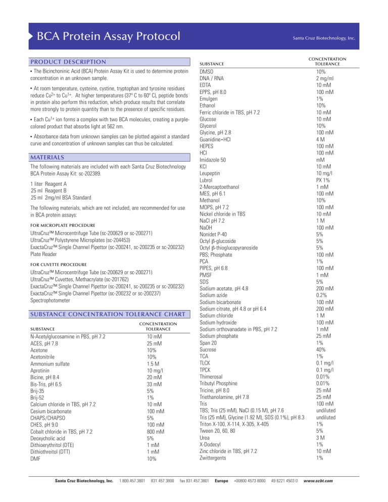
BCA Protein Assay Protocol PRODUCT DESCRIPTION The Bicinchoninic Acid (BCA) Protein Assay Kit is used to determine protein concentration in an unknown sample. ! At room temperature, cysteine, cystine, tryptophan and tyrosine residues reduce Cu2+ to Cu1+. At higher temperatures (37º C to 60º C), peptide bonds in protein also perform this reduction, which produce results that correlate more strongly to protein quantity than to the presence of specific residues. ! Each Cu1+ ion forms a complex with two BCA molecules, creating a purplecolored product that absorbs light at 562 nm. ! Absorbance data from unknown samples can be plotted against a standard curve and concentration of unknown samples can thus be calculated. MATERIALS The following materials are included with each Santa Cruz Biotechnology BCA Protein Assay Kit: sc-202389. 1 liter Reagent A 25 ml Reagent B 25 ml 2mg/ml BSA Standard The following materials, which are not included, are recommended for use in BCA protein assays: FOR MICROPLATE PROCEDURE UltraCruz™ Microcentrifuge Tube (sc-200629 or sc-200271) UltraCruz™ Polystyrene Microplates (sc-204453) ExactaCruz™ Single Channel Pipettor (sc-200241, sc-200235 or sc-200232) Plate Reader FOR CUVETTE PROCEDURE UltraCruz™ Microcentrifuge Tube (sc-200629 or sc-200271) UltraCruz™ Cuvettes, Methacrylate (sc-201762) ExactaCruz™ Single Channel Pipettor (sc-200241, sc-200235 or sc-200232) ExactaCruz™ Single Channel Pipettor (sc-200232 or sc-200237) Spectrophotometer SUBSTANCE CONCENTRATION TOLERANCE CHART CONCENTRATION TOLERANCE N-Acetylglucosamine in PBS, pH 7.2 ACES, pH 7.8 Acetone Acetonitrile Ammonium sulfate Aprotinin Bicine, pH 8.4 Bis-Tris, pH 6.5 Brij-35 Brij-52 Calcium chloride in TBS, pH 7.2 Cesium bicarbonate CHAPS/CHAPSO CHES, pH 9.0 Cobalt chloride in TBS, pH 7.2 Deoxycholic acid Dithioerythritol (DTE) Dithiothreitol (DTT) DMF Santa Cruz Biotechnology, Inc. CONCENTRATION TOLERANCE SUBSTANCE ! SUBSTANCE Santa Cruz Biotechnology, Inc. 10 mM 25 mM 10% 10% 1.5 M 10 mg/l 20 mM 33 mM 5% 1% 10 mM 100 mM 5% 100 mM 800 mM 5% 1 mM 1 mM 10% 1.800.457.3801 831.457.3800 DMSO DNA / RNA EDTA EPPS, pH 8.0 Emulgen Ethanol Ferric chloride in TBS, pH 7.2 Glucose Glycerol Glycine, pH 2.8 Guanidine • HCl HEPES HCl Imidazole 50 KCl Leupeptin Lubrol 2-Mercaptoethanol MES, pH 6.1 Methanol MOPS, pH 7.2 Nickel chloride in TBS NaCl pH 7.2 NaOH Nonidet P-40 Octyl β-glucoside Octyl β-thioglucopyranoside PBS; Phosphate PCA PIPES, pH 6.8 PMSF SDS Sodium acetate, pH 4.8 Sodium azide Sodium bicarbonate Sodium citrate, pH 4.8 or pH 6.4 Sodium chloride Sodium hydroxide Sodium orthovanadate in PBS, pH 7.2 Sodium phosphate Span 20 Sucrose TCA TLCK TPCK Thimerosal Tributyl Phosphine Tricine, pH 8.0 Triethanolamine, pH 7.8 Tris TBS; Tris (25 mM), NaCl (0.15 M), pH 7.6 Tris (25 mM), Glycine (1.92 M), SDS (0.1%), pH 8.3 Triton X-100, X-114, X-305, X-405 Tween 20, 60, 80 Urea X-Dodecyl Zinc chloride in TBS, pH 7.2 Zwittergents fax 831.457.3801 Europe +00800 4573 8000 49 6221 4503 0 10% 2 mg/ml 10 mM 100 mM 1% 10% 10 mM 10 mM 10% 100 mM 4M 100 mM 100 mM mM 10 mM 10 mg/l PX 1% 1 mM 100 mM 10% 100 mM 10 mM 1M 100 mM 5% 5% 5% 100 mM 1% 100 mM 1 mM 5% 200 mM 0.2% 100 mM 200 mM 1M 100 mM 1 mM 25 mM 1% 40% 1% 0.1 mg/l 0.1 mg/l 0.01% 0.01% 25 mM 25 mM 100 mM undiluted undiluted 1% 5% 3M 1% 10 mM 1% www.scbt.com 8 standard dilutions × # replicates = standard wells # unknown × 8 dilutions × # replicates = unknown wells (standard wells + unknown wells) × 0.2 = ml of Reagent AB MICROPLATE PROCEDURE The microplate procedure requires a smaller volume (200 µl) of protein sample, and because the sample to Reagent AB ratio is 1:8; substances in the Substance Concentration Tolerance Chart on the first page of this protocol are not diluted out as much as with the cuvette procedure. Example: 1 standard and 3 unknowns in triplicate: 8 dilutions × 3 = 24 wells of standard 3 unknown × 8 dilutions × 3 = 72 wells of unknown (24 + 72) * 0.2 = 19.2 ml for one full 96 well plate PREPARATION OF BSA STANDARD Use one of the tables below to prepare a set of BSA standards to make a standard curve. ! Select the set of dilutions that most closely corresponds to the expected concentration. ! In this example, at least 19.2 ml is needed: 20 ml Reagent A + 0.4 ml Reagent B ! ! Standard samples should be tested in triplicate and the results averaged. The tables below have volumes sufficient for all eight dilutions in triplicate, which are the standard for one plate. ! ! Stock BSA standard is 2 mg/ml. VIAL µl PBS µl and SOURCE A B C D E F G H 0 50 100 100 100 100 100 100 100 µl stock 150 µl stock 100 µl stock 100 µl Vial B 100 µl Vial C 100 µl Vial E 100 µl Vial F 0 VIAL µl PBS µl AND SOURCE A B C D E F G H 700 100 100 100 100 100 400 100 100 µl stock 400 µl Vial A 300 µl Vial B 100 µl Vial B 100 µl Vial D 100 µl Vial E 100 µl Vial F 0 CONCENTRATION 2,000 µg/ml 1,500 µg/ml 1,000 µg/ml 750 µg/ml 500 µg/ml 250 µg/ml 125 µg/ml 0 µg/ml Reagent AB must be prepared immediately before use. PREPARATION OF MICROPLATE CONCENTRATION Measure absorbance of all wells in a microplate reader at 562 nm. If using a microplate reader equipped with a curve-fitting algorithm, a four-parameter curve is most accurate. INTERFERING SUBSTANCES This table has volumes sufficient for all eight dilutions in triplicate. For each unknown sample CONCENTRATION stock 1/2 1/4 1/8 1/16 1/32 1/64 1/128 PREPARATION OF REAGENT AB Determine how much Reagent AB is required for 200 µl per well. a standard is required on each plate. 1.800.457.3801 ! ! Unknown samples should be tested in triplicate and the results averaged. µl AND SOURCE Incubate plate at 60º C for 15 minutes or 37º C for 30 minutes. If plotting the standard curve by hand, it is recommended to use a pointto-point linear approximation as opposed to an overall linear approximation, as this will produce more accurate results. ! 200 µl unknown sample 100 µl Vial A 100 µl Vial B 100 µl Vial C 100 µl Vial D 100 µl Vial E 100 µl Vial F 100 µl Vial G Add 200 µl of Reagent AB to every well containing a sample. ! ! 0 100 100 100 100 100 100 100 ! Plot a standard curve using the data from the standard sample dilutions and use the curve to determine the concentrations of each unknown. Below is a suggested serial dilution to perform on the unknown sample to get a broad range of concentrations. µl PBS In the next columns, repeat for unknown 2 and 3, if applicable. ! ! A B C D E F G H ! ! Remove plate from incubator and let cool at room temperature for 5 minutes. 250 µg/ml 200 µg/ml 150 µg/ml 100 µg/ml 50 µg/ml 25 µg/ml 5 µg/ml 0 VIAL ! Add 25 µl of unknown 1 into microplate wells, columns 4-6. Vial A goes into row A, Vial B goes into row B, and so on. ! PREPARATION OF UNKNOWN SAMPLES Santa Cruz Biotechnology, Inc. ! READING THE MICROPLATE 5-250 µg/ml NOTE: ! When Reagent A and B are mixed, the solution will turn light green. This is the expected result. ! Add 25 µl of each BSA standard into microplate wells, columns 1-3. Vial A goes into row A, Vial B goes into row B, and so on. 125-2,000 µg/ml ! Mix Reagent A with Reagent B in a 50:1 ratio 831.457.3800 Some substances are known to interfere with the BCA Assay, including those with reducing potential, chelating agents, and strong acids or bases. The following substances are known to interfere at even minute concentrations with accurate estimation of protein concentration: Ascorbic Acid Catecholamines Creatinine EGTA Hydrazides Hydrogen Peroxide Impure Glycerol Impure Sucrose Iron Lipids Melibiose Other Protein Phenol Red Tryptophan Tyrosine Uric Acid fax 831.457.3801 Europe +00800 4573 8000 49 6221 4503 0 www.scbt.com Example: 1 standard and 3 unknowns in triplicate: 8 × 3 = 24 vials 3 unknown × 8 dilutions × 3 = 72 unknown vials (24 + 72) = 96 ml CUVETTE PROCEDURE The cuvette procedure requires a larger volume (400 µl) of protein sample, and because the sample to Reagent AB ratio is 1:20; substances in the Substance Concentration Tolerance Chart on the first page of this protocol are diluted out more than with the microplate procedure. The cuvette procedure is favorable when high concentrations of intolerant substances are present in sample. PREPARATION OF BSA STANDARD Use one of the tables below to prepare a set of BSA standards to make a standard curve. ! Mix Reagent A with Reagent B in a 50:1 ratio In this example, at least 96 ml is needed: 100 ml Reagent A + 2 ml Reagent B ! When Reagent A and B are mixed, the solution will turn light green. This is the expected result. ! Select the set of dilutions that most closely corresponds to the expected concentration. ! ! Standard samples should be tested in triplicate and the results averaged. ! The tables below have volumes sufficient for all eight dilutions in triplicate. ! Stock BSA standard is 2 mg/ml. Reagent AB must be prepared immediately before use. PREPARATION OF CUVETTES ! Add 50 µl of each standard into labeled cuvettes. ! Add 50 µl of unknown into labeled cuvettes. ! Add 1 ml of Reagent AB to every cuvette. READING THE CUVETTES 125-2,000 µg/ml VIAL µl PBS µl and SOURCE A B C D E F G H 0 100 200 200 200 200 200 200 200 µl stock 300 µl stock 200 µl stock 200 µl Vial B 200 µl Vial C 200 µl Vial E 200 µl Vial F 0 VIAL µl PBS µl AND SOURCE A B C D E F G H 700 125 50 200 200 200 200 100 100 µl stock 500 µl Vial A 150 µl Vial B 200 µl Vial B 200 µl Vial D 200 µl Vial E 50 µl Vial F 0 2,000 µg/ml 1,500 µg/ml 1,000 µg/ml 750 µg/ml 500 µg/ml 250 µg/ml 125 µg/ml 0 µg/ml CONCENTRATION 250 µg/ml 200 µg/ml 150 µg/ml 100 µg/ml 50 µg/ml 25 µg/ml 5 µg/ml 0 PREPARATION OF UNKNOWN SAMPLES Below is a suggested serial dilution to perform on the unknown sample to get a broad range of concentrations. ! ! Unknown samples should be tested in triplicate and the results averaged. ! This table has volumes sufficient for all eight dilutions in triplicate. For each unknown sample VIAL µl PBS µl AND SOURCE A B C D E F G H 0 200 200 200 200 200 200 200 400 µl stock 200 µl Vial A 200 µl Vial B 200 µl Vial C 200 µl Vial D 200 µl Vial E 200 µl Vial F 200 µl Vial G ! Incubate cuvettes at 60º C for 15 minutes or 37º C for 30 minutes. ! Remove from incubator and let sit at room temperature for 5 minutes. ! Prepare a blank cuvette of PBS and zero the spectophotometer at 562 nm. CONCENTRATION 5-250 µg/ml CONCENTRATION stock 1/2 1/4 1/8 1/16 1/32 1/64 1/128 PREPARATION OF REAGENT AB ! ! Determine how much Reagent AB is required for 1 ml per cuvette. 8 standard dilutions × # replicates = standard vials # unknown × 8 dilutions × # replicates = unknown vials (standard vials + unknown vials) = ml of Reagent AB ! Measure absorbance of all samples at 562 nm. Samples must be measured within 10 minutes of each other, since BCA will continue to develop at a slow rate while at room temperature. NOTE: For best results, it is recommended that each set of triplicate sample dilutions be read together. For example, measure absorbance of each cuvette from the first set of triplicate dilutions, followed by the second set, and the third. This will minimize inaccuracy due to the BCA reaction continuing at room temperature. Plot a standard curve using the data from the standard sample dilutions and use the curve to determine the concentrations of each unknown. ! If plotting the standard curve by hand, it is recommended to use a pointto-point linear approximation as opposed to an overall linear approximation, as this will produce more accurate results. ! INTERFERING SUBSTANCES Some substances are known to interfere with the BCA Assay, including those with reducing potential, chelating agents, and strong acids or bases. The following substances are known to interfere at even minute concentrations with accurate estimation of protein concentration: Ascorbic Acid Catecholamines Creatinine EGTA Hydrazides Hydrogen Peroxide Impure Glycerol Impure Sucrose Iron Lipids Melibiose Other Protein Phenol Red Tryptophan Tyrosine Uric Acid INTERPRETING THE RESULTS STORAGE AND STABILITY By plotting an unknown sample’s absorbance on the standard curve and relating that absorbance to the standard, the concentration of the solution can be calculated. ! ! ! Store all BCA Protein Assay kit components at room temperature. ! Components remain stable for one year after date of shipment. RESEARCH USE Example standard curve of 2mg/ml BSA: For research use only, not for use in diagnostic procedures. ! Each dilution is averaged and plotted on a graph, and a trendline is added. ! Though the points may have some degree of deviation, they should be generally linear with a slight downward curve. Unknown concentrations are determined by relating their absorbances to the absorbances on the graph. ! Only use the unknown dilutions where the absorbance falls between the bounds of this graph (125-2,000 µg/ml or 5-250 µg/ml, depending on which set of standard dilutions were used). ! ! Plot the absorbance of each unknown dilution on the standard curve. Follow those points down to find the correlating concentration for each unknown dilution. ! Multiply those concentrations by the dilution of the sample to calculate the stock concentration. ! Average the calculated stock concentrations. If one of them is drastically different from the rest, it may be ignored. ! ! For example: This is an unknown concentration of BSA using the standard curve above, with these average BCA assay absorbances and the resulting conclusions: DILUTION stock 1/2 1/4 1/8 1/16 1/32 1/64 1/128 ABSORBANCE max max 2.674 1.567 1.009 0.560 0.322 0.193 CORRELATING CONCENTRATION CALCULATED STOCK CONCENTRATION max max 1.625 0.820 0.460 0.225 0.100 0.045 6.500 6.560 7.360 7.200 6.400 5.760 NOTES RE: THE EXAMPLE Stock and 1/2 dilution samples were too concentrated and no data was available from the plate reader. ! The 1/64 and 1/128 dilution samples have concentrations outside the effective range of this assay and must be discarded. ! Averaging the remaining 4 samples: (6.5 + 6.56 + 7.36 + 7.2) / 4 = 6.9 The stock concentration of unknown BSA is 6.9 mg/ml.
