Appendicular Skeleton Lab Part B: Pelvic Girdle and
advertisement

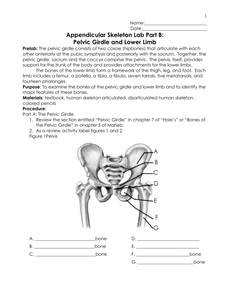
1 Name:____________________________ Date:_____________________________ Appendicular Skeleton Lab Part B: Pelvic Girdle and Lower Limb Prelab: The pelvic girdle consists of two coxae (hipbones) that articulate with each other anteriorly at the pubic symphysis and posteriorly with the sacrum. Together, the pelvic girdle, sacrum and the coccyx comprise the pelvis. The pelvis, itself, provides support for the trunk of the body and provides attachments for the lower limbs. The bones of the lower limb form a framework of the thigh, leg, and foot. Each limb includes a femur, a patella, a tibia, a fibula, seven tarsals, five metatarsals, and fourteen phalanges. Purpose: To examine the bones of the pelvic girdle and lower limb and to identify the major features of these bones. Materials: textbook, human skeleton articulated, disarticulated human skeleton, colored pencils Procedure: Part A: The Pelvic Girdle 1. Review the section entitled “Pelvic Girdle” in chapter 7 of “Hole’s” or “Bones of the Pelvic Girdle” in chapter 5 of Marieb. 2. As a review activity label figures 1 and 2 Figure 1Pelvis A. ___________________________bone D. ____________________________ B. ___________________________bone E. ____________________________ C. ___________________________bone F. ________________________bone G. _________________________bone 2 Figure 2 Split Pelvis A.____________________________ F._______________________________ B.____________________________bone G._______________________________ C.____________________________ H.______________________________bone D.____________________________ I.______________________________Bone E.____________________________bone 3. Examine the bones of the pelvic girdle and locate the following bones and features. acetabulum Ilium Iliac crest Greater sciatic notch Ishium 4. Complete part A of the conclusion. Ischial ramus Ischial spine Pubis Pubic symphysis Obturator foramen 3 Part B: the Lower Limb 1. Review the section entitled “Lower Limb” in chapter 7 of “Hole’s” and “Bones of the Lower Limb” in chapter 5 of Marieb. 2. As a review activity, label figures 3-6. Figure 3 The Femur A.____________________________ D.____________________________ B.____________________________ E.____________________________ C.____________________________ F._______________________________ 4 Figure 4 Lower leg A.____________________________bone E.____________________________ B.____________________________bone F._______________________________ C.____________________________ G._______________________________ D.____________________________ 5 Figure 5 Foot (superior) A. ____________________________ E. ____________________________ B. ____________________________ F. ____________________________ C. ____________________________ G. ____________________________ D. ____________________________ H. ____________________________ 6 Figure 6 Foot (Lateral) A.____________________________ E.____________________________bone B.____________________________ I.______________________________Bone C.____________________________ J.______________________________Bone D.____________________________bone 3. Examine the following bones and features of the lower limb: Femur Tibia Head Anterior crest neck Tibial tuberosity Greater trochanter Medial malleolus Lesser trochanter Tarsal bones Lateral condyle Talus Medial condyle Calcaneus Fibula Metatarsal bones head Phalanges Lateral malleolus Proximal middle distal 4. Use colored pencils to distinguish individual bones in each figure 1-6 Use different colors to represent the regions you are responsible for on the practical. 5. Complete part B of the conclusion 7 Conclusion: Part A: Complete the following statements. 1. The pelvic girdle consists of two ______________________________. 2. The head of the femur articulates with the ______________________________ of the coxae. 3. The __________________________________________ is the largest portion of the coxa. 4. The pubic bones come together anteriorly to form the joint called the _________________________________________. 5. The _______________________________________ is the portion of the ilium that causes the prominence of the hip. 6. When a person sits, the ____________________________________ of the ischium supports the weight of the body. 7. The ___________________________________ is the largest foramen of the skeleton. 8. The ilium joins the sacrum at the __________________________________ joint. Part B: Match the bones in column b with the features in column A. Place the letter of your choice in the space provided. Column A Column B ______1. Lesser trochanter ______2. medial malleolus ______3. Lateral condyle ______4. Calcaneus ______5. 7 bones of the ankle ______6. Talus ______7. lateral malleolus ______ 8. 5 bones that form the in step ______9. The big toe has only 2 ______10. Anterior crest ______11. neck ______12. Head (could be 2 bones) A. femur B. fibula C. metatarsals D. tarsals E. Tibia F. phalanges


