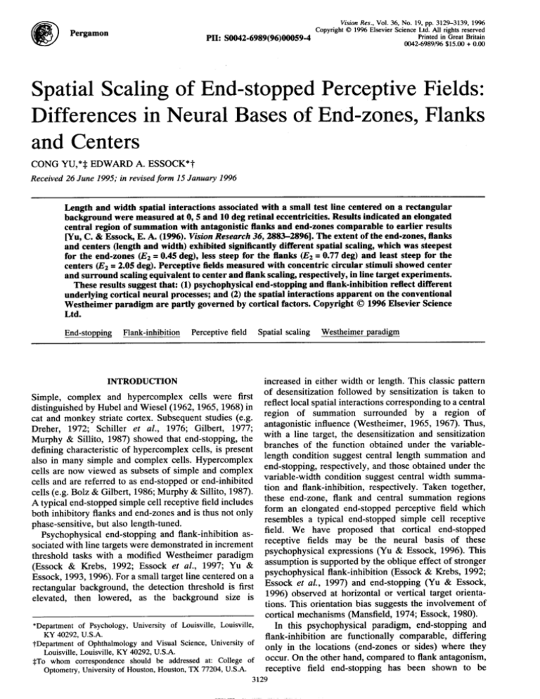
Pergamon
@
Vision Res., Vol. 36, No. 19, pp. 3129-3139, 1996
Copyright 01996 Elsevier Science Ltd. AU rights reserved
Printed in Great Britain
PII:S0042-6989(96)00059-4
0042-6989/96 $15.00 + 0.00
Spatial Scaling of End-stopped Perceptive Fields:
Differences in Neural Bases of End-zones, Flanks
and Centers
CONGYU,*$ EDWARD A. ESSOCK*T
Received 26 June 1995; in revisedform 15 January1996
Lengthand width spatialinteractionsassociatedwith a smalltest line centeredon a rectangular
backgroundweremeasuredat 0,5 and 10 deg retinaleccentricities.Resultsindicatedan elongated
centralregionof summationwith antagonisticflanksand end-zonescomparableto earlierresults
~u, C. & EssoclGE. A. (1996).VisionResearch36,2883-28961.
The extentof the end-zones,flanks
and centers(lengthand width)exhibitedsignificantlydifferentspatialscaling,whichwas steepest
for the end-zones(E2= 0.45deg), less steep for the flanks(E2= 0.77 deg) and least steep for the
centers(E2= 2.05 deg). Perceptivefieldsmeasuredwith concentriccircularstimulishowedcenter
andsurroundscalingequivalentto centerandflankscaling,respectively,in linetargetexperiments.
Theseresultssuggestthat: (1) psychophysicalend-stoppingandflank-inhibitionreflectdifferent
underlyingcorticalneuralprocesses;and (2) the spatialinteractionsapparenton the conventional
Westheimerparadigmare partlygovernedby corticalfactors.Copyright01996 ElsevierScience
Ltd.
End-stopping Flank-inhibition Perceptivefield Spatialscaling Westheimerparadigm
INTRODUCTION
increased in either width or length. This classic pattern
Simple, complex and hypercomplex cells were first of desensitization followed by sensitization is taken to
distinguishedby Hubel and Wiesel (1962, 1965, 1968)in reflectlocal spatialinteractionscorrespondingto a central
cat and monkey striate cortex. Subsequent studies (e.g. region of summation surrounded by a region of
Dreher, 1972; Schiller et al., 1976; Gilbert, 1977; antagonistic influence (Westheimer, 1965, 1967). Thus,
Murphy & Sillito, 1987) showed that end-stopping,the with a line target, the desensitization and sensitization
defining characteristic of hypercomplex cells, is present branches of the function obtained under the variablealso in many simple and complex cells. Hypercomplex length condition suggest central length summation and
cells are now viewed as subsets of simple and complex end-stopping,respectively,and those obtained under the
cells and are referred to as end-stopped or end-inhibited variable-width condition suggest central width summacells (e.g. Bolz & Gilbert, 1986;Murphy& Sillito, 1987). tion and flank-inhibition,respectively. Taken together,
Atypical end-stoppedsimple cell receptivefield includes these end-zone, flank and central summation regions
both inhibitoryflanksand end-zones and is thus not only form an elongated end-stopped perceptive field which
resembles a typical end-stopped simple cell receptive
phase-sensitive,but also length-tuned.
field.
We have proposed that cortical end-stopped
Psychophysical end-stopping and flank-inhibitionasreceptive
fields may be the neural basis of these
sociatedwith line targetswere demonstratedin increment
psychophysicalexpressions
(Yu & Essock, 1996). This
threshold tasks with a modified Westheimer paradigm
assumptionis
supportedby
the
oblique effect of stronger
(Essock & Krebs, 1992; Essock et al., 1997; Yu &
psychophysicalflank-inhibition(Essock
& Krebs, 1992;
Essock, 1993, 1996).For a small target line centered on a
Essock
et
al.,
1997)
and
end-stopping
(Yu & Essock,
rectangular background, the detection threshold is first
1996)
observed
at
horizontal
or
vertical
target orientaelevated, then lowered, as the background size is
tions. This orientation bias suggests the involvement of
cortical mechanisms(Mansfield,1974; Essock, 1980).
*Department
of Psychology,
Universityof Louisville,
Louisville, In this psychophysical paradigm, end-stopping and
KY40292,U.S.A.
flank-inhibition are functionally comparable, differing
~Department
of Ophthalmology
andVisualScience,University
of
only
in the locations (end-zones or sides) where they
Louisville,
Louisville,
KY40292,U.S.A.
occur.
On the other hand, compared to flank antagonism,
*To whomcorrespondence
shouldbe addressedat: Collegeof
Optometry,
University
ofHouston,
Houston,
TX77204,U.S.A. receptive field end-stopping has been shown to be
3129
3130
C.YUandE.A.ESSOCK
generated by distinct neural circuits, such as intracortical
inhibitionfrom cells with spatially offset receptive fields
(e.g. Hubel & Wiesel, 1965;Bolz & Gilbert, 1986).Bolz
and Gilbert (1986) demonstrated the disassociation of
end-zone and flank-inhibition by pharmacologically
abolishing end-inhibitionwhile preserving flank properties. Accordingly, if psychophysical end-stopping and
flank-inhibition are truly the behavioral expressions of
receptive field properties, they would have different
underlying neural mechanisms and therefore might
exhibit distinct features under appropriate psychophysical test circumstances. Thus, the psychophysical disassociation of end-stopping from flank-inhibition,as well
as from central summation, would be an important
criterion to evaluate the validity of our assumption.
Measuring the scale change of the extent of a spatial
property across various retinal eccentricitiescan provide
information about whether the processing is limited by
retinal or cortical factors (Levi et al., 1985;Wilson et al.,
1990; Drasdo, 1991). This spatial scaling is often
characterized by the value E2 defined by F = 1 + E/E2,
where F is the scaling factor indicating how a spatial
property or performance varies, E is the retinal
eccentricity, and E2 is the eccentricity at which the
measured value is equal to twice the foveal value. Levi et
al. (1985) and Wilson et al. (1990) suggested that the
spatial scaling across eccentricity of a variety of visual
tasks falls into two categories. Spatial scaling functions
for tasks such as hyperacuityand spatial interactionhave
an E2 value in the range 0.3-0.9 deg, which matches the
E2 values of cortical magnificationin human (Cowey &
Rolls, 1974) and monkey (Dow et al., 1981). It is
assumed that spatial abilities having E2 values comparable to that for cortical magnification (c. 0.8 deg) are
limited by cortical factors (e.g. Wilson et al., 1990). On
the other hand, spatial scaling functionsfor tasks such as
resolutionacuity and contrastsensitivityhave an E2 value
in the range 1.5-4 deg, which matches the E2 values of
cone and retinalganglioncell spacing(c. 2.5 deg) and are
presumed to be limited by retinal factors (Perry &
Cowey, 1985).These values also match the E2 values of
cortical receptivefield center size (Dow et al., 1981;Van
Essen et al., 1984), but the similar scaling of cortical
receptive field center size and cone and retinal ganglion
cell spacing suggeststhat cortical receptivefieldsreceive
their retinal input from a fixed number of neighboring
cones and ganglion cells at any retinal eccentricity
(Wilson et al., 1990) and thus their spatial scaling is
ultimately determined by retinal factors. Relatively
shallow spatial functions, with E2 values in the range
1.54 deg, are often regarded as indicating performance
being limited by retinal factors, and steep scaling
functions, with E2 values in the range 0.3-0.9 deg, as
reflecting a cortical limitation. For example, Toet and
Levi (1992) reported that theE2 values for resolutionof a
T-shaped target and for spatial interactionsbetween two
such targets were approximately2 deg and 0.2-0.4 deg,
respectively. The dramatic scaling difference was
attributed to retinal factors limiting the resolution of
these targets,and cortical factors limiting their spatial
interaction. Thus, spatial“scalingcan provide a way to
psychophysicallydetermine differences in neural limitations (i.e. retinal or cortical) of various visual processes.
Furthermore,although there is no solid psychophysical
evidence indicating that different cortical,.mechanisms
must necessarilylead to significantdifferencesof spatial
scaling among tasks they support, Drasdo (1991)
suggestedthat cortical magnificationin different cortical
areas, and cortical sampling by modular structures
unevenly distributed within these areas, theoretically
could create such differences.
In the present study,we extendedour earlier studieson
end-stopped perceptive fields (Yu ,& Essock, 1996) to
measure their spatial scaling across retinal eccentricity.
We anticipated that it would be possible to determine
whether the spatial interactions of the end-stopped
perceptive fields were ‘limited by retinal or cortical
factors, and also to differentiate spatial scaling among
end-stopping, flank-inhibition and central summation.
We measured spatial interactionsin both the length and
width dimensionsat O,5 and 10 deg retinal eccentricities
and determined the spatial scaling factors and E2 values
of the end-zone, flank and central summation regions of
the perceptive field. Our main purpose was to determine
whether the end-stopping and flank-inhibition demonstrated psychophysicallyappear to have different neural
bases, and thus support the assumptionthat they are the
psychophysicalcorrelates of cortical receptive field endstopping and flank-inhibition. A second goal was to
compare the mechanism underlying central summation
and those underlying psychophysical end-stopping and
flank-inhibition. As a control, we also measured the
spatial scaling of the center/surround organization of
circular perceptive fields associated with spot targets
(Westheimer, 1965, 1967). By using both line and spot
targets,we were able to compare the nature of the spatial
interactions obtained with line targets (Yu & Essock,
1996) to those obtained in the original spot-target
version. Brief reports of results in this paper were
presented earlier (Yu et al., 1995).
GENERALMETHODS
Observers
The same two subjects(one male and one female, both
30 yr old) served in all experiments.Both subjectswere
slightly myopic and wore appropriate lenses to correct
their vision to 20/20 or better. Subject YC (one of the
authors)was experiencedin psychophysicalobservation.
Subject HY had no prior psychophysicalexperience and
was naive as to the purpose of the study. She was given
considerable practice before the experiments formally
started.
Apparatus and stimuli
The stimuli were generated by a Vision Works
computer graphics system (Vision Research Graphics,
Inc.) and presented on a Nanao Flexscan 9080i color
SPATIAL
SCALING
OFEND-STOPPED
PERCEPTIVE
FIELDS
monitor. The resolution of the monitor was 1024x 512
pixels. Pixel size was 0.28 mm horizontalx 0.41 mm
vertical. The frame rate was 117 Hz. Luminance of the
monitorwas made linearby means of an eight-bitlook-up
table (LUT). Viewing distance was varied for testing at
the three retinal eccentricities to fit both fixation cross
and stimuli on the screen, yet maximize the resolutionof
stimuli. Subjectswere positionedby means of a chin rest
at 5.64 m from the screen for foveal viewing, half of the
foveal viewing distance (2.82 m) for 5 deg retinal
eccentricity viewing and a quarter of the foveal distance
(1.41 m) for 10 deg retinal eccentricityviewing. Viewing
was monocular by the dominant eye (right eyes for both
subjects) with a white translucent diffuser positioned
before the other eye.
An increment test field and a background field were
presented on the center of the monitor screen for foveal
viewing or at the 5 deg and 10 deg retinal eccentricities
on the temporal side of the horizontal meridian in the
visual field for peripheral viewing. The test field was a
target line centered on a rectangular background. In a
given experiment, only one dimension (e.g. length or
width) of the background field was varied and the other
dimension was fixed. The sides of the rectangular
background were parallel to the sides of the target line
in all experiments. The test line and background were
orientedvertically,except as notedbelow. The luminance
of the monitor screen was constant(6.85 cd/m2)throughout all experiments, as was the luminance of the
rectangular background (30 cd/m2). The luminance of
the target line was varied by a staircase procedure as the
dependent measure. Additional details are given in
correspondingsections.
Procedure
A successive two-alternative forced-choice (2AFC)
procedure was used. The background was presented in
each of the two intervals(1.1 sec each). In one of the two
intervals, the target line was also presented, starting
420 msec after the onset of the background, lasting for
420 msec, and disappearing 260 msec before the background offset. There was no interruption between two
intervals. In foveal viewing each trial was preceded by a
fixation cross which disappeared 100 msec before the
beginningof the trial. For peripheralviewing,the fixation
cross was present throughout testing. Intervals were
marked by toneswith differentfrequencies.Another tone
gave feedback on incorrect responses.
Each staircase consisted of four “practice” reversals
and six experimental reversals. Each correct response
lowered test field luminance by one step and each
incorrect response raised test luminance by three steps.
Step size was 3.6 cd/m2 at the first pair of Practice
reversals and 1.8 cd/m2 at the second pair. It was
().6cd/m2 throughout the experimentalphase. The mean
of six experimental reversals was used to estimate the
increment threshold which was defined as the difference
of target luminance at threshold and background
luminance on a log scale [log (AL+ L)–log JZ].
3131
Besidesthe practice at the beginningof the study,each
observeralso had two to three sessionsof practice before
each peripheral experiment. One experimental session
usually consisted of 9–13 background conditions presented in a random order and lasted for 50 to 60 min.
Each data point was the mean of the thresholdsfrom five
to six replication sessions, and the error bars represent
t 1 SEM.
EXPERIMENT
1: MEASUREMENT
OF LOCAL
SCALINGFACTORS
Sincevisual spatialsensitivitydeclineswith increasing
retinal eccentricity due to reduced neural sampling (e.g.
Rovamo & Virsu, 1979), it was desirable to equate the
visibility of peripheral and foveal targets before we
compared the perceptive fields at different retinal
eccentricities. It has been shown also that spatial
processing can be homogeneous across the visual field
if the stimuli are appropriately scaled (Rovamo et al.,
1978; Koenderinket al., 1978).Although estimating the
scalingfactor from cone or ganglioncell spacingwas first
thought to also reflect cortical magnification(Rovamo et
al., 1978),later studies suggestedthat retinal and cortical
scales are quite different(Levi et al., 1985;Wilson et al.,
1990; see Introduction).Therefore, it is inappropriateto
scale the peripheral stimuli either with the cone or
ganglioncell spacing data, or with cortical magnification
factors, before we know whether processingis limited by
retinal factors or by cortical factors. Alternatively,
Johnston (1987) and Watson (1987) suggested that any
particular aspect of visual processing can be equated at
any two visual field locationsby magnifyingthe stimulus
with a (local) scaling factor. This local scaling factor
(Watson, 1987) can be estimated by measuring the
sensitivityto a stimuluswhich has an identical form and
is varied only in its size. The estimationis independentof
any prior estimatesof cortical or retinal magnification,as
well as any presumptionof the neural basis of the visual
processing.
In this experiment, we applied the concept of local
scale and measured local scaling factors for line and spot
targets. These scaling factors were used later in the
following experiments to magnify peripheral stimuli to
equate their visibility. In this experiment, the detection
thresholds for a foveal 1 x 5’ line and a 1’diameter spot
(withoutthe backgroundfieldpresent)were measured,as
was a series of their magnifiedforms at the 5 and 10 deg
retinal eccentricities.The width and length of the foveal
line and the diameter of the foveal spot were magnified
by factorsof 1.33,2.00,2.66,3.33 and 4.00, respectively,
at the 5 deg retinaleccentricity,and 2.00, 2.66,3.33,4.00
and 4.66, respectively,at the 10 deg retinal eccentricity.
The luminance of the screen was 30 cd/m2,the same as
the background luminance in later experiments.
The peripheral data were fitted with an exponential
equation, T = ak#’, where T refers to threshold, M to
magnificationfactor, and a and b are free parameters.The
magnification factors that produced thresholds which
matched the foveal thresholds were taken as the local
3132
C.
() a
YU and E. A. ESSOCK
0.36
0.36-
HY
0.32 0
d 0.28 I
~ 0.24
K
~ 020 .
,&
~
~ 0.16
w
2 0.12
u
K
(J 0.08
z
z—
- 0.04l“”””””””””””””
0.00
1
0
:...k..............k
0.040,00
2
3
4
5
6
I
0
24.
3
4
5
,
6
MAGNIFICATIONFACTOR
MAGNIFICATIONFACTOR
(b)
2
Yc
HY
21
$j18
-
!315 .
L
Q12 z
i
z
W
9 6 3 e’-’-0
o
- +.----—--5
ECCENTRICITY(deg)
:
10
‘;L
0
5
ECCENTRICITY(deg)
10
FIGURE1. Local scaling factors used to magnify the sizes of peripheral stimuli for equal retinal sampling across retinal
eccentricity,
(a) An example of data fitting and local scaling factor derivation. Data were measured at the 10 deg retinal
eccentricity for’the line target. The raw dat;(filled circles) are-firstfitted by an exponentialequation(see text). The fi;ed data
(solid curve) are then matched with foveal threshold (indicated by the dotted horizontal line). The x value of the intersection
point of the dottedhorizontalline (foveal threshold)and solid curve (fitteddata) is taken as the local scaling factor (indicatedby
the dotted vertical line). (b) Local scaling factors [as obtained in (a)] plotted as a function of the retinal eccentricity. Leastsquares regression lines are plotted for line-only ( x ) and spot-only( + ) targets.
scaling factors. Examples of this procedure are shown in
Fig. l(a). The E2 values in this and later experiments
were calculated from the equation F = 1 + E/E2 given
earlier. As seen from this function, Ez is actually the
inverse of the slope of the eccentricityfunction, and thus
is independentof any specificeccentricity.Local scaling
factors plotted as a function of retinal eccentricity are
shown in Fig. l(b). For subject HY, the local scaling
factor for the line target is 2.11 (E2 = 4.51 deg) at the
5 deg retinal eccentricity and 3.52 (E2 = 3.97 deg) at the
10 deg retinal eccentricity,and for the spot target is 2.02
(E2 = 4.90 deg) at the 5 deg retinal eccentricity and 3.06
(E2 = 4.85 deg) at the 10 deg retinal eccentricity. For
subject YC, the local scaling factor for the line target is
2.06 (E2 = 4.72 deg) at the 5 deg retinal eccentricity and
3.06 (E2 = 4.85 deg) at the 10 deg retinal eccentricity,
and for the spot target is 2.32 (E2 = 3.79 deg) at the 5 deg
retinal eccentricityand 2.82 (E2 = 5.49 deg) at the 10 deg
retinal eccentricity.As Fig. l(b) indicates,each subject’s
spatial scaling functions for line and spot targets are
linear and essentially identical. The E2 values from the
two subjects fall into a range 3.79–5.49deg, with an
overall mean value of 4.64 deg (the overall slope of the
psychometric functions is about 0.22). These E2 values
are about equal to Watson’s (1987) estimation of local
spatial scale in a contrast sensitivity function measurement using a similar procedure (E2 = 4.17 deg, recalculated from Watson, 1987).
EXPERIMENT
2: LENGTHSUMMATION
AND
END-STOPPING
ACROSSRETINALECCENTRICITY
Length summationand end-stoppingwere measured first
at the Odeg retinal eccentricity for a 1 x 5’ line
superimposed on a 3’-wide rectangular background of
various lengths. This was a replication of an earlier
experiment (Experiment 2, Yu & Essock, 1996) and
served as the baseline for later 5 and 10 deg retinal
eccentricity length experiments. Because data collected
from seven subjectsin the earlier measurementhad been
very consistent,only five-six critical background length
conditions were selected. Increment threshold as a
fimction of background length is shown in Fig. 2(a).
The length of central summationregion (i.e. background
length at which the peak threshold occurs) is about 11’
3133
SPATIALSCALINGOF END-STOPPEDPERCEPTIVEFIELDS
()
a
0.30
0.30-
HY
0.27[
0.27 n
~
0.24
:
u
:
0.21
+
+
3
0.15 -
Yc
n
J 0.24
0
r
(J 0.21
w
: 0,18
0,18
+
k~
z
w
IY
;
—
0,12
L
a 0.09
E
~ oo5 .
.
0.03 ,,,() ~
O
3
6
9 12 15 18 21 24 27 30 33 36 39
0.03 -
L
0,00 ~
O 3
6
0.15
(),, 2
0.09
o,06
9 12 15 18 21 24 27 30 33 36 39
0.600.54 n
~
n
~
0.48
0.48
~ 0.42
w
: 0.36
+
tfi
>
w
K
>
—
!:
;w.
s
w 0.18
w
: 0,12
—
0.06 0.00
0.24
0.18
0,1 ‘2
o,oo~
0.06 -
J
0,
0.30 -
20
40
60
0
80 100120140160180200
20
40
60
80 100120140160180200
k
‘:j—____—
0
40
80
120
160
200
240
280
~ 0.28
2
w 0.21
K
S 0,14
—
0.07 0.00
320
o
24-
24 -
21
21
160
200
240
280
!
320
&18
~15
$
:12
i
-
-----
--- 0 center
“’6
.-.
0
()
5
ECCENTRICITY (deg)
----
0 center
3
3 0
120
end–zone
~18 +
!/15
L
:12
i
‘6
80
BACKGROUND LENGTH (rein arc)
BACKGROUND LENGTH (rein arc)
(d)
40
10
0
5
10
ECCENTRICITY (deg)
FIGURE 2. (a)--(c)Increment threshold plotted as a function of the length of the backgroundfield at O,5 and 10 deg retinal
eccentricities. The rising portion of the function is taken to reflect length summation, and the declining portion reflects endstopping.Note the scale ofx andy abscissas are different amongfigures(also in Figs 3 and4). (d) Spatialscalingfactors (ratio of
peripheral data: foveal data) for the lengthsof the center region and end-zoneregionof the perceptivefield plotted as a function
of the retinal eccentricity.
3134
C. YU and E. A. ESSOCK
0.30-
0.30 -
HY
0.27
0,27 0.24 -
0.24
0.21
0.18
0.15
0.12
0.09
:
#yX%__
0.09
0.06
0.06 -
p%
0.03
0.03 0.00
O
0.70
0.63
3
6
9
12
15
18
’21
t
24
0
3
6
9
12
15
18
0.70-
E = 50
HY
0.00
,
24
21
E = 5.
0.63 -
[
:
:+$
0.28
‘
0.21
0.14 -
0.14
0.07
0.07 I
0.00
I
0
15
30
45
60
75
0.00
90 105120135150
,
0
15
30
45
60
75
90 105120135150
0.70 -
0.70
0.63
,
E = 1 0“
HY
[
0.63
:
i<
0.28
0.21
0.14
1
:;:~
ii___
o
20
40
60
80
o
100 120 140 160 180
BACKGROUND WIDTH(rein arc)
24
24
20
40
60
80
100 120 140 160 180
BACKGROUND WIDTH(rein arc)
Yc
HY
21
21 [
L
g18
515
flank
-
L
flank
g12
i
39
i
“’6
-,
.z ---
---
0
center
●
---0
‘6
3
3
0
0
0
5
ECCENTRICITY(deg)
center
10
o
5
10
ECCENTRICHY(deg)
FIGURE3. Spatial interactionfunctionsand spatial scaling functionsfor a backgroundof variable width plotted as in Fig. 2 for
variable-lengthbackground.(a)-(c) Width summationand flank-inhibitionat O,5 and 10 deg retinal eccentricities. (d) Spatial
scaling factors for the width of the flank region and center region of tbe perceptive field plotted as a function of the retinal
eccentricity.
SPATIALSCALINGOF END-STOPPEDPERCEPTIVEFIELDS
long, and the length of the end-stopping region (half of
the peak-to-plateau distance in terms of background
length)* is about 4.5’ long, respectively, for the two
subjects. These data are comparable to those reported in
earlier measurements(Yu & Essock, 1996).
This test was then performed at the 5 and 10 deg retinal
eccentricities. For each subject, the width and length of
the target line and the width of the rectangular background were magnified by hiw’hercorresponding local
scaling factors of the line target determined in Experiment 1. Therefore, the stimulusconfigurationat the 5 deg
eccentricity was a 2.11 x 10.55’line centered on a 6.33’
wide backgroundfor HY and a 2.06 x 10.3’line centered
on a 6.18’wide backgroundfor YC. At the 10 deg retinal
eccentricity, it was a 3.52 x 17.60’line on a 10.56’wide
background for HY and a 3.06x 15.30’line on a 9.18’
wide background for YC. Data collected at the 5 deg
retinal eccentricity are plotted in Fig. 2(b). The length of
the central summation region is 32’ (F= 2.91, Ez = 2.62
deg) for HY and 40’ (F= 3.64, E2 = 1.90 deg) for YC
(where F is the ratio of peripheral data to fovea] data and
E2 is calculated from F based on the equation F = 1 + E/
E2). The length of the end-stopping region is 59’
(F= 13.11,E,= 0.41 deg) for HY and 55’ (F= 12.22,
E2 = 0.45 deg) for YC. Data collected at the 10 deg
retinal eccentricity are plotted in Fig. 2(c). The length of
the central summation region is 61’ (F= 5.55,
Ez = 2.20deg) for HY and 63’ (F= 5.73, E2 = 2.12 deg)
for YC. The length of the end-stopping region is 100’
(F= 22.11, E2 = 0.47 deg) for HY and 102’ (F= 22.67,
E2 = 0.46 deg) for YC.
Figure 2(d) plots the scaling factor as a function of
retinal eccentricity. Both subjects’ data show the same
trend. Spatial scaling factors for the end-zone and center
both increase linearly with retinal eccentricity, but the
increase in scaling for the end-zone size is much steeper
than that for the center region. The average E2 value is
about 0.45 deg for the end-zone (slope = 2.23) and
2.21 deg for the center (slope= 0.45). This scaling
difference suggests that end-stopping and central summation may depend on different neural mechanisms.
EXPERIMENT
3: WIDTHSUMMATION
AND
FLANK-INHIBITION
ACROSSRETINAL
ECCENTRICITY
The extent of width summationand flank-inhibitionwere
first measured at the Odeg retinal eccentricityfor a 1 x 5’
line superimposed on a 6’-long rectangular background
with various widths. This was also a replication of an
earlier experiment (Experiment 1, Yu & Essock, 1996)
and set the baseline for later periphery experiments.
Results are shown in Fig. 3(a). The widths of the central
summation region (background width at which the peak
threshold occurs) and the flank-inhibitionregion (half of
the peak-to-plateau distance in terms of background
*The peak-to-plateaudistance is halved to provide the length of each
end-zone on the assumptionof symmetrical end-zones.
3135
width) are about 6’ and 3’ for HY, and 6’ and 4’ for YC,
respectively.
The same conditions were then tested at the 5 and
10 deg retinal eccentricities.The width and length of the
target line and the length of the rectangular background
were also magnified by each subjects’ local scaling
factors of line target. The line sizes were the same as in
Experiment2. The backgroundlength at the 5 deg retinal
eccentricitywas 12.66’for HY and 12.36’for YC. At the
10 deg retinal eccentricityit was 21.12’for HY and 18.36
for YC. However,both the target line and the background
were set to horizontalin this measurement.The width of
the background was thus varied vertically so that the
retinal eccentricity would remain fairly constant, particularly when the background was very wide. Previous
data (Yu & Essock, 1996)demonstratedthat results from
horizontal and vertical conditionsdo not differ.
Data collected at the 5 deg retinal eccentricity are
plotted in Fig. 3(b). The width of the central summation
region is 18’ (F= 3.00, E2 = 2.50 deg) for HY and 24’
(F= 4.00,E2 = 1.67 deg) for YC. The width of the flankinhibitionregion is 28’ (F= 9.33, E2 = 0.60 deg) for HY
and 33’ (F = 8.25, E2 = 0.69 deg) for YC. Data collected
at the 10 deg retinal eccentricity are plotted in Fig. 3(c).
The width of the central summation region is 37’
(F= 6.17, E2 = 1.94 deg) for HY and 38’ (F= 6.33,
E2 = 1.88deg) for YC. The width of the flank-inhibition
region is 43’ (F= 14.17,E2 = 0.76 deg) for HY and 44’
(F= 10.88,E2 = 1.01 deg) for YC.
Figure 3(d) plots the scaling factor as a function of the
retinal eccentricity.Similar to the length experimentdata
(Experiment2), a linearspatialscalingcan also be seen in
the flank-widthand center-widthfunctions,althoughthis
relationfor the flankfunctionis less clear than that for the
other data. The average E2 value is about 0.77 deg for
flanks (slope = 1.31) and 2.00 deg for centers
(slope = 0.50). That the flank function is much steeper
than the center function suggests that flank antagonism
and central summation may also depend on different
neural mechanisms. The average E2 value for the
summation center is 2.21 deg (Experiment 2) in the
length dimension and 2.00 deg in the width dimension
(current experiment), indicating that the summation
center is homogeneous across both dimensions with
respect to scaling.
EXPERIMENT
4: CENTEWSURROUND
SPATIAL
INTERACTION
FORA SPOTTARGETACROSS
RETINALECCENTRICITY
Westheimer (1965, 1967) noted that the spatial interactions associated with a small spot target centered on a
circular background appear to reflect center/surround
organizationcomparableto that of a retinal ganglioncell
receptive field. Numerous studies performed with both
human and animal subjects using a variety of behavioral
methods (e.g. Westheimer, 1965, 1967; Enoch & Sunga,
1969; Spillmann et al., 1987) as well as single-unit
recordings of retinal ganglion cells (Essock et al.,
unpublished data) all support this assumption of a
3136
C. YU and E. A. ESSOCK
0.60
0.60
HY
0.54 [
0.54
Yc
[
0.48
0.42
0.36
0.30
0.24
0.18
:%
0.12
0.06 I
0.00
o
3
6
9
I2
I5
I8
21
iL--0
24
3
6
9
12
15
18
21
24
15
30
45
60
75
90
105
,
120
E = 50
HY
0.4 :$
0.3 0.2 0.1
,,o~
o
0.0
15
1.20
30
45
60
75
105
120
0
Yc
E = 100
HY
1.08
90
E = 100
0.96
0.84 I -
n
iL
0
15
30
45
60
75
90 105 120 135 150
0,00 ~
0
15
30
45
60
75
90 105120135150
BACKGROUND DIAMETER(rein arc)
BACKGROUND DIAMETER(rein arc)
24-
Yc
21
~18
k
015
~
012
z
i
:9
:12
i
759
“’6
----
-0
“’6
center
3 -
3
0
center
surround
n
0
5
ECCENTRICITY(deg]
10
“o
5
ECCENTRICITY(deg)
10
FIGURE4. Spatial interaction functionsand spatial scaling functionsobtained for a circular spot target centered on a circular
backgroundof variable diameter. (a)--(c)Center/surroundspatial interactionsat 0,5 and 10deg retinal eccentricities. (d) Spatial
scaling factors for the sizes of center and surroundregionof the perceptivefield plotted as a functionof the retinal eccentricity.
SPATIALSCALINGOF END-STOPPEDPERCEPTIVEFIELDS
relation of psychophysicalspatial interactionsand retinal
ganglion cell receptive field properties. Several experiments measured the spatial interactions at different
retinal eccentricities (Westheimer, 1967; Enoch, 1978;
Spillmann et al., 1987).Westheimer(1967)measured the
spatial scaling of only the center of the perceptive field
and found it to be about the same as the spatial scaling of
resolution acuity. However, in his measurementthe spot
target was not magnified to equate its effective size at
each retinal eccentricity, which may have resulted in
retinal under-sampling and made the data less accurate.
Spillmann et al. (1987) reported the spatial scaling of
perceptivefieldsin both human and monkey. They found
that sizes of the center and surround both increase with
retinal eccentricity, and that the slope of the surround
function is steeper,
In this experiment, we first measured central summation and surround antagonism at the Odeg retinal
eccentricity for a If-diameter spot centered on a circular
background.Resultsare shown in Fig. 4(a). The diameter
of the summation center (backgrounddiameter at which
peak threshold occurs) and inhibitory surround on each
side (half of the peak-to-plateau distance in terms of
backgrounddiameter)are about 6 and 3’,respectively,for
HY, and 6 and 4’, respectively, for YC.* The same
functionswere then measured at the 5 and 10 deg retinal
eccentricities. For each subject, the diameter of the spot
target was magnifiedby his/her local scaling factor of the
spot target (Experiment 1). This factor was 2.02’for HY
and 2.32’for YC at the 5 deg retinal eccentricityand 3.06’
for HY and 2.82’for YC at the 10 deg retinal eccentricity.
Data collected at the 5 deg retinal eccentricityare plotted
in Fig. 4(b). The size of the central summation region is
17’in diameter (F = 2.90, E2 = 2.63 deg) for HY and 23’
(F= 3.87, E2 = 1.74 deg) for YC. The size of the
surround-inhibition region is 26’ (F = 8.70, E2 = 0.65
deg) for HY and 23’ (F= 5.80, E2 = 1.04 deg) for YC.
Data collected at the 10 deg retinal eccentricity are
plotted in Fig. 4(c). The size of the central summation
region is 28’in diameter(F= 4.67, Ez = 2.73 deg) for HY
and 32’(F= 5.33, E2 = 2.31 deg) for YC. The size of the
surround-inhibition region is 43’ (F= 14.17, E2 = 0.76
deg) for HY and 42’ (F= 10.50,E2 = 1.05 deg) for YC.
Figure 4(d) plots the scaling factors as a function of
retinal eccentricity. It shows that the spatial scaling
factors for the surround and the center both increase
linearly with retinal eccentricity, and that the surround
function is steeper than the center function. The average
E2 value is 2.35 deg for center functions (slope= 0.43)
and 0.88 deg for surround functions (slope = 1.14). The
general trend of spatial scaling is comparable to
Spillmann and colleagues’ human and monkey data,
which also showed steeper scaling in the surround
function.
3137
5
0
10
ECCENTRICITY (deg)
24
21
~lg
Yc
Z
[
/
-
L
g12
i
<9
8
end-zone
,
flank
6-
o
5
10
ECCENTRICITY (deg)
FIGURE5. Summaryof spatial scaling functions in each experiment
replotted from earlier figures [Figs l(b), 2(d), 3(d) and 4(d)]. The
scaling functions fall into four groups: (1) end-zone scaling (filled
circles); (2) antagonisticflank regions for a line target (filled squares)
and antagonisticsurroundfor a spot target (tilled diamonds);(3) width
(triangles) and length (circles) of center region for a line target and
diameter (diamonds) of center region for a spot target; and (4) local
scaling factors for line ( x ) and spot ( + ) targets.
GENERALDISCUSSION
In this study, the spatial scaling of spatial interactions
was measuredfor elongatedand circularperceptivefields
across retinal eccentricity. Scaling for components of
elongated perceptive fields (center width, center length,
flank width and end-zone length) and components of
circularly symmetric perceptive fields (center and
surroundsizes) were measured. When the spatial scaling
functions in each experiment [Fig. l(b), 2(d), 3(d) and
4(d)] are plotted together (Fig. 5), four categories of
spatial scaling can be seen. The spatial scaling of endzones is the steepest and standsout from the others. Next
steepest is the spatial scaling of flanks (line target) and
*To be consistent with the values reported for rectilinear stimuli in
Experiments 2 and 3, these values are reported as the full width surrounds (spot target), which are very similar to each
(diameter) of the center and the extent of the surroundon one side other and form a second category.The spatial scaling for
(i.e. the “thickness” of an annulus).
center regions is the next steepest and forms a third
3138
C. YU and E. A. ESSOCK
category, with equivalentscaling for length and width of
elongated centers, and for diameter of circular centers.
Center scaling is close to, but consistently steeper than,
local scaling functionsfor increment threshold of line or
spot stimuli (i.e. targets with no background present).
These line and spot local scalingfunctionsare identicalto
each other, the least steep, and form the fourth category.
Both the psychophysical end-stopping and flankinhibition are most likely limited by cortical factors.
The E2 values of 0.45 deg for end-stoppingand 0.77 deg
for flank-inhibition fall squarely into the 0.3-0.9 deg
range (Levi et al., 1985; Wilson et al., 1990), corresponding to human cortical magnification,and cannot be
explained by the much slower increase of cone and
ganglion cell spacing across eccentricity. That these
inhibitory processes reflect cortical organization is also
supported by the earlier demonstrations of orientation
anisotropies in end-stopping (Yu & Essock, 1996) and
flank-inhibition(Essock & Krebs, 1992; Essock et al.,
1997). In addition, the large scaling difference between
psychophysical end-stopping (E2 = 0.45 deg, slope=
2.23) and flank-inhibition (E2 = 0.77 deg, slope= 1.31)
indicates that these two types of antagonism may
themselves be based on different cortical mechanisms,
a conclusion consistent with the neurophysiological
differencesbetween receptive field end-zonesand flanks
(see Introductionsection), and further supportedby more
recent evidencethat psychophysicalend-stoppingis more
severely impaired than flank-inhibition in amblyopic
eyes (Yu & Levi, 1996). Thus, we conclude that
psychophysicalend-stoppingand flank-inhibitionreflect
two different types of cortical inhibitoryprocesseswhich
appear to be receptive field end-stopping and flankinhibition.The scaling difference between psychophysical end-stopping and flank-inhibitiondemonstrates that
measurement of psychophysical spatial scaling may be
able not only to differentiate retinal and cortical visual
processing, but also to distinguish visual functions
constrained by different cortical mechanisms. Why
psychophysicalend-stoppinghas a steeper spatialscaling
than flank-inhibitionis not yet known. It might be due to
the fact that the population of end-stopped cells is
relatively small and thus a larger sampling or higher
magnification factor (lower E2) would be required to
equate the foveal and peripheral performances on tasks
related to end-stopping.
The scaling of central summationshows functionsthat
are much less steep in,comparisonto psychophysicalendstopping and flank-inhibition. This difference clearly
indicates that the factors limiting central summation are
different from those limiting end-stopping and flankinhibition. However, whether central summation is
limited by retinal or cortical factors cannot be decided
by the spatial scaling function alone, since the width and
length E2 values (2.21 and 2.00 deg) fall into the range
(1.5-4 deg) correspondingto the spatial scaling of either
cones, ganglion cells, or cortical receptive field center
sizes (Levi et al., 1985; Wilson et al., 1990). This issue
might be clarified by further dichoptic testing.
These findings indicate that even center/surround
spatial interactions observed with circular stimuli are
partly based on post-retinal processing. First, the center
and surround spatial scaling functions obtained with a
spot target are essentiallyidenticalto center (eitherwidth
or length) and flank functions, respectively, measured
with line targets, suggesting a correspondencebetween
the center mechanisms and between the flank and
surround mechanisms whether measured with spot or
rectilinear stimuli. Since the E2 value of surround
antagonism,like that of flank-inhibition,matches the E2
value of cortical magnification, a role of cortical
processing is indicated. Second, both Spillmann and
colleagues’ and our data indicate that the size of the
surround increases with retinal eccentricity at a higher
rate than does the size of the center, whereas recent
single-unit recordings of P and M macaque ganglion
cells (Croner & Kaplan, 1995) indicate that center and
surroundsizes of neuronsincrease at the same rate. Thus,
a post-retinalfactor appears to affect the scaling factor of
the surroundsobserved on the conventionalWestheimer
paradigm. Based on these findings,we conclude that the
weighting functions of the center/surroundmechanisms
inferred with the Westheimer paradigm include modification by some cortical, probably inhibitory process.
That is, the exact shape of the Westheimer paradigm
functions reflects some cortical influence in addition to
retinal center/surroundorganization.
An alternativeaccount of differencesin spatial scaling
has been presented by Whitaker et al. (1992a, b) who
measured spatial scaling in a number of position and
movement acuity tasks, including vernier acuity, bisection acuity, spatial interval discrimination, and referenced and unreferenced displacement detection. The
enormous differences of E2 values across these tasks
(over 100-fold)led them to proposethatE2 valuesmaybe
primarily decided by a task-dependent scale selection
mechanismin the visual system, rather than by the locus
of the visual system (e.g. retinal or cortical) or the
particular neurological pathways (e.g. a particular cell
type or subset of cells). In the current study, the role of
task-dependence was obviated since functions (center,
flank and end-zone) were measured in the same
increment threshold task with an identical target. The
dramatic scaling differences that we report for these
differentspatial interactionsprovidestrong evidencethat
differences in scaling between different neural levels or
pathways is an important factor in determining the
psychophysical spatial scaling performance and E2
values.
REFERENCES
Bolz, J. & Gilbert, C. D. (1986). Generation of end-inhibitionin the
visual cortex via inter Iaminar connections.Nature, 320, 362–365.
Cowey, A. & Rolls, E. T. (1974). Humancortical magnificationfactor
and its relation to visual acuity. Experimental Brain Research, 21,
447-454.
Croner, L. J. & Kaplan, E. (1995). Receptive fields of P and M
ganglioncells across the primate retina. Vision Research, 35, 7–24.
Dow, B. M., Snyder, A. Z., Vautin, R. G. & Bauer, R. (1981).
SPATIALSCALINGOF END-STOPPEDPERCEPTIVEFIELDS
Magnificationfactor and receptive field size in foveal striate cortex
of the monkey. Experimental Brain Research, 44, 213–228.
Drasdo, N. (1991). Neural substrates and threshold gradients of
peripheral vision. In Vision and visual dysjimction (Vol. 5, Limits of
vision). London: Macmillan.
Dreher, B. (1972). Hypercomplex cells in the cat’s striate cortex.
Investigative Ophthalmology, 11, 355-356.
Enoch, J. (1978). Quantitative layer-by-layer perimetry. Investigative
Ophthalmology and Visual Science, 17, 208-257.
Enoch,J. & Sunga, R. (1969).Developmentof quantitativeperimetric
tests. Documenta Ophthalmology, 26, 215-229.
Essock, E. A. (1980). The oblique effect of stimulus identification
consideredwith respect to two classes of obliqueeffects. Perception,
9, 37-46.
Essock, E. A. & Krebs, W. K. (1992). Sensitization of a line target
depends on orientation and temporal modulation. Investigative
Ophthalmology and Visual Science (Suppl.), 33, 1349.
Essock, E. A., McCarley,J. S., Sinai, M. J., Khang,B. G., Lehmkuhle,
S., Krebs, W. K. & Yu, C. (1997). Extensionsof the sustained-like
and transient-like effects. In LakshminarayananV. (Ed.), Basic and
clinical applications of vision science, Dordrecht, Netherlands:
KfuwerAcademic Press.
Gilbert, C. D. (1977).Laminar differences in receptive field properties
of cells in cat primary visual cortex. Journal of Physiology, 268,
391+21.
Hubel, D. H. & Wiesel, T. N. (1962). Receptive fields, binocular
interaction and functional architecture in the cat’s striate cortex.
Journal of Physiology, 160, 106-154.
Hubel, D. H. & Wiesel, T. N. (1965). Receptive fields and functional
architecture in two nonstriate visual areas (18 and 19) of the cat.
Journal of Neurophysiology, 28, 229–289.
HubeI, D. H. & Wiesel, T. N. (1968). Receptive fields and functional
architecture of monkey striate cortex. Journal of Physiology, 195,
215-243.
Johnston,A. (1987). Spatial scaling of central and peripheral contrastsensitivityfunctions.Journal of the Optical Society ofAmerica A, 4,
1583–1593.
Koenderink, J. J., Bourman, M. A., Bueno de Mesquita, A. E. &
Slappendel,S. (1978). Perimetry of contrast detection thresholdsof
moving spatial sine wave patterns. III. The target extent as a
sensitivity controlling parameter. Journal of the Optical Socie~ of
America, 61, 1530-1537.
Levi, D. M., Kfein, S. A. & Aitsebaomo,A. P. (1985).Vernier acuity,
crowdingand cortical magnification.Vision Research, 25, 963-977.
Mansfield, R. J. (1974). Neural basis of orientation perception in
primate vision. Science, 186, 1133–1135.
Murphy, P. C. & Sillito, A. M. (1987). Corticofugal feedback
influences the generation of length tuning in the visual pathway.
3139
Rovamo,J. & Virsu, V. (1979). An estimation and application of the
human cortical magnificationfactor. Experimental Brain Research,
37,495-510.
Rovamo, J., Virsu, V. & Nasanen, R. (1978). Cortical magnification
factor predicts the photopiccontrast sensitivity of peripheralvision.
Nature, 271,54-56.
Schiller, P. H., Finlay, B. L. & Volman, S. F. (1976). Quantitative
studies of single-cell properties in monkey striate cortex. I.
Spatiotemporal organization of receptive fields. Journal of
Neurophysiology, 39, 1288-1319.
Spillmann,L., Ransom-Hogg,A. & Oehler, R. (1987). A comparison
of perceptive and receptive fields in man and monkey. Human
Neurobiology, 6, 51-62.
Toet, A. & Levi, D. M. (1992). The two-dimensionalshape of spatial
interactionzones in the parafovea. Vision Research, 32, 1349-1357.
Van Essen, D. C., Newsome, W. T. & Maunsell,J. H. R. (1984). The
visual field representationin striate cortex of the macaque monkey:
Asymmetries, anisotropies, and individual variability. Vision
Research, 24, 429448.
Watson, A. B. (1987).Estimationof local spatial scale. Journal of the
Optical Society of America A, 4, 1579–1582.
Westheimer, G. (1965). Spatial interaction in the human retina during
scotopic vision. Journal of Physiology, 181, 812-894.
Westheimer, G. (1967). Spatial interaction in human cone vision.
Journal of Physiology, 190, 139-154.
Whitaker, D., Makela, P., Rovamo, J. & Latham, K. (1992). The
influence of eccentricity on position and movement acuities as
revealed by spatial scaling. Vision Research, 32, 1913-1930.
Whitaker, D., Rovamo,J., MacVeigh,D. & Makela, P. (1992). Spatial
scaling of vernier acuity tasks. Vision Research, 32, 1481–1491.
Wilson,H. R., Levi, D., Maffei, L., Rovamo,J. & DeValois,R. (1990).
The perception of form. In Spillmann, L. & Werner, J. S. (Eds),
Visual perception: the neurophysiological foundations. San Diego:
Academic Press.
Yu, C. & Essock, E. A. (1993). Psychophysicalend-zone inhibition
demonstratedwith the Westheimerparadigm.Investigative Ophfhalmology and Visual Science (Suppl.), 34, 418.
Yu, C. & Essock, E. A. (1996). Psychophysical end-stopping
associated with line target. Vision Research, 36, 2883–2896.
Yu, C., McCarley, J. S. & Essock, E. A. (1995). Psychophysicalendstopping, flank-inhibition, and central summation of perceptive
fields are based on different neural substrates. Investigative
Ophthalmology and Visual Science (Suppl.), 36, 2146.
Yu, C. & Levi, D. M. (1996). Psychophysicalend-stoppingand flankinhibition: a consequence of intracortical inhibition. Investigative
Ophthalmology and Visual Science (Suppl.),37, 1334.
Naturej 329, 727-729.
Perry, V. H. & Cowey, A. (1985). The ganglion cell and cone
distributions in the monkey retina: Implications for central
magnificationfactors. Vision Research, 25, 1795–1810.
Acknowledgement—This research was supported by Fight-For-Sight
grant GA90095.


![is a polynomial of degree n > 0 in C[x].](http://s3.studylib.net/store/data/005885464_1-afb5a233d683974016ad4b633f0cabfc-300x300.png)
