Simultaneous measurement of the TCA cycle and respiration
advertisement

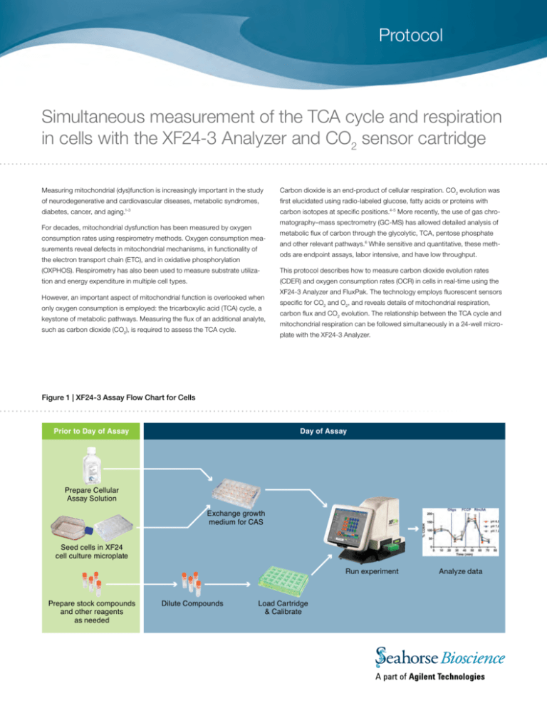
Protocol Simultaneous measurement of the TCA cycle and respiration in cells with the XF24-3 Analyzer and CO2 sensor cartridge Measuring mitochondrial (dys)function is increasingly important in the study Carbon dioxide is an end-product of cellular respiration. CO2 evolution was of neurodegenerative and cardiovascular diseases, metabolic syndromes, first elucidated using radio-labeled glucose, fatty acids or proteins with diabetes, cancer, and aging.1-3 carbon isotopes at specific positions.4-5 More recently, the use of gas chromatography–mass spectrometry (GC-MS) has allowed detailed analysis of For decades, mitochondrial dysfunction has been measured by oxygen consumption rates using respirometry methods. Oxygen consumption measurements reveal defects in mitochondrial mechanisms, in functionality of metabolic flux of carbon through the glycolytic, TCA, pentose phosphate and other relevant pathways.6 While sensitive and quantitative, these methods are endpoint assays, labor intensive, and have low throughput. the electron transport chain (ETC), and in oxidative phosphorylation (OXPHOS). Respirometry has also been used to measure substrate utiliza- This protocol describes how to measure carbon dioxide evolution rates tion and energy expenditure in multiple cell types. (CDER) and oxygen consumption rates (OCR) in cells in real-time using the However, an important aspect of mitochondrial function is overlooked when only oxygen consumption is employed: the tricarboxylic acid (TCA) cycle, a keystone of metabolic pathways. Measuring the flux of an additional analyte, such as carbon dioxide (CO2), is required to assess the TCA cycle. XF24-3 Analyzer and FluxPak. The technology employs fluorescent sensors specific for CO2 and O2, and reveals details of mitochondrial respiration, carbon flux and CO2 evolution. The relationship between the TCA cycle and mitochondrial respiration can be followed simultaneously in a 24-well microplate with the XF24-3 Analyzer. Figure 1 | XF24-3 Assay Flow Chart for Cells Prior to Day of Assay Day of Assay Prepare Cellular Assay Solution Exchange growth medium for CAS Seed cells in XF24 cell culture microplate Run experiment Prepare stock compounds and other reagents as needed Dilute Compounds Load Cartridge & Calibrate Analyze data Materials Reagents XF24-3 Assay Cartridge Preparation Special Notes: The CO2 sensor in the XF24-3 Assay Cartridge has unique characteristics and requires strict adherence to the recommended cartridge preparation and assay conditions described. 1. Uridine [Sigma-Aldrich] 2. Pen/Strep [Sigma-Aldrich] 3. Hepes [Sigma-Aldrich] 1. Assays must be run at a fixed pH of 6.8, 7.0 or 7.2. Cellular Assay 4. Trypsin [Invitrogen] Solution (CAS) should be buffered as noted above for the pH selected. 5. FBS [Hyclone] Shifts in absolute pH should NOT exceed 0.10 pH units during the assay. 2. Phenol red interferes with the function of the CO2 sensor and must not Cellular Assay Solution (CAS) Component be used in the assay medium. Concentration 3. The Assay Cartridge sensor must be constantly hydrated. Hydration is maintained by the cap-mat securely attached to the cartridge. At least DMEM [Sigma D5030] Glucose [Sigma-Aldrich] 25 mM Glutamine [Sigma-Aldrich] 2 mM Sodium Pyruvate [Sigma-Aldrich] 1 mM Buffering Agent, pH PIPES [Sigma-Aldrich], pH 6.8 25 mM BES [Sigma-Aldrich], pH 7.0 25 mM MOPS [Sigma-Aldrich], pH 7.2 25 mM 2 hours before the assay (and preferably overnight), remove the capmat and transfer the cartridge to a utility plate pre-loaded with 1.0 ml of XF Calibration Solution per well. Incubate at 37°C, in a CO2-free environment, for at least 2 hours. 4. Due to CO2 sensor gain decay, the shelf life of CO2 sensor cartridge is 2 months from date of manufacture. FluxPaks will be shipped with a minimum of 1 month of shelf life remaining. 5. Carbon Dioxide Evolution Rate (CDER) is reported as a relative rate, based on changes in fluorescence of the CO2 sensor. Cells 6. The minimum measurement time for each measurement period is 2.5 1. 143b wild-type and ΔCytb mutant cells [generous gift from Dr. Naveep Chandel, Northwestern University] 2. C2C12 cells [ATCC] Consumables 1. XF24-3 FluxPak [Seahorse Bioscience #102070-001] 2. XF Cell Mito Stress Test Kit [Seahorse Bioscience #101706-100] minutes, and 3-4 minute measurement times are recommended. Note: The XF24-3 FluxPak is provided with standard polystyrene (PS) cell culture plates. In certain situations, where additional CO2 sensitivity is required, polyethylene terephthalat (PET) cell culture plates may be used. PET cell culture plates exhibit lower gas permeability, and therefore may reduce background for XF24-3 assays. However, care must be taken, as cell types frequently exhibit distinct morphological, proliferation, and metabolic characteristics when seeded in a PET plate. Methods Preparation of cells C2C12 cells: Cells were maintained in DMEM, 10% FBS + 25 mM glucose. 143b wild-type cells and ΔCytb mutant cells (posessing a defective The cells were maintained in 75-cm2 T-flasks in a controlled incubator at mitochondrial Complex III): Cells were maintained in DMEM with 25 mM 37°C, 95% humidity, and 10% CO2. glucose, 2 mM glutamine, 10% FBS, 0.1 mM sodium pyruvate, 100 µg/ml Every 2–3 days, C2C12 cultures were detached from the flasks using a 0.25% solution of trypsin and subcultured at an initial seeding density of 1.0x10⁵ cells/ml (for a 2-day culture) or 5×10⁴ cells/ml (for a 3-day culture). All uridine, 1% of Pen/Strep and 1 mM HEPES (pH 7.4). The cells were maintained in 75-cm2 T-flasks in a controlled incubator at 37°C, 95% humidity, and 10% CO2. cultures were maintained at less than 80% confluence at the time of Cell culture medium was refreshed every 2 days. Every 3 to 4 days, the cells subculture. were detached from the flasks using a 0.25% solution of trypsin and subcul- For XF24-3 assays, cells were seeded at 30,000 cells/well in an XF24 cell culture microplate. Cells were seeded in a 100 µl volume of growth media, tured at an initial seeding density of 2.5x10⁵ cells/ml. All cultures were maintained at less than 80% confluence at the time of subculture. allowed to attach for 3 hours, then an addition 100 µl of growth media was For XF24-3 assays, cells were seeded at 30,000 cells/well for wild-type and added to each well. 40,000 cells/well for ΔCytb mutant cells, in an XF24 cell culture microplate, using the same cell seeding method as described above. 2 www.seahorsebio.com XF Bioenergetic Analysis Day 1 Day 2 1. Remove cap-mat from the XF24-3 Assay Cartridge and hydrate overnight in Seahorse XF Calibrant at 37°C in a CO2-free environment. 1. Prepare CAS and injection compounds at desired concentrations. 2. Perform the medium exchange from growth medium to CAS. If the cell 2. Seed cells in an XF24 cell culture microplate at the appropriate density. culture plate was exposed to a CO2 environment (e.g. a growth 3. Prepare stock CAS buffered to the proper pH. incubator), then incubate the plate at 37°C in a CO2-free environment 4. Prepare stock reagents. for 90 minutes. Note: pH of the injection reagents should match the pH of the running medium. 5. Create an assay template in the XF software using the Assay Wizard. Table 1 describes typical mix and measurement cycle times cell-based experiments with the XF24-3 FluxPak. 3. Load injection ports with compounds to be injected during the assay. 4. Open the assay template in the XF software, and press Start. Insert the XF24-3 cartridge when prompted. As the cartridge barcode is scanned, you will see the following screen: Table 1 | Typical mix and measurement cycle times for XF24-3 assays Command Time (min) Port # Repeat Calibrate Equilibrate Mix 2 Wait 2 Measure 4 Inject 3 A Mix 2 Wait 2 Measure 4 Inject 2-3 5. Ensure that the indicator buttons for all three analytes (O2, pH, CO2) B Mix 2 Wait 2 Measure 4 Inject are engaged (green). 2-3 6. Press Continue to start the calibration. C Mix 2 Wait 2 Measure 4 Inject Note: This screen will only appear if the instrument is set to 2 analytes and a XF24-3 cartridge is detected. If XF24-3 cartridges are run in sequence this screen will not appear and the assay will proceed as normal. 7. After the calibration is complete, you will be prompted to load the 2-3 assay plate. D Mix 2 Wait 2 Measure 4 2-3 Note: The Mix-Wait-Measure settings used in this protocol are specific for studies using cell populations, and are different than the optimal settings for other studies with the XF24-3 Analyzer. www.seahorsebio.com 3 OCR and CDER profiles of C2C12 cells using the XF Cell Mito Stress Test Kit and XF24-3 FluxPak Note: Optimization assays, including cell seeding density titration and compound injection titration, were run prior to these experiments to determine optimal experimental conditions (data not shown). Optimization assays should always be performed when starting with a new biological model or sample type (e.g. cell line), or going from a 2-analyte to a 3-analyte experiment. Figure 2 | OCR and CDER profiles of C2C12 cells using the XF Cell Mito Stress Test Kit Figure 2 illustrates the relative OCR and CDER responses of C2C12 cells at pH 6.8, 7.0 and 7.2. To demonstrate the utility of the XF24-3 FluxPak for cell-based experiments, C2C12 cells were tested under three different pH levels, using the XF Cell Mito Stress Test Kit. The XF Cell Mito Stress Test Kit reveals information about the key parameters of mitochondrial function: basal respiration, ATP turnover, proton leak, and maximal respiration.7 The assay consists of 3 basal rate measurements followed by sequential injections of oligomycin, FCCP, and rotenone plus antimycin A. Two rate measurements were performed after each injection. XF Cell Mito Stress Test Compound Injections: Injection Injection Final Volume Concentration Concentration Oligomycin 50 μl 10 μg/ml 1 μg/ml FCCP 55 μl 10 μM 1 μM 1 μM 0.1 μM 20 μM 2 μM Port Compound A B C Rotenone Antimycin A 60 μl A. Relative CDER response under different fixed pH levels. As the pH of the running media decreases, the solubility of total CO2 in solution in the assay well, and consequently the relative rates of CO2 evolution reported, increases [Figure 2A]. Reported rates of oxygen consumption are not affected by changing pH [Figure 2B]. However, when data is converted to percent of baseline [Figure 2C], the relative CO2 response rates of C2C12 cells are identical under the three pH conditions. For this reason, it is recom- B. Relative OCR response under different fixed pH levels. mended that CDER data be reported as a relative change between experimental groups or treatments. The data also indicates that in this cellular context, the TCA cycle and ETC/ OXPHOS systems are working in tandem, as manipulation of the OXPHOS system (oligomycin injection) and ETC (FCCP and rotenone/antimycin A injections) produces similar changes in O2 consumption and CO2 evolution through the TCA cycle [Figure 2D]. pH level data is shown in Figure 3. In all conditions, the criteria for constant pH (Δ 0.1 pH units during the assay) were met. Figure 3 | pH Level Data of C2C12 cells C. Relative CDER response, expressed as a percent response from baseline (third basal measurement, before oligomycin injection). D. Relative CDER and OCR responses at pH 6.8, expressed as a percent response from baseline (third basal measurement, before oligomycin injection). 4 www.seahorsebio.com OCR and CDER profiles of 143b osteosarcoma cells using the XF Cell Mito Stress Test Kit and XF24-3 FluxPak Note: Optimization assays, including cell seeding density titration and compound injection titration, were run prior to these experiments to determine optimal experimental conditions (data not shown). Optimization assays should always be performed when starting with a new biological model or sample type (e.g. cell line), or going from a 2-analyte to a 3-analyte experiment. Figure 4 | Relative OCR and CDER responses in wild-type and ΔCytb mutant 143b cells The XF Cell Mito Stress Test Kit was used to examine O2 consumption and CO2 evolution in wild-type and ΔCytb mutant 143b cells. The assay consists of 3 basal rate measurements followed by sequential injections of oligomycin, FCCP, and rotenone plus antimycin A. Two rate measurements were performed after each injection. XF Cell Mito Stress Test Compound Injections: Injection Injection Final Volume Concentration Concentration Oligomycin 50 μl 10 μg/ml 1 μg/ml FCCP 55 μl 10 μM 1 μM 1 μM 0.1 μM 20 μM 2 μM Port Compound A B C Rotenone Antimycin A 60 μl In this cellular context, the TCA cycle and ETC/OXPHOS systems were shown to be working in tandem, as illustrated by the XF Cell Mito Stress Test profiles for wild-type and mutant populations, respectively. In the wild-type population, manipulation of the OXPHOS system (oligomycin injection) and ETC (FCCP and rotenone/antimycin A injections) produced expected changes in both O2 consumption and CO2 evolution through the TCA cycle. XF Cell Mito Stress Test OCR [A], and CDER [B] profiles of wild- In the ΔCytb mutant population, both OCR and CDER responses were type and Complex III-deficient 143b osteosarcoma cells. severely attenuated, consistent with the Complex III-deficiency phenotype. Through the simultaneous measurement of OCR and CDER, the XF24-3 Analyzer provided functional validation of the genetic modifications present in the ΔCytb mutant cell line, in a single experiment and microplate. Note: Absolute rates of CO2 evolution should only be used to determine relative response changes within an assay plate. For comparisons across assay plates or across experiments, absolute rates should be converted to relative percent responses (% baseline). Summary In summary, this protocol introduces a novel method for simultaneous measurement of O2 consumption (OCR) and CO2 evolution (CDER) in a microplate format. The CDER component is reported as a relative rate, based on changes in fluorescence of the CO2 sensor, and strict control of pH is required during the assay. Additionally, absolute rates of CO2 evolution should only be used to determine relative response changes within an assay plate; for comparisons across assay plates or across experiments, data must be converted to relative percent response (% baseline). In spite of these limitations, the simultaneous measurements provide a powerful tool for measuring mitochondrial dysfunction. In a cellular context, simultaneous measurement of OCR and CDER can be utilized to perform mechanistic studies of mitochondrial function, through biochemical manipulations, and distinguish differential treatment effects on the TCA cycle and ETC/OXPHOS systems. Changes in OCR and CDER can also reveal and validate underlying mechanisms contributing to the metabolic phenotype. www.seahorsebio.com 5 References 1. Tseng, Y.H., Cypess, A.M., Kahn, C.R. (2010) Cellular bioenergetics as a target for obesity therapy. Nat. Rev. Drug Discov. 9, 465-82 2. Moreira, P.I., Cardoso, S.M., Pereira, C.M., Santos, M.S. and Oliveira, C.R. (2009) Mitochondria as a therapeutic target in Alzheimer‘s disease and diabetes. CNS Neurol. Disord.Drug Targets 8, 492-511 3. Schon, E.A., DiMauro, S., Hirano, M. and Gilkerson, R.W. (2010) Therapeutic prospects for mitochondrial disease. Trends Mol. Med. 16, 268-76 4. Bloom, B and Stetten Jr, D (1955), The Fraction of Glucose Catabolized via the Glycolytic Pathway. J. Biol. Chem. 212, 555-563. 5. Katz, J and Wood HG. (1960) The Use of GlucoseC14 for the Evaluation of Pathways of Glucose Metabolism. J. Biol. Chem., 235, 2165-2177 6. Metallo, C. Walther, J. and Stephanopoulos G. (2009) Evaluation on 13C isotopic tracers for metabolic flux analysis in mammalian cells. J. Biotech., 144, 167-174. 7. Brand, MD and Nicholls, DG (2011) Assessing mitochondrial dysfunction in cells. Biochem J. 435, 297-312 Corporate Headquarters European Headquarters Asia-Pacific Headquarters Seahorse Bioscience Inc. 16 Esquire Road North Billerica, MA 01862 US Phone: 1.978.671.1600 Seahorse Bioscience Europe Fruebjergvej 3 2100 Copenhagen DK Phone: +45 31 36 98 78 Seahorse Bioscience Asia 199 Guo Shou Jing Rd, Suite 207 Pudong, Shanghai 201203 CN Phone: 0086 21 33901768 201205149 www.seahorsebio.com


