BIOL 347 General Physiology Lab The Special
advertisement
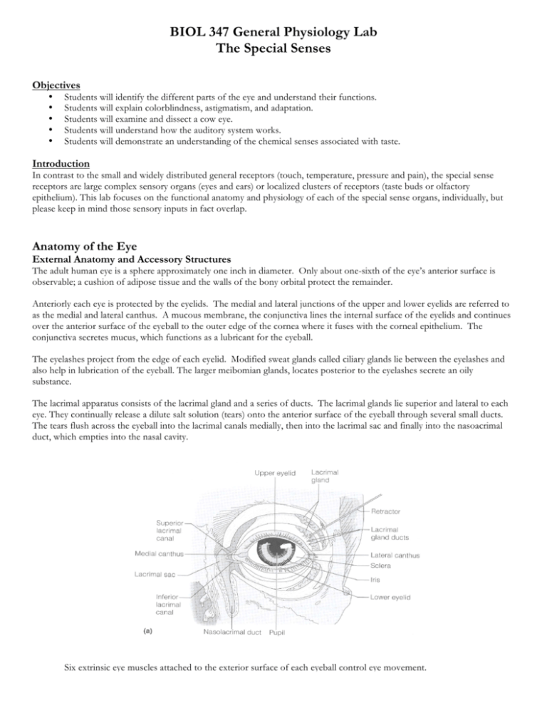
BIOL 347 General Physiology Lab The Special Senses Objectives • • • • • Students will identify the different parts of the eye and understand their functions. Students will explain colorblindness, astigmatism, and adaptation. Students will examine and dissect a cow eye. Students will understand how the auditory system works. Students will demonstrate an understanding of the chemical senses associated with taste. Introduction In contrast to the small and widely distributed general receptors (touch, temperature, pressure and pain), the special sense receptors are large complex sensory organs (eyes and ears) or localized clusters of receptors (taste buds or olfactory epithelium). This lab focuses on the functional anatomy and physiology of each of the special sense organs, individually, but please keep in mind those sensory inputs in fact overlap. Anatomy of the Eye External Anatomy and Accessory Structures The adult human eye is a sphere approximately one inch in diameter. Only about one-sixth of the eye’s anterior surface is observable; a cushion of adipose tissue and the walls of the bony orbital protect the remainder. Anteriorly each eye is protected by the eyelids. The medial and lateral junctions of the upper and lower eyelids are referred to as the medial and lateral canthus. A mucous membrane, the conjunctiva lines the internal surface of the eyelids and continues over the anterior surface of the eyeball to the outer edge of the cornea where it fuses with the corneal epithelium. The conjunctiva secretes mucus, which functions as a lubricant for the eyeball. The eyelashes project from the edge of each eyelid. Modified sweat glands called ciliary glands lie between the eyelashes and also help in lubrication of the eyeball. The larger meibomian glands, locates posterior to the eyelashes secrete an oily substance. The lacrimal apparatus consists of the lacrimal gland and a series of ducts. The lacrimal glands lie superior and lateral to each eye. They continually release a dilute salt solution (tears) onto the anterior surface of the eyeball through several small ducts. The tears flush across the eyeball into the lacrimal canals medially, then into the lacrimal sac and finally into the nasoacrimal duct, which empties into the nasal cavity. Six extrinsic eye muscles attached to the exterior surface of each eyeball control eye movement. Name Controlling Cranial Nerve Action Lateral retcus Medial rectus Superior rectus Inferior rectus Inferior oblique Superior oblique VI (abducens) III (oculomotor) III (oculomotor) III (oculomotor) III (oculomotor) IV (trochlear) Moves eye laterally Moves eye medially Elevates eye or rolls it superiorly Depresses eye or rolls it inferiorly Elevates eye and turns it laterally Depresses eye and turns it laterally Activity: Gross Examination of the Eye Observe the eyes of another student and identify as many structures as possible. Ask the student to look to the left. What extrinsic muscles produce this reaction for the: Right eye:_____________________________ Left eye:______________________________ Now ask your subject to look superiorly. What two extrinsic muscles of each eye can bring about this motion? Right eye:_____________________________ Left eye:______________________________ Internal Anatomy of the Eye The outermost fibrous tunic of the eye is a protective layer composed of dense connective tissue. This can be divided into two identifiable regions: the white sclera, also known as the white of the eye, makes up the majority of the fibrous tunic and the transparent cornea. The middle tunic is referred to as the uvea or vascular tunic. The posterior region of the tunic, known as the choroid, is rich in blood and contains pigments that prevent the scattering of light within the eye. The choroid is modified to form the ciliary body to which the lens is attached. The iris is pigmented and the pupil is the rounded opening through which light passes. The innermost sensory tunic of the eye is the retina. It contains the photoreceptors, rods and cones, which starts the electrical chain of events from photoreceptors to bipolar cells and then to ganglion cells. The photoreceptors are located all over the retina, except for where the optical nerve leaves the eyeball. This site is referred to as the optical disc or the blind spot. The lens divides the eye into two segments. The anterior segment, found anterior to the lens, contains a clear watery fluid called the aqueous humor. The posterior segment behind the lens is filled with a more viscous fluid and is referred to as the vitreous humor. Lateral to the each blind spot is the macula lutea, an area of high cone density. In its center is the fovea centralis, a minute pit approximately .5mm in diameter, which contains only cones and is the area of highest visual acuity. Activity: Dissection of the Cow Eye The cow eye is a typical mammalian eye, but it has specific adaptations characteristic to the grazing animals that have been domesticated. The eyes are located well to the side of the head, providing a total field of vision of approximately 350 degrees with a small binocular field of only 25 degrees. This arrangement allows for maximal alertness for monocular but not binocular vision. In humans, the frontal location of the eyes provides a total visual field of approximately 200 degrees with a binocular overlap of about 110 degrees. The eyes of the human are thus specialized for binocular depth perception of objects relatively close to the eyes. The cow eye that has been provided for you has been fixed in Carosafe, a non-toxic version of formalin. Fixation of the specimen arrests decomposition and hardens the tissues by the coagulation of the protein material. The fixed eye is easier to handle and retains its form better, the unfixed eye being soft and partially collapsed owing to the loss of intraocular pressure. Fixation however, causes color changes in the tissues and causes loss of transparency of the cornea. The dissection of the cow eye should be preformed by the guidelines in the accompanying handout. Activity: Visual Tests and Experiments The Blind Spot Using the blind spot diagram, hold it approximately eighteen inches from your eyes. Close your left eye and focus your right eye on the plus sign, which should be positioned so that it is directly lined-up with your right eye. Move the diagram very slowly towards your face, always keeping your right eye focused on the plus sign. When the dot focuses on the blind spot, the region without photoreceptors, it will disappear from your view. Have your lab partner record the metric distance at which this occurs. Repeat with the other eye. Right eye:_____________________________ Left eye:______________________________ + Adaptation of the Retina If your retinas are exposed to strong light over a period of time, your eyes become sensitive to light. Fix your eyes on the white dot in the center of the peace symbol for at least thirty seconds. Then look at the black dot to the right. What happens when you stare at the black dot? Explain what is happening. Visual Acuity Have your partner stand 20 feet from the posted Snellen eye chart and cover one eye. As your partner reads the lines, check for their accuracy. Record the line with the smallest-sized letters read. If itis 20/20 then the subject vision is normal. If the vision is recorded as any thing with a ratio less than one, 20/40 for example, then the vision acuity is recorded as less than normal. If the visual acuity ration is greater than one, then the subject has better than normal vision. Record your observations. Right eye:_____________________________ Left eye:______________________________ Astigmatism The astigmatism chart tests for defects in the refracting surfaced of the lens and/or cornea. View the chart first with one eye, then the other. Focus on the center of the chart. If all the radiating lines appear equally dark and distinct, your refracting surface surfaces are not distorted. If some of the lines are blurred and appear less dark than others, then some degree of astigmatism is present. Record your observations. Right eye:_____________________________ Left eye:______________________________ Anatomy of the Ear The ear contains the sensory receptors for hearing and equilibrium. It can be divided into three compartments: the outer ear, the middle ear and the inner ear. The outer and middle ear function in hearing only, while the inner ear functions in both equilibrium and hearing reception. The outer ear is composed of the pinna and the external auditory canal. The pinna is the skin-covered cartilage region encircling the auditory opening. The sound waves that enter the external auditory canal eventually hit the tympanic membrane, commonly called the eardrum. The eardrum separates the outer ear from the middle ear. The middle ear is a small fluid-filled chamber known as the tympanic cavity. Within the cavity are three small bones collectively known as the ossicles ( malleus, incus and stapes). These bones transmit the vibratory motion of the eardrum to the fluids of the inner ear via the oval window. The inner ear is composed of many chambers referred to as the bony labyrinth. The three subdivisions of the bony labyrinth are the cochlea, vestibule and the semicircular canals. Activity: Auditory Tests and Experiments Acuity Test Have your lab partner pack one ear with cotton and quietly sit with their eyes closed. Obtain a watch or ticking clock and hold it to your partners unpacked ear. Then slowly move the clock away from the ear until your partner indicates that he/she can no longer hear the ticking. Record the distance at which the ticking in inaudible. Right ear:_____________________________ Left ear: ______________________________ Weber Test Obtain a tuning fork and rubber mallet. Strike the tuning fork with the mallet and place the handle of the tuning fork medially on your partner’s head. Is the tone equally loud in both ears? If it is equally loud in both ears then the subject has equal hearing or equal hearing loss. If sensineural deafness is heard in present in one ear, the tone will be heard in the unaffected ear but not in the ear with sensineural deafness. If conduction deafness is present, the sound will be heard more strongly in the ear in which there is hearing loss. Rinne Test Strike the tuning fork and place its handle on your partner’s mastoid process, the bone behind the ear and slightly lower. Have your partner indicate when he/she can no longer hear the sound. At that point move the fork with the still vibrating prongs close to his/her auditory canal. If your partner can hear the fork again (by air conductance) then hearing is not impaired. Repeat for both ears. Record your observations. Right ear:_____________________________ Left ear: ______________________________ Repeat this test, except do the air conductance first and the bone conductance second. If the subject hears the tone after hearing by air conductance is lost, then there is some conductive deafness. Repeat for both ears. Right ear:_____________________________ Left ear: ______________________________ Does the subject hear better by bone or by air conductance? Anatomy of the Tongue The superior tongue surface is covered with small projections known as papillae. There are three major forms of papillae: filiform, fungiform and circumvallate. The taste buds, specific receptors for the sense of taste are distributed throughout the oral cavity; however, the majority are located on the tongue. Each taste bud consists of a globular arrangement of two types of epithelial cells: the gustatory (taste cells) that are the actual receptors and the supporting cells. Activity: Stimulating the Taste Buds With the paper towels provided, dry the superior surface of the tongue. Place a few sugar crystals on your dried tongue. Do NOT close your mouth. Time how long it takes for you to taste the sugar. Time:_________________ Why couldn’t you taste the sugar immediately? Activity: Individual Differences in Taste There is an inherited preference and/or disgust for certain tastes. Use the four test papers (control, PTC, Thiourea and Sodium benzoate) and record your reaction and taste. Paper Control PTC Thiourea Sodium Benzoate Taste Activity: Effects of Olfactory Stimulation There is no question that what is commonly tasted depends heavily on the sense of smell, particularly in the case of heavy scented substances. These following experiments should illustrate that. Obtain paper cups with labeled ingredients. Ask the subject to sit so that he/she cannot see which sample is being used. Have the subject dry the superior surface of their tongue and hold their nose closed. Apply one drop of the substance on the subject’s tongue. Can the subject distinguish the flavor? Now have the subject open their nose and record the change in sensation. Have the subject rinse their mouth out and dry their tongue. Prepare two swabs of differing substances. Hold one swab under the subject’s nose and hold the other on their tongue. Record your observations. This lab has been adapted from E. N. Marieb’s Essential of HumanAnatomy and Physiology Seocnd Edition. Experimenting with the Sense of Taste: the Jelly Bean Lab “I was most unfortunate in my youth to come across a vomit-flavored one. But I suppose I'll be safe with a nice toffee. (Eats candy.) Alas. Ear wax.” -Albus Dumbledore in the 2001 movie Harry Potter and the Sorcerer's Stone about Bertie Bott's Every Flavor Beans Question: Does Vision Influence Taste? For each subject you test, you will need pairs of jelly beans. For example, get 2 cherry jelly beans, 2 lime jelly beans, 2 lemon jelly beans and 2 orange jelly beans. Each jelly bean flavor must have its own unique color: (for example) red for cherry, green for lime, yellow for lemon and orange for orange. Divide the jelly beans into two groups: each group should have one of each flavor. Label small containers or napkins with the numbers 1 through 4. Place the jelly beans from the first group into a container or on a napkin - one jelly bean into each container or on each napkin. Place the jelly beans in the second group in a cup so that your subjects cannot see them. Label these cups with the numbers 1 through 4. Make sure that the flavors of the second group have different numbers than the flavors in the first group. Now you are ready to start the experiment. Tell the subject to look at the jelly bean in container #1 of the first group and then taste the jelly bean. After they have tasted the jelly bean, tell the subject to write down its flavor. Do the same thing with jelly beans #2-#4. The next part of the experiment is a bit more difficult. You must keep the color of the jelly beans in group 2 hidden from your subjects. You can blindfold your subjects or have them close their eyes while they taste the jelly beans. Keep track of the flavors that your subjects say each jelly bean tastes like. Test Subject: ____________________________________________________________________ Jelly Bean Actual Flavor Group 1 Number Subject’s Guess Group 2 Number Subject’s Guess Did you subjects make any mistakes when they could not see the color of the jelly bean? If they did, what was the most common mistake? Does vision influence taste? What would happen if you used an unusual flavor? What would happen if you found a jelly bean with an abnormal color...for example a red-colored lemon-flavor jelly bean? Question: Does Smell Influence Taste? Identify a test subject. Choose one flavor of jellybean. Do not allow the subject to know the actual flavor or see the color. Have the subject hold his or her nose closed while they put the jellybean in their mouth. The subject should chew the jellybean for 10 seconds while holding the nose closed. Ask the subject the flavor of the jellybean. Now have the subject release his or her nose. Now ask them to indentify the flavor of the jellybean. Repeat this experiment making sure that every group member has a chance to be the subject. Record your observations. Jelly Bean Actual Flavor Subject Subject’s Guess (no smell) Subject’s Guess (with smell) Could the subject(s) taste the jellybean while holding their nose(s)? Was the subject(s) able to taste the jellybean once they regained their sense of smell? Were any jellybean flavors so strong that the subject(s) could identify them without a sense of smell? Why does food often seem tasteless when you have a cold? Scientists have concluded that our taste buds on our tongues can distinguish five basic taste sensations -- sweet, sour, bitter, salty, and umami. Would the five basic taste sensations be sufficient to distinguish all the different flavors of food? Could you identify the different flavors of jellybeans just by taste? Or do you need additional characteristics to distinguish the different flavors?
