PERGAMON
Micron 31 (2000) 355–362
www.elsevier.com/locate/micron
Valence state mapping of cobalt and manganese using near-edge fine
structures
Z.L. Wang a,*, J. Bentley b, N.D. Evans b
a
School of Materials Science and Engineering, Georgia Institute of Technology, Atlanta, GA 30332-0245, USA
b
Metals and Ceramics Division, Oak Ridge National Laboratory, Oak Ridge, TN 37831-6376, USA
Abstract
The properties of transition metal oxides are related to the presence of elements with mixed valences. The spectroscopy analysis of the
valence states is feasible experimentally, but a spatial mapping of valence states of transition metal elements is a challenge to existing
microscopy techniques. In this paper, with the use of valence state information provided by the white lines and near-edge fine structures
observed using the electron energy-loss spectroscopy (EELS) in a transmission electron microscope (TEM), a novel experimental approach
is demonstrated to map the valence state distributions of Mn and Co using the ratio of white lines in the energy-filtered TEM. The valence
state map is almost independent of specimen thickness in the thickness range adequate for quantitative EELS microanalysis. An optimum
spatial resolution of ,2 nm has been achieved for a two-phase Co oxides. q 2000 Elsevier Science Ltd. All rights reserved.
Keywords: Electron energy-loss spectroscopy; Transmission electron microscope; Transition metal oxides
1. Introduction
Many properties of smart and functional materials are
determined by the elements with mixed valences in the
structure (Wang and Kang, 1998), by which we mean an
element that has two or more different valences while forming a compound. In the perovskite structured La12xAxMnO3
A Ca; Sr, or Ba) colossal magnetoresistance (CMR)
materials (Jin et al., 1994; Wang and Zhang, 1996), for
example, the residual charges induced by a partial substitution of trivalent La 31 by divalent element A 21 are balanced
by the conversion of Mn valence states between Mn 31 and
Mn 41 (or Co 31 and Co 41 for Co) and the creation of oxygen
vacancies as well, and the ionic structure of La12xAxMnO32y
is
21
31
41
22
O
La31
12x Ax Mn12x12y Mnx22y O32y Vy
provided there is no residual charge trapped in the vacancy
sites, where VO
y stands for the fraction of oxygen vacancies.
Valence transition between Mn 31 and Mn 41 is the key that
leads to the transition from insulator to conductor and possibly the magnetoresistance.
Electron energy-loss spectroscopy (EELS) has been
widely used in chemical microanalysis and the studies of
solid state effects (Egerton, 1996). In EELS, the L ionization
* Corresponding author. Tel.: 1404-894-8008; fax: 1404-894-9140.
E-mail address: zhong.wang@mse.gatech.edu (Z.L. Wang).
edges of transition-metal and rare-earth elements usually
display sharp peaks at the near edge region which are
known as white lines. For transition metals with unoccupied
3d states, the transition of an electron from 2p state to 3d
levels leads to the formation of white lines. Numerous EELS
experiments have shown that a change in valence state of
cations introduces a dramatic change in the ratio of the
white lines, leading to the possibility of identifying the
occupation number of 3d orbital using EELS (Pease et al.,
1986; Krivanek and Paterson, 1990; Kurata and Colliex,
1993; Pearson et al., 1993; Yuan et al., 1994; Yin and
Wang, 1997; Wang et al. 1997a,b; Wang and Yin, 1998;
Zhang et al., 1998). The spectroscopy analysis of the white
line intensity is feasible for a region selected by the electron
beam. In this paper, with the use of energy-filtered transmission electron microscopy (EF-TEM), a new experimental
approach is introduced for mapping the distribution of a
cation valences, allowing a direct identification of cations
with different valence states. The valence state map is
almost independent of the specimen thickness and it can
be used to directly read out the local valence state from the
image. A spatial resolution of ,2 nm has been demonstrated.
2. Experimental approach for mapping the valence
states of Co and Mn
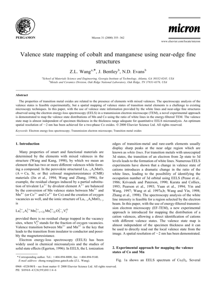
Fig. 1a shows an EELS spectrum of Co3O4. Several
0968–4328/00/$ - see front matter q 2000 Elsevier Science Ltd. All rights reserved.
PII: S0968-432 8(99)00114-6
356
Z.L. Wang et al. / Micron 31 (2000) 355–362
Fig. 1. (a) An EELS spectrum acquired from Co3O4, showing the fivewindow technique used to extract the intensities of the white lines and
the three-window technique for O-K edge. (b) The Co-L edge after subtraction of the background, illustrating the background underneath the L3 and
L2 lines.
techniques have been proposed to correlate the observed
EELS signals with the valence states, the ratio of white
lines the normalized white line intensity in reference to
the continuous state intensity located ,50–100 eV beyond
the energy of the L2 line, and the absolute energy shift of the
white lines. In this study, we use the white line intensity
ratio that is calculated using a method demonstrated in Fig.
1b (Pearson et al., 1993). The background intensity was
modeled by step functions in the threshold regions, as
schematically shown in Fig. 1b. A straight line over a
range of approximately 50 eV was fit to the background
intensity immediately following the L2 white line. This
line was then modified into a double step of the same
slope with onsets occurring at the white-line maxima. The
ratio of the step heights is chosen to be 2:1 in accordance
with the multiplicity of the initial states (four 2p3/2 electrons
and two 2p1/2 electrons). Although there exist some
disagreements in the literature about the calculation of the
normalized white line intensity because the theory behind
the white lines and their continuos background is rather
complex (Thole and van der Laan, 1988), it appears,
based on our experience, that this ratio of the white line
intensities is likely to be a reliable and sensitive approach.
This background subtraction procedure is followed consistently for all of the acquired spectra. The calculated L3/L2 is
rather stable and is not sensitive to the specimen thickness.
EELS analysis of valence state is carried out in reference
to the spectra acquired from standard specimens with known
cation valence states. Since the intensity ratio of L3/L2 is
directly related to the valence state of the corresponding
element, a series of EELS spectra were acquired from
several standard specimens with known valence states, an
empirical plot of these data serves as the reference for determining the valence state of the element present in a new
compound. The L3/L2 ratios of Co and Mn have been
presented in our recent paper (Wang et al., 1999), which
will be used as the standards in the analysis described
below.
The information provided by EF-TEM is mostly about the
elemental distribution in a thin section of a specimen
(Reimer, 1995). To map the distribution of ionization states,
an energy window of ,10 eV in width is required to isolate
the L3 from L2 white lines. A five-window technique is
introduced (see Fig. 1a): two images are acquired at the
energy-losses prior to the L ionization edges, and they are
to be used to subtract the background for the characteristic L
edge signals; two images are acquired from the L3 and L2
white lines, respectively, and the fifth image is recorded
using the electrons right after the L2 line that will be used
to subtract the continuous background underneath the L3
and L2 lines. To extract the L3/L2 image that is most sensitive to the valence state of Mn or Co, a background subtraction procedure illustrated in Fig. 1b needs to be carried out.
This procedure can be easily done in the EELS spectrum, but
for the energy-filtered image acquired in TEM under parallel
illumination a different approach has to be taken, as given by
L3 =L2
I
L3 2 aI
post-lines
;
I
L2 2 bI
post-lines
1
where I(L3), I(L2) and I(post-lines) are the images recorded by
positioning the energy selection window at the L3, L2 and the
post L2 line energy-losses, respectively, after subtracting the
conventional background as ascribed by A exp
2rDE; a and
b are the adjustable parameters that represent the fractions of
the continuous background below the L3 and L2 lines, respectively, as contributed by the single atomic scattering (as illustrated in Fig. 1b). The choices of the a and b factors may
depend on the specimen thickness because the I(post-lines)
image is strongly affected by the multiple scattering effect,
while I(L3) and I(L2) are less affected. Eq. (1) represents the
optimum choice for L3/L2 mapping in EF-TEM under the data
collection conditions allowed by the Gatan Imaging Filter. A
more accurate data treatment can be adopted in STEM.
To confirm the information provided by the L3/L2 images,
the specimen composition is mapped from the integrated
intensities of the O-K and Mn-L2,3 ionization edges by
following the routine procedure of EELS microanalysis
(Egerton, 1996)
nO
I
D sMn
D
;
O
nMn
IMn
D sO
D
2
where IO(D ) and IMn(D ) are the integrated intensities of the
Z.L. Wang et al. / Micron 31 (2000) 355–362
O-K and Mn-L edges for an energy window D , respectively,
above the ionization thresholds; s Mn(D ) and s O(D ) are the
integrated ionization cross-sections for the corresponding
energy window D , and they can be calculated by the
sigmak2 and sigmal2 programs in the hydrogen-like atomic
model. From the energy-filtered images, the distribution
map of the atomic ratio O/Mn or O/Co can be calculated.
The EF-TEM experiments were performed using a
Philips CM30 (300 kV) TEM, equipped with a Gatan
image filtering (GIF) system. This TEM provides a high
beam current needed for chemical imaging. The energy
window width was selected to be 10 eV for Mn or 12 eV
for Co, and it took 10–30 s (depending on specimen) exposure to acquire a single raw data image with satisfactory
signal-to-noise ratio. The selection of the energy window
width depends on the energy separation between the L3 and
L2 lines. It took 2–4.5 min. to acquire a complete set of
images. Specimen drift between different images was
corrected after the acquisition, but it was important to
ensure the least drift of the specimen during data acquisition.
3. Results
3.1. Mapping the valence states of Co using the white line
ratio
The first specimen selected for illustrating the experimental approach is a directionally solidified eutectic ZrO2/CoO
(Bentley et al., 1993), which is composed of trilayer structures of ZrO2, Co3O4 and CoO after heat treatment in a high
oxygen partial pressure, with ideal geometry for studying
CoO–Co3O4 interfaces. The differences in crystal structure,
the coordination configuration of cations and the valence
states result in dramatic differences in EELS spectra of
CoO and Co3O4 (Bentley and Anderson, 1996). Shown in
Fig. 2 is an one-dimensional spatially dispersed EELS spectra across a CoO–Co3O4 interface. The valence-loss spectra
of the two phases are distinctly different. The O-K edge
exhibits a double split peak for Co3O4 while no split for
CoO. The white lines of the two structures are different
not only in their relative intensity, but also in having a slight
chemical shift ( < 1.5 eV). It was critical to select the width
of the energy window and position it at the correct energies
for acquired each group of data. The peak-to-peak energy
between L3 and L2 is 15 eV and the full width of the line at
10% intensity cut-off is 7–8 eV, a choice of energy window
width D 12 eV is adequately to separate the two lines as
well as to ensure the signal-to-noise ratio.
Fig. 3 shows a group of energy-filtered image from a
triple point in the CoO–Co3O4 specimen. The bright-field
TEM image (Fig. 3a) shows one of the grains has a strong
diffraction contrast. The energy-filtered images using the L3
and L2 lines (Fig. 3b and c) shows distinctly difference in
contrast distribution due to a difference in the relative white
357
line intensities (see Wang et al., 1999). The finally analyzed
L3/L2 image clearly displays the distribution of cobalt
oxides having different valence states (Fig. 3d), where the
diffraction contrast disappears. The region with lower
oxidation state (Co 21) shows stronger contrast, and the
ones with high oxidation states show darker contrast.
Although the energy-filtered O-K edge image exhibits
some diffraction contrast, the O/Co compositional ratio
image greatly reduces the effect. The residual contrast
seen in Fig. 3f might be due to the specimen drift during
the 10 s exposure. The O/Co image was calculated from the
images recorded from the O-K edge and the L3 1 L2 white
lines for an energy window width of D 24 eV: The high
intensity region in the O/Co image indicates the relative
high local concentration in oxygen (e.g. higher Co
oxidation states), the low intensity region contains relatively less oxygen (e.g. lower Co valence state), entirely
consistent with the information provided by the L3/L2
image.
Under the single scattering approximation, the intensities
of the L3 and L2 lines scale up in proportional to the specimen thickness, thus, their ratio L3/L2 has little dependence
on the specimen thickness. This result holds even for
slightly thicker specimens because the near edge structure
is less affected by the multiple plasmon scattering effect and
the energies of the characteristic plasmon peaks are larger
than the energy split between the L3 and L2 lines. Therefore,
in the conventional thickness range for performing EELS
microanalysis of t=L , 0:8; where t is the specimen thickness and L is the mean-free-path length of electron inelastic
scattering, the L3/L2 image truly reflects the distribution of
valence state across the specimen.
Fig. 4 shows another group of images recorded from the
CoO–Co3O4 specimen. The L3 and L2 images display some
contrast across the phases, while the image recorded from
the post-line energy-loss region shows a small contrast
variation (Fig. 4c) possibly because that the post-line region
is dominated by the single atomic scatting properties and it
is less affected by the solid state effects, provided the thickness-projected density of Co atoms is fairly uniform across
the grains. The image from the O-K edge show some variation due to diffraction contrast as well as specimen thickness
(Fig. 4d). In contrast, the L3/L2 image (Fig. 4e) and the O/Co
image (Fig. 4f) show little dependence on the specimen
thickness and the diffracting condition. This is uniquely
suited for mapping the valence state distribution across
the specimen.
To determine the optimum spatial resolution achieved in
the L3/L2 image, a line scan across the CoO–Co3O4 interface
at a 30 pixel average in width is displayed in Fig. 4g. The
half width of the profile at the interface is about 2 pixels,
which correspond to a resolution of ,1.8 nm. The image
across the interface in the O/Co image shows a half width of
3 pixels, which correspond to a resolution of ,2.8 nm. It
must be pointed out that a better resolution achieved in the
L3/L2 image is likely due to the smaller width of the energy
358
Z.L. Wang et al. / Micron 31 (2000) 355–362
Fig. 2. Zero-loss bright-field TEM image of CoO–Co3O4 interface, showing the selection area aperture for forming the EELS spectra; spectrum lines for the
low-loss region, the O-K edge, and the Co-L edge, exhibiting distinct differences in the intensities and energy positions of the characteristic peaks between CoO
and Co3O4.
Fig. 3. A group of images recorded from the same specimen region using signals of (a) the zero-loss bright-field, (b) the Co-L3 edge, (c) the Co-L2 edge, (d) the
L3/L2 ratio, (e) the O-K edge, and (f) the atomic concentration ratio of O/Co. The continuous background contributed from the single atom scattering has been
removed from the displayed Co-L3 and Co-L2 images. The L3/L2 ratio image was calculated by taking a 0:3 and b 0:7: The O/Co image is normalized in
reference to the standard composition of CoO for the low portion of the image in order to eliminate the strong influence on the ionization cross-section from the
white lines. Each raw image was acquired with an energy window width of D 12 eV except for O-K at D 24 eV:
Z.L. Wang et al. / Micron 31 (2000) 355–362
359
Fig. 4. A group of images recorded from the same specimen region using signals of: (a) the Co-L2 edge; (b) the Co-L3 edge; (c) the post Co-L2 continuous
energy-loss region; (d) the O-K edge; (e) the L3/L2 ratio; and (f) the atomic concentration ratio of O/Co. The continuous background contributed from the single
atom scattering has been removed from the displayed Co-L3 and Co-L2 images. The L3/L2 ratio image was calculated by taking a 0:3 and b 0:8: (g) A line
scan across the CoO–Co3O4 interface in the L3/L2 image after making a 30 pixels of average in width parallel to the interface, showing the local average white
line ratio. A comparison of the displayed numbers with the values measured from standard specimens (Wang et al., 1999), the regions corresponding to CoO
and Co3O4 are apparent. (h) A line scan across the CoO–Co3O4 interface in the O/Co ratio image after making a 30 pixels of average in width parallel to the
interface, from which the local compositions match very well to CoO and Co3O4. Each raw image was acquired with an energy window width of D 12 eV
except for O-K at D 24 eV:
window
D 12 eV than the D 24 eV used for O/Co
image as well as the sharp shape of the white lines.
3.2. In situ observation of valence state transition of Mn
For demonstrating the application of the technique for a
more complex case, a reduced MnOx powder was prepared
by in situ annealing (Wang et al., 1997a). A Gatan TEM
specimen heating stage was employed to carry out the in situ
EELS experiments, and the specimen temperature could be
increased continuously from room temperature to 10008C.
The column pressure was kept at 3 × 1028 Torr or lower
during the in situ analysis. Due to the reduction of the
oxide, multi-valences will be developed in the system. A
360
Z.L. Wang et al. / Micron 31 (2000) 355–362
Fig. 5. A group of energy-filtered images acquired from the same specimen region of mixed phases of MnO2 and Mn3O4: (a) The conventional bright-field TEM
image; (b) the energy-filtered TEM images of Mn-L3 white line; (c) the calculated Mn L3/L2 ratio image; and (d) the distribution of O/Mn in the region
calculated according to Eq. (2) using the energy-filtered images from the O-K and Mn-L edges. The complimentary contrast of (c) to (d) proves the
experimental feasibility of valence state mapping using the white line ratio. Each raw image was acquired with an energy window width of D 10 eV
except for O-K at D 20 eV:
detailed analysis of this reduction process by EELS has been
reported previously (Wang et al., 1997a, 1999). The results
indicated that the reduction of MnO2 occurs at 3008C. As the
temperature reaches 4508C, the specimen is dominated by
Mn 21 and Mn 31 and the composition is O=Mn 1:3 ^ 0:5;
corresponding to Mn3O4.
To obtain a specimen with multi-valences, the in situ
annealing of MnO2 was carried out up to 3508C and then
cool back to room temperature, and the resulting reduced
phases were a mixture of oxides of Mn with valences of
2 1 , 3 1 and 4 1 . This is a model system to be used for
mapping the valence state distribution of Mn. Fig. 5 shows a
group of images recorded from an agglomeration of MnOx
with different valences. The bright-field image hardly indicate any information about the valence states of Mn. The
EF-TEM Mn-L3 image reflects the distribution of Mn
phases, but its contrast is approximately proportional to
the local projected thickness of the specimen. The L3/L2
ratio image (Fig. 5c) directly gives the distribution of
Mn 41, Mn 21 and Mn 31. The low intensity regions are
Mn 41, and the high intensity regions are the mixed valences
of Mn 21 and Mn 31, in correspondence to the formation of
Mn3O4. To confirm this results, the atomic ratio O/Mn
image is calculated from the images acquired from the OK and Mn-L edges, and the result is given in Fig. 5d. The
image clearly indicates that the regions with Mn 41 have
higher O atomic concentration because of the balance of
the cation charge. This is an excellent proof of the information provided by the L3/L2 image.
3.3. Phase separation using the near edge fine structure
The near-edge fine structure observed in EELS is closely
related to the solid state effect and it is most sensitive to the
bonding and near-neighbor coordination configurations.
Phase and bonding mapping using the near-edge structure
have been carried out for diamond (Batson et al., 1994;
Mayer and Plitzko, 1996), in which the images were formed
using the p p and s p peaks in the C-K edge and the distribution of diamond bond was retrieved. The O-K edge
displayed in Fig. 2 clearly shows the difference in the near
edge structure of CoO from Co3O4. The first peak observed
in the O-K of Co3O4 is separated by 12 eV from the main
peak. Using an energy selection window of 6 eV in width it
is possible to map the Co3O4 regions that generate this peak.
Fig. 6b shows an energy-filtered TEM image from a region
whose bright-field image is given in Fig. 6a. It is clear that
the Co3O4 regions show stronger intensity, just as expected.
4. Discussion
There are several questions that need to be discussed
relating to the L3/L2 image. The L3/L2 image has little
dependence on either the specimen thickness or diffraction
Z.L. Wang et al. / Micron 31 (2000) 355–362
Fig. 6. (a) The bright-field TEM image and (b) the energy-filtered image
recorded by selecting the sharp peak located at 532 eV in the O-K, displaying the distribution of the Co3O4 phase. Energy window width D 6 eV:
361
effect in the thickness range adequate for EELS microanalysis
t=L , 0:8: For thicker specimens, the intensities of
the L2 line and the continuous post-line would be strongly
affected by the specimen thickness. However, the L3/L2 ratio
is much less sensitive to the specimen thickness, because the
multiple scattering mainly affects the background underneath the L3 and L2 lines (see Fig. 7). Retrieving a single
scattering profile would be possible by deconvolution if the
entire spectra for each pixel were recorded. Unfortunately,
the GIF system only captures the integrated data points and
the deconvolution procedure cannot be carried out.
The calculation of the L3/L2 image is not critically sensitive to the selection of the a and b factors. This is possibly
due to the flatter contrast of the post-line image, which
depends mainly on the thickness-projected atom density
and the diffraction effect. The best choices of the a and b
factors would be to match the local average L3/L2 value with
the standard data measured using spectroscopy in the region
whose phase might be known. The choices of a and b
should not result in large negative counts in the image due
to over subtraction. One should avoid capture in a single
image regions of large differences in thickness because the
a and b factors might also depend on specimen thickness.
The subtraction of the continuous component beneath the
white lines (see Fig. 1b) may not be trivial because its spectrum shape may be a broad peak rather than a smooth decay
curve. One is advised to check the shape of the EELS spectra to ensure the procedure of data analysis.
The quality of the L3/L2 image is strongly affected by the
quality of the L2 image because of the dramatic magnification to the noise level. The critical challenge is the signal-tonoise ratio because of the finite beam current and the small
energy window. A very thin region is inadequate for EFTEM imaging because of the lack of the inelastic scattering
signal, while a too thick region results in complexity in data
quantification.
Finally, the O/Mn map displayed in this paper may not be
the true atomic ratio in the specimen because the accuracy
of EELS microanalysis strongly depends on the choice of
the energy window. The most reasonable choice of energy
window width is 50–100 eV in order to minimize the contribution from the near-edge fine structure (Colliex, 1985). In
L3/L2 imaging, the width of the energy window is required
to be no more than the energy split between the white lines,
typically 10–15 eV. On the other hand, the heights of the L3
and L2 lines scale up in proportional to the local density of
atoms. The O/Mn image can be used as a reliable reference
for examining the information provided by the L3/L2 image.
5. Summary
Fig. 7. A comparison of the Co-L edge before and after deconvoluting the
low-loss spectrum, exhibiting the minor effect of multiple scattering on the
intensities of white lines. The specimen thickness t 0:5L:
For characterizing advanced materials that usually
contain cations with mixed valences, EELS is a very powerful approach with a spatial resolution higher than any other
spectroscopy techniques available. Based on the intensity
362
Z.L. Wang et al. / Micron 31 (2000) 355–362
ratio of white lines, a new experimental approach has been
demonstrated for mapping the valence states of Co and Mn
in oxides using the energy-filtered transmission electron
microscopy. The L3/L2 image has little dependence on either
the specimen thickness or diffraction effects, and it is reliable for mapping the distribution of cation valences. An
optimum spatial resolution of ,2.0 nm has been attained
experimentally. This is a remarkable application of the
EF-TEM for characterizing the electronic structure of
magnetic oxides.
Acknowledgements
Research sponsored by the US NSF grant DMR-9733160,
the Outstanding Oversea Young Scientist Award of China
NSF (59825503), and by the Division of Materials Sciences,
US Department of Energy, under contract DE-AC0596OR22464 with Lockheed Martin Energy Research
Corp., and through the SHaRE Program under contract
DE-AC05-76OR00033 with Oak Ridge Associated Universities. Thanks to Drs A. Revcolevschi, S. McKernan and
C.B. Carter for kindly providing the oxidized CoO
specimen.
References
Batson, P.E., Browning, N.D., Muller, D.A., 1994. EELS at Buried Interfaces: Pushing Towards Atomic Resolution, MSA Bulletin, 24. Microscopy Society of America, pp. 371–374.
Bentley, J., Anderson, I.M., 1996. Spectrum Lines Across Interfaces by
Energy-Filtered TEM. In: Bailey, G.W., Corbett, J.M., Dimlich,
R.V.W., Michael, J.R., Zaluzec, N.J. (Eds.). Proc. Microscopy and
Microanalysis, San Francisco Press, San Francisco, pp. 532–533.
Bentley, J., McKernan, S., Carter, C.B., Revcolevschi, A., 1993. Microanalysis of Directionally Solidified Cobalt Oxide-Zirconia Eutectic. In:
Armstrong, J.T., Porter, J.R. (Eds.). Microbeam Analysis, 2. pp. S286–
287 suppl.
Botton, G.A., Appel, C.C., Horsewell, A., Stobbs, W.M., 1995. Quantification of the EELS near-edge structures to study Mn doping in oxides.
J. Microsc. 180, 211–216.
Colliex, C., 1985. An illustrative view on various factors governing the high
spatial resolution capabilities in EELS microanalysis. Ultramicroscopy
18, 131–150.
Egerton, R.F., 1996. Electron Energy-Loss Spectroscopy in the Electron
Microscope, 2. Plenum Press, New York.
Jin, S., Tiefel, T.H., McCormack, M., Fastnacht, R.A., Ramech, R., Chen,
L.H., 1994. Thousandfold change in resistivity in magnetoresistive La–
Ca–Mn–O films. Science 264, 413–415.
Krivanek, O.L., Paterson, J.H., 1990. ELNES of 3d transition-metal oxides:
I. Variations across the periodic table. Ultramicroscopy 32, 313–318.
Kurata, H., Colliex, C., 1993. Electron-energy loss core-edge structures in
manganese oxides. Phys. Rev. B 48, 2102–2108.
Mayer, J., Plitzko, J.M., 1996. Mapping of ELNES on a nanometre scale by
electron spectroscopic imaging. J. Microsc. 183, 2–8.
Pearson, D.H., Ahn, C.C., Fultz, B., 1993. White lines and d-electron occupancies for the 3d and 4d transition metals. Phys. Rev. B 47, 8471–
8478.
Pease, D.M., Bader, S.D., Brodsky, M.B., Budnick, J.I., Morrison,
T.I., Zaluzec, N.J., 1986. Anomalous L3/L2 white line ratios and spin
pairing in 3d transition metals and alloys: Cr metals and Cr20Au80. Phys.
Lett. 114A, 491–494.
Reimer, L. (Ed.), 1995. Energy-Filtering Transmission Electron Microscopy Springer Series in Optical Sciences, 71. Springer, Berlin.
Thole, B.T., van der Laan, G., 1988. Branching ratio in X-ray absorption
spectroscopy. Phys. Rev. B 38, 3158–3169.
Wang, Z.L., Kang, Z.C., 1998. Functional and Smart Materials—Structural
Evolution and Structure Analysis, Plenum Press, New York.
Wang, Z.L., Zhang, J., 1996. Tetragonal domain structure magnetoresistance of La12xSrxCoO3. Phys. Rev. B 54, 1153–1158.
Wang, Z.L., Yin, J.S., 1998. Cobalt valence and crystal structure of
La0.5Sr0.5CoO2.25. Philos. Mag. B 77, 49–65.
Wang, Z.L., Yin, J.S., Zhang, J.Z., Mo, W.D., 1997a. In-situ analysis of
valence conversion in transition metal oxides using electron energy-loss
spectroscopy. J. Phys. Chem. B 101, 6793–6798.
Wang, Z.L., Yin, J.S., Jiang, Y.D., Zhang, J., 1997b. Studies of Mn valence
conversion and oxygen vacancies in La12xCaxMnO32y using electron
energy-loss spectroscopy. Appl. Phys. Lett. 70, 3362–3364.
Wang, Z.L., Yin, J.S., Jiang, Y.D., 1999. EELS analysis of cation valence
states and oxygen vacancies in magnetic oxides. Micron, in press.
Yin, J.S., Wang, Z.L., 1997. Ordered self-assembling of tetrahedral oxide
nanocrystals. Phys. Rev. Lett. 79, 2570–2573.
Yuan, J., Gu, E., Gester, M., Bland, J.A.C., Brown, L.M., 1994. Electron
energy-loss spectroscopy of Fe thin-films on GaAs(001). J. Appl. Phys.
75, 6501–6503.
Zhang, Z.J., Wang, Z.L., Chakoumakos, B.C., Yin, J.S., 1998. Temperature
dependence of cation distribution and oxidation state in magnetic Mn–
Fe ferrite nanocrystals. J. Am. Chem. Soc. 120, 1800–1804.


