Neurology Board Review Questions
Ischemic Stroke: Pathophysiology and
Principles of Localization
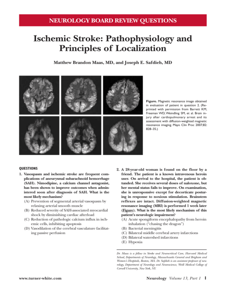
Matthew Brandon Maas, MD, and Joseph E. Safdieh, MD Figure. Magnetic resonance image obtained
in evaluation of patient in question 2. (Reprinted with permission from Barrett KM,
Freeman WD, Weindling SM, et al. Brain injury after cardiopulmonary arrest and its
assessment with diffusion-weighted magnetic
resonance imaging. Mayo Clin Proc 2007;82:
828–35.)
QUESTIONS
1. Vasospasm and ischemic stroke are frequent complications of aneurysmal subarachnoid hemorrhage
(SAH). Nimodipine, a calcium channel antagonist,
has been shown to improve outcomes when administered soon after diagnosis of SAH. What is the
most likely mechanism?
(A)Prevention of segmental arterial vasospasm by
relaxing arterial smooth muscle
(B)Reduced severity of SAH-associated myocardial
shock by diminishing cardiac afterload
(C)Reduction of pathologic calcium influx in ischemic cells, inhibiting apoptosis
(D)Vasodilation of the cerebral vasculature facilitating passive perfusion
2. A 28-year-old woman is found on the floor by a
friend. The patient is a known intravenous heroin
user. On arrival to the hospital, the patient is obtunded. She receives several doses of naloxone, but
her mental status fails to improve. On examination,
she is unresponsive except for decorticate posturing in response to noxious stimulation. Brainstem
reflexes are intact. Diffusion-weighted magnetic
resonance imaging (MRI) is performed 1 week later
(Figure). What is the most likely mechanism of this
patient’s neurologic impairment?
(A)Acute spongiform encephalopathy from heroin
inhalation (“chasing the dragon”)
(B)Bacterial meningitis
(C)Bilateral middle cerebral artery infarctions
(D)Bilateral watershed infarctions
(E) Hypoxia
Dr. Maas is a fellow in Stroke and Neurocritical Care, Harvard Medical
School, Departments of Neurology, Massachusetts General and Brigham and
Women’s Hospitals, Boston, MA. Dr. Safdieh is an assistant professor of neurology, Department of Neurology and Neuroscience, Weill Medical College of
Cornell University, New York, NY.
www.turner-white.com
Neurology Volume 13, Part 1 Ischemic Stroke: Pathophysiology and Principles of Localization
3. A 64-year-old man collapses in a public place. Emergency medical service (EMS) personnel arrive after
approximately 7 minutes, begin cardiopulmonary resuscitation, and apply a defibrillator successfully to
treat ventricular fibrillation. Due to the quick EMS response and excellent inpatient care for his cardiac
disease, the patient achieves a good recovery. If the
patient were to be examined 6 months after the
event, what neurologic deficit attributable to the cardiac arrest is most likely to be encountered?
(A)Ataxia
(B)Lower extremity spasticity (spastic diplegia)
(C)Peripheral large fiber sensory neuropathy
(D)Short-term memory impairment
Questions 4 and 5 refer to the following case.
4. A 38-year-old man presents to the emergency department complaining of an episode of left arm heaviness and clumsiness that lasted for approximately
5 minutes. The patient is no longer concerned
about the event. However, he explains that he has
not felt well for the past 2 days and stayed awake
with chills the night before. He is sure the event was
caused by being tired. The neurologic examination
is normal. The general examination is notable for
a mild heart murmur, which the patient states has
been present since childhood, and some scarring on
the forearms and antecubital areas. The patient reports no family history of neurologic disease. Based
on the information presented, which mechanism of
cerebral ischemia appears to be most plausible?
(A)Cardioembolism
(B) Cerebral autosomal dominant arteriopathy with
subcortical infarcts and leukoencephalopathy
(CADASIL)
(C)Cerebral vasculitis
(D)Lipohyalinosis
(E) Mitochondrial encephalomyopathy lactic acidosis and stroke-like episodes (MELAS)
5. The patient is diagnosed with infectious endocarditis, and an appropriate antibiotic regimen is started.
The next day, he reports that he is experiencing
some abnormal difficulty reading the newspaper,
which had not been a problem for him 5 minutes
prior when he set the paper down to use the bathroom. A second evaluation reveals that he has no
problem speaking or understanding speech or writing, but he is unable to read. He also has a right
homonymous hemianopia. What is the likely location of his lesion?
Hospital Physician Board Review Manual
(A)Left anterior choroidal territory
(B)Left inferolateral frontal lobe
(C)Left occipital lobe extending to the splenium of
the corpus callosum
(D)Left superior temporal lobe
Questions 6–9 refer to the following case.
A 45-year-old woman presents to the emergency
department complaining of sudden vision loss in her
right eye. She was recently diagnosed with hypertension and takes hydrochlorothiazide. She reports no
other active medical problems and has no history suggestive of transient ischemic attack or stroke. Her concerned friend reports that the patient has been struggling at work lately. Her coworkers have complained
that she is uncharacteristically rude and abrupt, and
her job performance has been erratic. The last audit
she performed in her capacity as an accountant contained several errors. The patient has begun seeing a
therapist and has blamed her recent problems on feeling depressed.
6. Several cerebrovascular disease syndromes include
cognitive and psychiatric findings. Which of the following conditions does not have cognitive dysfunction as a prominent characteristic?
(A)Behçet’s disease
(B)CADASIL
(C)Fibromuscular dysplasia
(D)Lupus cerebritis
(E) Primary angiitis of the central nervous system
(CNS)
7. Evaluation by an ophthalmologist reveals a branch
retinal artery occlusion. On mental status examination, she shows mild memory impairment and is unable to perform serial 7s. Neurologic examination is
significant for decreased hearing bilaterally. She has
no family history of similar neurologic problems
and no abnormal skin findings. Which of the following diagnoses is most consistent with this patient’s
clinical presentation?
(A)CADASIL
(B)Giant cell arteritis
(C)MELAS
(D)Sneddon’s syndrome
(E) Susac’s syndrome
www.turner-white.com
Ischemic Stroke: Pathophysiology and Principles of Localization
8. An MRI of the brain is obtained to provide confirmatory evidence for this patient’s condition. Which
of the following findings is most consistent with
Susac’s syndrome?
(A)Diffuse white matter hyperintensity on fluid attenuation inversion recovery (FLAIR) images
sparing the subcortical U fibers
(B)Hyperintensity on FLAIR images in the central
portion of the corpus callosum
(C)Punctate areas of hypointensity on gradient
echocardiography images
(D)Sulcal hyperintensity on T2 images
(E) White matter hyperintensity on FLAIR images
predominantly in the occipital lobes
hyperintensity of the cortex. All cortical surfaces are
involved, and involvement is not limited to middle
cerebral artery or watershed distributions. Although
purulence from bacterial meningitis can cause hyperintensity on diffusion-weighted imaging, the signal
change extends throughout the subarachnoid space
including the sulci, whereas in these images the
cortex itself is involved. Spongiform encephalopathy
from heroin inhalation is a leukoencephalopathy
with spongiform vacuolar degeneration of oligodendroglia, occasionally with pallidal damage.1 The MRI
finding shown in this figure is consistent with laminar
necrosis, which is seen in conjunction with hypoxic
ischemia injury. Many different pathologic processes
can trigger hypoxic ischemia injury, including cardiac
arrest, respiratory arrest, strangulation, near drowning, status epilepticus, and hypoglycemia. Respiratory arrest is a consequence of opiate intoxication,
suggested by the patient’s history of heroin use. The
third layer of cortex (lamina III) is quite susceptible
to ischemia, and grey matter is more vulnerable to
ischemia than white matter. When blood flow is globally interrupted for a brief period, the cerebral white
matter may survive the insult despite infarction of the
cortex. Histologically, a band of necrosis can be seen
in the middle layers of the cortex. This finding correlates to a poor prognosis for meaningful recovery.2,3
In cases of severe hypoxic ischemia injury, cellular damage is widespread, while in milder cases
damage may be subtle and limited. Not all neuronal
populations are equally susceptible to ischemic
damage. Brief episodes of global brain oxygen deprivation reveal an underlying pattern of vulnerability. Knowledge of the variable susceptibility is
important for recognizing or predicting sequelae
of events such as cardiac arrest. In question 3, the
clinical consequences of hypoxic ischemia injury in
a patient who survives such an event are considered.
9. Research on Susac’s syndrome indicates a pathologic role of antiendothelial cell antibodies. Several
autoimmune conditions can lead to cerebral infarction. A pathologic self-recognizing antibody has
been identified for which of the following presumably autoimmune stroke-causing conditions?
(A)Behçet’s disease
(B)Giant cell arteritis
(C)Primary angiitis of the CNS
(D)Sneddon’s syndrome
(E) Wegener’s granulomatosis
ANSWERS
1. The correct answer is (C), reduction of pathologic
calcium influx in ischemic cells, inhibiting apoptosis. Numerous trials have demonstrated the efficacy
of nimodipine in treatment of SAH. Although nimodipine can reduce systemic vascular resistance
(and thereby blood pressure) at higher doses and
may also act on arterial smooth muscle in the cerebral vasculature, studies of nimodipine in SAH have
shown no significant reduction in the incidence of
symptomatic vasospasm or change in vessel caliber.1
Nimodipine most likely acts to diminish the early
pathologic calcium influx due to cellular ischemia,
thereby functioning as a neuroprotectant that inhibits the apoptosis cascade.
Reference
1.Mayberg MR, Batjer HH, Dacey R, et al. Guidelines for
the management of aneurysmal subarachnoid hemorrhage. A statement for healthcare professionals from a
special writing group of the Stroke Council, American
Heart Association. Stroke 1994;25:2315–28.
2. The correct answer is (E), hypoxia. The diffusionweighted images in the figure show an abnormal
www.turner-white.com
References
1.Hill MD, Cooper PW, Perry JR. Chasing the dragon—
neurological toxicity associated with inhalation of heroin
vapour: case report. CMAJ 2000;162:236–8.
2.Barrett KM, Freeman WD, Weindling SM, et al. Brain injury after cardiopulmonary arrest and its assessment with
diffusion-weighted magnetic resonance imaging. Mayo
Clin Proc 2007;82:828–35.
3.McKinney AM, Teksam M, Felice R, et al. Diffusionweighted imaging in the setting of diffuse cortical laminar necrosis and hypoxic-ischemic encephalopathy.
AJNR Am J Neuroradiol 2004;25:1659–65.
Neurology Volume 13, Part 1 Ischemic Stroke: Pathophysiology and Principles of Localization
3. The correct answer is (D), short-term memory impairment. Several populations of neurons are particularly
sensitive to ischemia, the most prominent of which
are the CA1 layer of the hippocampus, basal ganglia, thalamus, Purkinje cells of the cerebellum, and
layer III neurons of the cerebral cortex. Hippocampal
damage appears to be the most likely, and most isolated complaints following cardiac arrest pertain to the
memory domain. Careful examination may uncover
subtle motor coordination deficits in many patients
with isolated memory complaints, although overt
ataxia or extrapyramidal findings are uncommon.1
Reference
1.Lim C, Alexander MP, LaFleche G, et al. The neurological and cognitive sequelae of cardiac arrest. Neurology
2004;63:1774–8.
4. The correct answer is (A), cardioembolism. With no
family history of neurologic disease, CADASIL and
MELAS are less probable. Furthermore, CADASIL
and MELAS patients show prominent cognitive
dysfunction. MELAS is a clinically diverse syndrome
but usually presents at a much younger age and is
accompanied by muscle fatigue, diabetes, and other
signs. Many patients with vasculitis have systemic
signs and symptoms, and the course is subacute with
prominent encephalopathy. Lipohyalinosis is associated with older individuals with a history of diabetes
and hypertension. This patient has signs of infection. Considering the scarring on his forearms and
antecubital areas suggestive of intravenous drug use
and the heart murmur, infectious endocarditis is of
prominent concern. Up to 30% of patients with endocarditis develop neurologic symptoms.1
Reference
1.Heiro M, Helenius H, Mäkilä S, et al. Infective endocarditis in a Finnish teaching hospital: a study on 326
episodes treated during 1980–2004. Heart 2006;92:
1457–62.
5. The correct answer is (C), left occipital lobe extending to the splenium of the corpus callosum. This
patient has alexia without agraphia, a syndrome
caused by a lesion of the left occipital lobe that
extends to the splenium of the corpus callosum.
The corpus callosum involvement prevents visual
information from the right occipital lobe from
reaching the receptive language areas in the left
hemisphere. Infarcts of the left anterior choroidal
Hospital Physician Board Review Manual
territory classically cause a triad of contralateral
hemiparesis, hemisensory impairment, and visual
field loss, although visual field loss is infrequent in
most cases. Lesions in the left inferolateral frontal
lobe, which involves Broca’s area, cause prominent
expressive aphasia. The left superior temporal lobe
corresponds to Wernicke’s area, and infarcts there
manifest as receptive aphasia.
6. The correct answer is (C), fibromuscular dysplasia.
Behçet’s disease, primary angiitis of the CNS, lupus
cerebritis, and CADASIL all involve microvascular
ischemia that may manifest as an encephalopathy
with cognitive dysfunction, executive behavioral
abnormalities, and psychiatric symptoms. Fibromuscular dysplasia is a large vessel vasculopathy, and
diffuse cognitive and psychiatric symptoms are not
characteristic.
7. The correct answer is (E), Susac’s syndrome. This
patient’s clinical presentation is most consistent
with Susac’s syndrome, a microvascular ischemic
disease characterized by the clinical triad of encephalopathy, branch retinal artery occlusions, and hearing loss. A further description of the other diseases
listed is provided in part 1 of this review.1
Reference
1.Maas MB, Safdieh JE. Ischemic stroke: pathophysiology
and principles of localization. Hospital Physician Neurology Board Review Manual. Volume 13, Part 1. February
2009.
8. The correct answer is (B), hyperintensity on FLAIR images in the central portion of the corpus callosum. Virtually all patients with Susac’s syndrome
demonstrate involvement of the central white matter tracts of the corpus callosum on T2-weighted
MRI images. Punctate hypointensities on gradient
echocardiography images can be seen with amyloid
angiopathy due to microhemorrhages. Sulci are
always hyperintense on T2 images; the benefit of
FLAIR sequences is suppression of cerebrospinal
fluid (CSF) signal to allow for identification of lesions near CSF-containing spaces (ie, juxtacortical
lesions). Occipital predominant FLAIR hyperintensity can be seen in a variety of other cerebrovascular
diseases, such as posterior reversible encephalopathy syndrome. Diffuse leukoencephalopathy sparing the subcortical U fibers is the classic finding in
metachromatic leukodystrophy.
www.turner-white.com
Ischemic Stroke: Pathophysiology and Principles of Localization
9. The correct answer is (E), Wegener’s granulomatosis. Wegener’s granulomatosis is associated with
cytoplasmic-staining antineutrophil cytoplasmic antibodies (c-ANCA). In the appropriate clinical context, a positive c-ANCA test and absent antinuclear
antibodies is highly diagnostic. Behçet’s disease,
Sneddon’s syndrome, giant cell arteritis, and primary angiitis of CNS are diagnosed by the presence
of the appropriate clinical features, with supporting evidence from nonspecific clinical tests such as
erythrocyte sedimentation rate and biopsies.1
Reference
1.Siva A. Vasculitis of the nervous system. J Neurol 2001;
248:451–68.
Copyright 2009 by Turner White Communications Inc., Wayne, PA. All rights reserved.
www.turner-white.com
Neurology Volume 13, Part 1

