Injection technique Posterior Superior Alveolar N. Block (PSA) PSA
advertisement
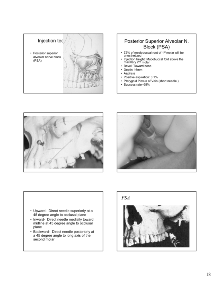
Injection technique • Posterior superior alveolar nerve block (PSA) Posterior Superior Alveolar N. Block (PSA) • 72% of mesiobuccal root of 1st molar will be anesthetized • Injection height: Mucobuccal fold above the maxillary 2nd molar • Bevel: Toward bone • Depth: 16mm • Aspirate • Positive aspiration: 3.1% • Pterygoid Plexus of Vein (short needle ) • Success rate>95% PSA • Upward- Direct needle superiorly at a 45 degree angle to occlusal plane • Inward- Direct needle medially toward midline at 45 degree angle to occlusal plane • Backward- Direct needle posteriorly at a 45 degree angle to long axis of the second molar 18 Middle Superior Alveolar N. Block (MSA) Disadvantages • • • • Risk of hematoma No bony landmarks Second injection may be required Positive aspiration: 3.1% Injection Technique • Infra-orbital nerve block Intraoral approach Extraoral approach • Insertion of needle: 2nd premolar mucobuccal fold • Bevel: Toward bone • Anesthetized: 1st, 2nd premolar, mesial root of the 1st molar Anterior Superior Alveolar N. Block (ASA) (Infraorbital N. Block) • • Anesthesia : Pulp of the maxillary central incisor through the canine on the injection side 72% of patients, pulp of Mx. premolars and mesiobuccal root of the 1st molar – • • • • (only 28% of population present MSA nerve) Area of Insertion : over the first premolar Target : infraorbital foramen Needle length : 16 mm Deposit : 0.9 – 1.2 mL Infraorbital Nerve Block 19 Palatal Anesthesia Traumatic experience Pain – discomfort Topical anesthesia Pressure anesthesia Cotton applicator stick Handle of a mouth mirror Ischemia (blanching) Deposit solution slowly Finger rest. Elbow rest. Nasopalatine N. Block • Through the palate • Through the labial Nasopalatine N. Block Two approaches: i) Palatal: lateral to the incisive palpilla Anesthetized : anterior portion of the hard palate (soft & hard tissues)from the both sides of mesial of 1st premolar Advance needle toward foramen (5mm) Continue to deposit small amount of anesthetic throughout the procedure (any injections) Deposit : 0.45 mL ii) Labial approach (2-3 injections) a) inject labial frenum b) interdental papilla c) possible lateral to the incisive papilla 20 Greater Palatine N. Block • • • • • • Extended neck Open wide Find the depression (distal to the 2nd molar) Depth < 10mm 0.45 – 0.6mL Anesthetized: distal of 1st premolar (Hard and Soft tissue) Maxillary Nerve Block (2nd Division Nerve Block) Area anesthetized : Hemimaxilla Deposit : 1.8ml Two approaches 1) High-tuberosity approach pterygopalatine fossa depth: 30mm 1) Greater palatine canal approach through the greater palatine foramen depth:30mm 21 Pain and anxiety Control Mandibular Injection Technique Mandibular Injection Techniques • • • • • • • • Inferior Alveolar N. Block (IAN) Lingual Nerve Block Gow-Gates (V3) Vazirani-Akinosi Mental N. Block Incisive N. Block Long Buccal N. Block Infiltration 22 Anatomy Anatomy Injection technique The Needle • Gauge: the larger the gauge the smaller the internal diameter of the needle -25g red cap -27g yellow cap -30g blue cap Long Needle:32mm Short Needle:20mm • Inferior alveolar nerve block Differences by manufacturer Inferior Alveolar N. Block (IAN) • Most frequently used • Positive aspiration 10 – 15% • Height of injection: 6 – 10mm above the occlusal plane • Landmark:coronoid notch, – Pterygomandibular raphe – Occlusal plane etc Inferior Alveolar N. Block (IAN) cont. • Target area : Before alveolar N. enter into the foramen • • • • Depth: 20 – 25mm If bone is contacted too soon: If bone is not contacted: Lingual N:Deposit small amount of anesthetic upon withthrouing to anesthtized lingual N. Remember lower incisor region overlaps of sensory fibers from the contralateral side. 23 Inferior Alveolar N. Block(IAN) Clinical failure rate : 15-20% (anatomical variation, depth of soft tissue)height of mandibula foramen Avoid, if possible, bilateral IAN Anesthetized area: Position of patient: supine or semisupine Location of needle tip:superior to the mandibular foramen Deposit = 1.5mL Signs and Symptoms IAN • Tingling and numbness of lower lip • Tingling and numbness of tongue • Elimination of pain 24 Injection technique • Remember – Always aspirate before injection! Complications of IANB 1). Hematoma 2). Trismus 3). Transient facial paraylsis Mandibular Nerve Block (Gow – Gates technique) 1973 : George Gow-Gates from Australia described true mandibular n. block Success rate : >95% (IAN:80-85%) Aspiration rate: < 2%(IAN 10-15%) Failure of Anesthesia (IANB) 1).Deposition of anesthetic too low, too anteriorly 2).Accessory innervation - Mylohyoid Nerves - Overlapping fibers of the contralateral alveolar nerve Injection Technique • Gow-Gates Block Gow-Gates Technique Distribution of V3 Target area: Lateral side of the condylar neck Landmark: Intertragic notch, corner of the mouth, mesiolingual cusp of maxillary 2ndmolar Penetration:Distal to the Mx 2nd or 3rd molar Height:Mesiolingual cusp of Mx 2nd molar (10 – 25mm from occlusal plane) Depth: 25mm Deposit: 1.8ml Time of onset:5-10”(IAN 3-5”) Bone is not contact:no deposit anesthetics move the syringe distally Keep the mouth open:1-2” 25 Gow-Gates Varizani-Akinosi Closedmouth Mandibular Block Gow-Gates Akinosi Trismus:Extraoral mandibular block 1960 : Varizani described technique 1977 : Dr. Joseph Akinosi – Useful for patient with trismus Insertion : height of the mucogingival junction adjacent to the maxillary 3rd molar Depth :25mm Deposit : 1.5-1.8mL 26 Akinosi Akinosi Injection technique • Long buccal block Long Buccal Nerve Block Anesthetized: Soft tissue and periosteum buccal to the mandibular molar teeth Indications:Scaling,curettage,the use of rubberdam clamp,subgingival tooth preparation,place of matrix band Insertion: Distal,Buccal of last molar Length of needle penetration : 1-2 mm Deposit : 0.3mL Vevel : Toward the bone Landmark:Mucobuccal fold 27 Long Buccal Injection Technique • Mental nerve block Mental Nerve Block • Indications:when buccal soft tissue anesthesia is necessary for procedures in the mandible anterior to the mental foramen • Area anesthetized:buccal mucous membrane anterior to the mental foramen, Lower lip and chin. • Technique:25-27 gauge short needle • Least frequently employed 28 Mental Nerve Block Mental Nerve Block Area of insertion:mucobuccal fold at or just anterior to the mental foramen Target area: between the apices of the two premolars Patient`s mouth: partially clsed Located the mental foramen Radiograph Clinical exam Deoth:5-6mm Deposit:0.6ml Bevel:Toward the bone Indications Incisive Nerve Block • Pulpal anesthesia to teeth anterior to mental foramen • When inferior alveolar nerve block is not indicated Incisive N. Bloc Supplemental Injection Techniques Lingual soft tissue are not anesthetized Local infiltration through the interdental papilla or partial lingual N. blick Not necessary for the needle to enter into the foramen Area anesthetized : buccal mucosa, lower lip, pulp of the teeth Deposit = 0.6 mL Depth of penetration : 5-6mm Periodontal ligament injection (PDL) Intraseptal Intraosseous (IO) technique Intrapulpal injection 29 Chart Notation for Local Anesthsia • • • • • COMPLICATIONS OF LOCAL ANESTHETICS Give drug name Give volume Give dosage Give location of injection Give concentrations – local anesthetic agent – vasoconstrictor Lack of profound anesthesia • Regional block – Attempt Gow Gates or higher level IAN • Deposit more local anesthetic – overdose precautions!! • Use of articaine, prilocaine – contraindicated in regional blocks • PDL, intrapulpal injections • Consider adjuncts – nitrous oxide – sedation • Terminate procedure – consider conscious sedation/ GA LOCAL complications TRISMUS • Cause (IAN/Akinosi/Gow Gates) – – – – intramuscular injection (Med. Pterygoid, temporalis) Hemorrhage Barbed needle Contamination by alcohol or sterilant • Treatment – – – – Moist towel 20mins/hr Physiotherapy Analgesics &/or muscle relaxants Rule out infection LOCAL complications Needle breakage • Cause – Unexpected movement by patient – Needle size – Needle manipulation • Treatment – DO NOT PANIC ! – visible Æ remove – Not visible Æ refer to OMFS LOCAL complications Hematoma Cause Arterial or venous disruption Less common in palate Treatment KNOW your anatomy (esp. PSA, IAN, mental nerve) Pressure application to site Analgesics Heat application (>6 hours postinjection à vasodilatory) 30 LOCAL complications Pain on injection • Causes – – – – pH of local anesthetic (~pH 5.0) Temperature of local anesthetic Rapid injection technique Contamination with alcohol/ sterilant • Treatment – Careful administration – Transient discomfort • Cause (up to 22% incidence in select cases) – Trauma to neural sheath – Perineural hemorrhage – Articaine and Prilocaine in regional blocks LOCAL complications Persistent anesthesia/ Paresthesia • Treatment – Prevent self-inflicted injury (stickers, short LA) – Resolution in 8wks – Persistent (>2 months) • document degree and extent • Refer to OMFS within 3 months for consultation LOCAL complications CNVII Paralysis • Cause – Deposition into parotid gland – Branches of CN VII • • • • • Temporal Zygomatic Buccal Marginal mandibular Cervical • Treatment – Transient paralysis – Protect cornea LOCAL complications Epithelial Desquamation • Cause – Prolonged use of topical anesthetic – High concentration of vasoconstrictors – Predominantly in palatal mucosa • Treatment – Resolution in 7-10 days – Analgesics – Na bicarbonate, saline or Peridex mouthrinse LOCAL complications Edema • Cause – – – – Trauma Infection Hemorrhage Angioedema (allergy) 31 SYSTEMIC COMPLICATIONS LOCAL complications Treatment Analgesics Antibiotics (infection) Oral antihistamines (allergy) Airway compromise Basic life support Activate emergency medical service (911) Allergy to local anesthetic • Cause – PABA in esters – Metabisulfite preservatives in vasoconstrictorcontaining local anesthetics – Sulfa allergy (articaine) – Latex • Treatment – Obtain accurate history – 1% diphenhydramine c 1:100,000 epi – Consider GA Causes of LA toxicity • Overdose MAXIMUM DOSES IN PATIENTS WITH CORONARY ARTERY DISEASE vasoconstrictor concentration/ type – 2% lidocaine c 1:100,000 epi • 7mg/kg adult • 70kg adult = 490mg max • 490 / 36mg per 1.8cc carpule = 13.6 carpules • Intravascular injection • Altered metabolism/ excretion – Hepatic insufficiency – Renal dysfunction Symptoms of LA Toxicity Initial symptoms (doses approximately 6.0 mcg/mL) vasoconst dental Max in CAD rictor cartridge (mg) (mg/ml) (mg/1.8c c) 1:20,000 levonordefrin 0.05 mg 0.09 mg 1:50,000 epinephrine 0.02 mg 0.036 mg 0.04 mg epinephrine 1 1:100,000 epinephrine 0.01 mg 0.018 mg 0.04 mg epinephrine 2 1:200,000 epinephrine 0.005 mg 0.009 mg 0.04 mg epinephrine 4 2 DEGRADATION OF CATECHOLAMINES: • selective presynaptic reuptake mechanism • Catechol-O-methyltransferase (COMT) inactivation • Monoamine oxidase (MAO) inactivation Symptoms of LA Toxicity Higher dose (>10 mcg/mL) • Lightheadedness initial CNS excitation followed by a rapid CNS depression • Dizziness Bradycardia • Visual and auditory disturbances Convulsions / seizures • Disorientation 0.20 mg levonordefrin max cartridges • Syncope • Coma • Respiratory depression and arrest • Cardiovascular depression and collapse • Drowsiness • Tachycardia 32 Management of LA toxicity • Reassure patient • 5mg diazepam or 1mg midazolam IV (5mg midazolam IM) for anxiety and convulsions • Supplemental oxygen • Continuous monitoring of vital signs • Cardiopulmonary resuscitation and activation of EMS in unconscious patients MALIGNANT HYPERTHERMIA 1:15,000 children and 1:50,000 adults, AD Pathophysiology: • Acute elevation in Ca++ levels • continuous release of Ca++ • dysfunction in reuptake mechanism Symptoms: • tachycardia, fever, cardiac dysrhythmia, muscle rigidity, cyanosis, death Associated agents: succinylcholine (77% of cases), halothane (60% of cases), lidocaine, mepivacaine, bupivicaine Judicious use of amide local anesthetics, blocks better than infiltrations Judicious use of vasoconstrictor (not more than 0.04mg per pt) Treatment: Dantrolene sodium 2-3mg/kg IV bolus Journal (Canadian Dental Association). 61(4):319-20, 323-6, 329-30, 1995 Apr METHEMOGLOBINEMIA Oxidation of Fe++ to Fe +++ à inability of Hb to release O2 (methemoglobin) Symptoms: respiratory depression, syncope, cyanosis, chocolate brown arterial blood Drugs: prilocaine lidocaine large dose benzocaine Treatment: 1% methylene blue (1.5 mg/kg) Congenital methemoglobinemia= relative contraindication Haas DA and Lennon D A retrospective study of paresthesia following the injection of local anesthetic in dentistry was conducted by examining every report of paresthesia recorded by Ontario's Professional Liability Program from 1973 to 1993, inclusive. Only those cases where surgery was not conducted were considered in this study. The parameters examined included patient age and gender, needle gauge, site of injection, area affected, report of pain or any additional symptoms, and the type of local anesthetic used. From 1973 to 1993, there were 143 reports of paresthesia not associated with surgery. There were no significant differences found with respect to patient age, patient gender, or needle gauge. All reports involved anesthesia of the mandibular arch, with the tongue most frequently reported to be symptomatic, followed by the lip. Pain was reported in 22 per cent of the cases. Paresthesia was reported most often following the injection of articaine and prilocaine. In 1993 alone, there were 14 reports of paresthesia not associated with surgery. This can be projected to an incidence of 1:785,000 injections. Articaine was administered in 10 of these cases or prilocaine in the other four. The observed frequencies of paresthesia following the administration of articaine (p < 0.002) or prilocaine (p < 0.025) were significantly greater than the expected frequencies for these agents, based on the distribution of local anesthetic use in Ontario in 1993. These results are consistent with the suggestion that local anesthetic formulations may have the potential for mild neurotoxicity. Further studies are needed to investigate the mechanisms for this, and to determine whether similar findings would be found elsewhere. Management of complications Rule #1 always document complications no matter how complicated they are Rule #2 reassure patient and make them aware of these complications (informed consent) Rule #3 always document complications to CYA Rule #4 monitor patient and vital signs 33 Thank You 34
