organic papers
Redetermination of p-ethoxyacetanilide (phenacetin)
Acta Crystallographica Section E
Structure Reports
Online
ISSN 1600-5368
Lars Kr. Hansen,a* German L.
Perlovichb,c and Annette
Bauer-Brandlb
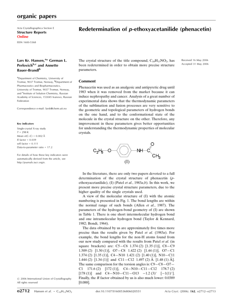
The crystal structure of the title compound, C10H13NO2, has
been redetermined in order to obtain more precise structure
parameters.
Received 16 May 2006
Accepted 31 May 2006
a
Department of Chemistry, University of
Tromsø, 9037 Tromsø, Norway, bDepartment of
Pharmaceutics and Biopharmaceutics,
University of Tromsø, 9037 Tromsø, Norway,
and cInstitute of Solution Chemistry, Russian
Academy of Sciences, 153045 Ivanovo, Russian
Federation
Correspondence e-mail: larsk@chem.uit.no
Key indicators
Single-crystal X-ray study
T = 298 K
Mean (C–C) = 0.002 Å
R factor = 0.039
wR factor = 0.111
Data-to-parameter ratio = 17.2
Comment
Phenacetin was used as an analgesic and antipyretic drug until
1983 when it was removed from the market because it can
induce nephropathy and cancer. Analysis of a great number of
experimental data shows that the thermodynamic parameters
of the sublimation and fusion processes are very sensitive to
the geometric and topological parameters of hydrogen bonds
on the one hand, and to the conformational state of the
molecule in the crystal structure on the other. Therefore, any
improvement in these parameters gives better opportunities
for understanding the thermodynamic properties of molecular
crystals.
For details of how these key indicators were
automatically derived from the article, see
http://journals.iucr.org/e.
# 2006 International Union of Crystallography
All rights reserved
o2712
Hansen et al.
C10H13NO2
In the literature, there are only two papers devoted to a full
determination of the crystal structure of phenacetin (pethoxyacetanilide), (I) (Patel et al., 1983a,b). In this work, we
present more precise crystal structure parameters, due to the
higher quality of the single crystals used.
A view of the molecular structure of (I) with the atomic
numbering is presented in Fig. 1. The bond lengths are within
the normal range of such bonds (Allen et al., 1987). The
parameters of the hydrogen-bond geometry of (I) are shown
in Table 1. There is one short intermolecular hydrogen bond
and one intramolecular hydrogen bond (Taylor & Kennard,
1982; Bondi, 1964).
The data obtained by us are approximately five times more
precise than the results given by Patel et al. (1983a). For
example, the bond lengths for the non-H atoms found from
our new study compared with the results from Patel et al. (in
square brackets) are: C5—C6 1.374 (2) [1.35 (1)], C8—C9
1.509 (2) [1.50 (1)], O7—C8 1.422 (2) [1.44 (1)], O7—C1
1.374 (2) [1.35 (1)], C4—N10 1.421 (2) [1.40 (1)], N10—C11
1.444 (2) [1.34 (1)] and C11—C12 1.497 (2) Å [1.48 (1) Å].
The same comparison for the torsion angles is: C9—C8—O7—
C1 171.6 (2) [172 (1)], C4—N10—C11—C12 178.7 (2)
[178 (1)] and C4—N10—C11—O13
1.2 (3) [ 1(1) ].
Finally, the R factor obtained by us is also much lower: 0.0389
[0.088].
doi:10.1107/S1600536806020551
Acta Cryst. (2006). E62, o2712–o2713
organic papers
Table 1
Hydrogen-bond geometry (Å, ).
D—H A
D—H
H A
D A
D—H A
C3—H3 O13
N10—H10 O13i
0.93
0.86
2.42
2.09
2.915 (2)
2.9426 (18)
114
175
Symmetry code: (i)
Figure 1
A view of the molecular structure of (I), with the atomic numbering
scheme. Displacement ellipsoids are drawn at the 30% probability level.
Experimental
Crystals of (I) were grown by slow evaporation of an ethanol solution.
Crystal data
C10H13NO2
Mr = 179.21
Monoclinic, P21 =c
a = 13.3236 (14) Å
b = 9.6159 (15) Å
c = 7.7331 (15) Å
= 103.992 (14)
V = 961.4 (3) Å3
Z=4
Dx = 1.238 Mg m 3
Mo K radiation
= 0.09 mm 1
T = 298 (2) K
Block, colourless
0.50 0.40 0.30 mm
Data collection
Enraf–Nonius CAD-4
diffractometer
!/2 scans
Absorption correction: scan
[ABSCALC in OSCAIL
(McArdle & Daly, 1999) and
North et al. (1968)]
Tmin = 0.958, Tmax = 0.975
2299 measured reflections
2083 independent reflections
1056 reflections with I > 2(I)
Rint = 0.015
max = 27.0
3 standard reflections
frequency: 120 min
intensity decay: 1%
Refinement
Refinement on F 2
R[F 2 > 2(F 2)] = 0.039
wR(F 2) = 0.111
S = 1.00
2083 reflections
121 parameters
H-atom parameters constrained
Acta Cryst. (2006). E62, o2712–o2713
w = 1/[ 2(Fo2) + (0.0541P)2
+ 0.0361P]
where P = (Fo2 + 2Fc2)/3
(/)max = 0.009
max = 0.16 e Å 3
min = 0.14 e Å 3
Extinction correction: SHELXL97
(Sheldrick, 1997)
Extinction coefficient: 0.021 (3)
x; y
1
2;
z
1
2.
All H atoms were positioned geometrically and refined using a
riding model, fixing the aromatic C—H distances at 0.93 Å, the N—H
distance at 0.86 Å and the CH2 C—H distances at 0.97 Å, with
Uiso(H) = 1.3Ueq(C,N). The CH3 C—H distances were fixed at 0.96 Å,
with Uiso(H) = 1.4Ueq(C). In addition, the methyl groups were
allowed to rotate but not to tip.
Data collection: CAD-4-PC Software (Enraf–Nonius, 1992); cell
refinement: CELDIM in CAD-4-PC Software; data reduction:
XCAD4 (McArdle & Higgins, 1995); program(s) used to solve
structure: OSCAIL-X for Windows (McArdle, 2005) and SHELXS97
(Sheldrick, 1997); program(s) used to refine structure: OSCAIL-X for
Windows and SHELXL97 (Sheldrick, 1997); molecular graphics:
ORTEX (McArdle, 1993) and ORTEPIII (Burnett & Johnson,
(1996); software used to prepare material for publication: OSCAIL-X
for Windows.
References
Allen, F. H., Kennard, O., Watson, D. G., Brammer, L., Orpen, A. G. & Taylor,
R. (1987). J. Chem. Soc. Perkin Trans. 2, pp. S1–19.
Bondi, A. (1964). J. Phys. Chem. 68, 441–451.
Burnett, M. N. & Johnson, C. K. (1996). ORTEPIII. Report ORNL-6895. Oak
Ridge National Laboratory, Tennessee, USA.
Enraf–Nonius (1992). CAD-4-PC Software. Version 1.1. Enraf–Nonius, Delft,
The Netherlands.
McArdle, P. (1993). J. Appl. Cryst. 26, 752.
McArdle, P. (2005). OSCAIL-X for Windows. Version 1.0.7. Crystallography
Centre, Chemistry Department, NUI Galway, Ireland.
McArdle, P. & Daly, P. (1999). ABSCALC. PC version. National University of
Ireland, Galway, Ireland.
McArdle, P. & Higgins, T. (1995). XCAD. National University of Ireland,
Galway, Ireland.
North, A. C. T., Phillips, D. C. & Mathews, F. S. (1968). Acta Cryst. A24, 351–
359.
Patel, U., Patel, T. C. & Singh, T. P. (1983a). Acta Cryst. C39, 1445–1446.
Patel, U., Patel, T. C. & Singh, T. P. (1983b). Curr. Sci. 52, 20–21.
Sheldrick, G. M. (1997). SHELXL97 and SHELXS97. University of
Göttingen, Germany.
Taylor, R. & Kennard, O. (1982). J. Am. Chem. Soc. 104, 5063–5070.
Hansen et al.
C10H13NO2
o2713


![(6 S*)-6-[(1S*,2R*)-1,2-Dihydroxypentyl]- Experimental](http://s2.studylib.net/store/data/013550387_1-701baf9290191754761def53644827d5-300x300.png)
