AAN.COM
Chapter 1 - The Neurologic Examination
HOME
AAN.COM
©2013 American Academy of
Neurology - All Rights Reserved
Section 2
In this section the following systems will be reviewed in greater detail:
• Cranial Nerves
• Cerebellum
• Mental Status Examination
• Peripheral Nervous System
• Sensory System
• Individual Muscle Testing
• Extrapyramidal System
• Evaluation of Speech and
Language
• Pyramidal System
System Review
Cranial Nerves
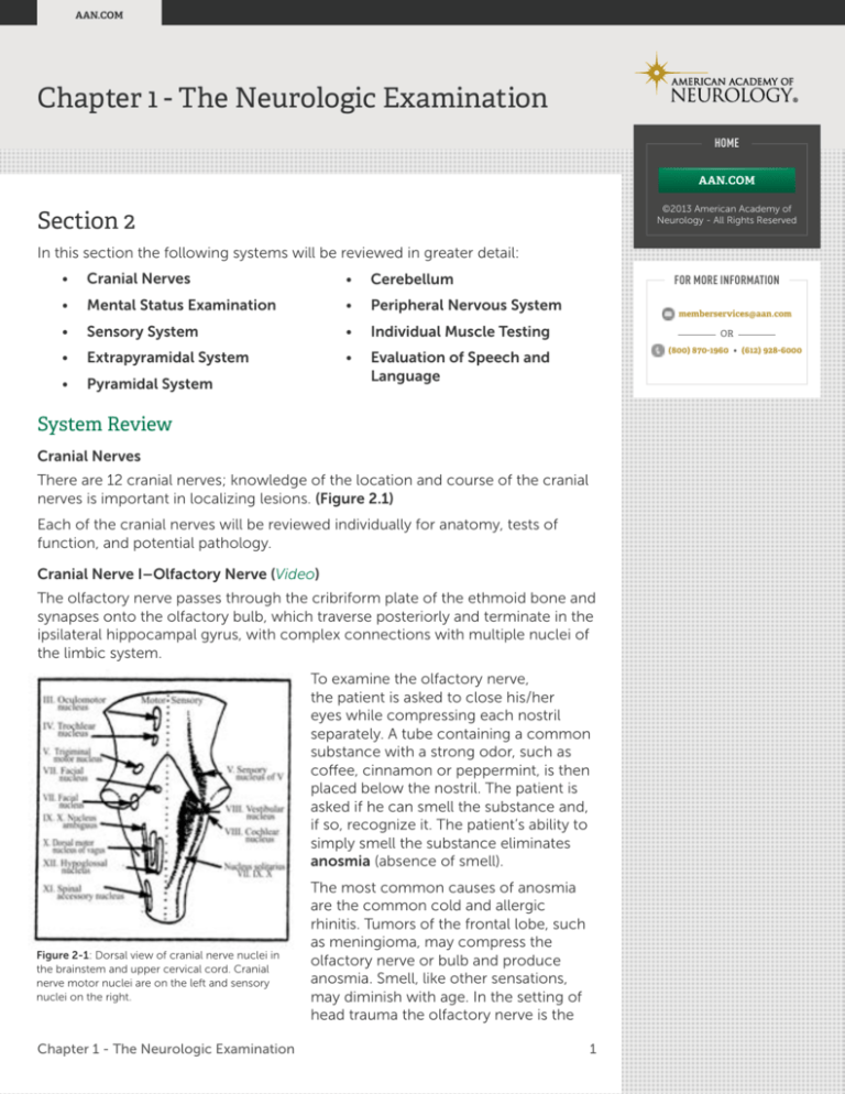
There are 12 cranial nerves; knowledge of the location and course of the cranial
nerves is important in localizing lesions. (Figure 2.1)
Each of the cranial nerves will be reviewed individually for anatomy, tests of
function, and potential pathology.
Cranial Nerve I–Olfactory Nerve (Video)
The olfactory nerve passes through the cribriform plate of the ethmoid bone and
synapses onto the olfactory bulb, which traverse posteriorly and terminate in the
ipsilateral hippocampal gyrus, with complex connections with multiple nuclei of
the limbic system.
To examine the olfactory nerve,
the patient is asked to close his/her
eyes while compressing each nostril
separately. A tube containing a common
substance with a strong odor, such as
coffee, cinnamon or peppermint, is then
placed below the nostril. The patient is
asked if he can smell the substance and,
if so, recognize it. The patient’s ability to
simply smell the substance eliminates
anosmia (absence of smell).
Figure 2-1: Dorsal view of cranial nerve nuclei in
the brainstem and upper cervical cord. Cranial
nerve motor nuclei are on the left and sensory
nuclei on the right.
The most common causes of anosmia
are the common cold and allergic
rhinitis. Tumors of the frontal lobe, such
as meningioma, may compress the
olfactory nerve or bulb and produce
anosmia. Smell, like other sensations,
may diminish with age. In the setting of
head trauma the olfactory nerve is the
Chapter 1 - The Neurologic Examination1
FOR MORE INFORMATION
memberservices@aan.com
OR (800) 870-1960 • (612) 928-6000
AAN.COM
most commonly injured cranial nerve due to shearing injuries that may or may
not be associated with fractures of the cribriform plate. If rhinorrhea occurs after
head trauma, nasal drip should be checked for the presence of glucose with
a Dextrostix? or urine test strip. A positive test for glucose suggests cribriform
plate fracture with cerebrospinal fluid leak as discharge from nasal mucosa does
not contain glucose.
Cranial Nerve II–Optic nerve
(see Chapter on the Visual System for a separate discussion)
Cranial nerve II, the optic nerve, is composed of axons that originate in the
ganglion cell layer of the retina. The optic disk of the fundus corresponds to
the attachment of the optic nerve to the retina. The absence of rods and cones,
the fundamental organs of sight, at the optic disk accounts for the blind spot in
one’s visual field. The optic nerve traverses posteriorly from the orbit through
the optic foramen (which also contains the ophthalmic artery) and merges with
the contralateral optic nerve to form the optic chiasm. A partial decussation of
the optic nerves at the optic chiasm results in the formation of the optic tracts.
Each tract contains axons from both retina, and project around the cerebral
peduncles to synapse at the lateral geniculate body. Some fibers from the
lateral geniculate body project to the midbrain to participate in the pupillary
light reflex. From the lateral geniculate
body arise the optic radiations which
hug the lateral ventricles as they traverse
posteriorly and then medially to the
primary visual cortex in the occipital
lobe.
Figure 2-2: Examination of the patient’s left eye
visual fields by confrontation. The patient is asked
to identify the number of fingers, which the
examiner raises in each quadrant while centering
his gaze on the examiner’s right eye.
The optic nerve is a special sensory
nerve that can be assessed by testing
for visual acuity, visual fields, and
funduscopic examination of the retina.
Visual acuity reflects central vision or
vision subserved by the macula where
cones are in highest concentration.
Monocular vision is tested by having the
patient cover one eye, hold a pocketsized Snellen chart at arm’s length, and
read the smallest numbers on the chart
that can be read. Visual acuity is graded
from 20/20 to 20/800. Corrective lens,
if available, should be worn during
testing. In the event that visual acuity
is so severely impaired that a miniature
Snellen chart is not useful, ask the
patient to count fingers placed about
14 inches in front. Failing this, check for
perception of movement, then light.
Poor visual acuity may be associated
with lesions involving the lens (cataracts),
anterior optic chamber (glaucoma),
retina (macular degeneration) or optic
nerve (optic neuritis).
Chapter 1 - The Neurologic Examination2
HOME
AAN.COM
©2013 American Academy of
Neurology - All Rights Reserved
FOR MORE INFORMATION
memberservices@aan.com
OR (800) 870-1960 • (612) 928-6000
AAN.COM
Visual field testing assesses the integrity
of the optic pathways as it comes from
the retina, optic nerves, optic chiasm,
optic tracts, and optic radiations to the
primary visual cortex. It is most commonly
performed by confrontation (Figure 2-2).
The patient faces the examiner while
covering one eye so that he fixates on the
opposite eye of the examiner, directly in
front of him.
Testing is performed by covering one
of the patient’s eyes and having the
patient fixate on the examiner’s nose.
One to three fingers are then shown
to the patient in each of the four visual
quadrants of each uncovered eye and
the patient asked to state the number of
fingers seen. Lack of vision in quadrants
can then be detected and mapped out to
various types of field defect.
Figure 2-3: Lesions of the optic nerve, optic
chiasm, optic tract, optic radiations and primary
visual cortex produce characteristic visual field
deficits.
If the patient is uncooperative, visual
field examination may be grossly tested
by asserting a threatening hand to half
of a visual field (while cautiously avoiding movement of air that can result in a
corneal blink reflex) and observing for a blink to threat.
Monocular visual field deficits are often due to lesions anterior to the optic
chiasm, ipsilateral to the field cut as may be seen with lens dislocation, or retinal
infarction from occlusion of the ophthalmic artery. Homonymous visual field
deficits (toward the same side, e.g., left temporal, right nasal = left homonymous
hemianopsia) imply a lesion posterior to the optic chiasm (Figure 2-3). The
more congruous, (looks the same for each eye), the homonymous field cut; the
more posterior the lesion is along the optic radiations. If macular sparing, or
sparing of the center of vision, is detected with a homonymous hemianopsia,
the lesion is most likely in the occipital lobe, as the macular area of the visual
cortex is kept viable after a posterior cerebral artery infarct by terminal branches
of the middle cerebral artery.
Figure 2-4: The action and nerve supply of the
extraocular muscles is demonstrated.
Funduscopy is performed with an
ophthalmoscope. The patient is asked
to fixate on an object in the distance
while the examiner uses his right eye to
examine the patient’s right eye and the
left eye for examination of the patient’s
left eye. Once the fundus is visualized,
systematic examination of the optic disk,
with attention to color and definition
of disk margins, arterial supply, venous
pulsations, and surrounding retina is
conducted.
Swelling of the optic disk may be due to inflammation of the optic nerve, optic
neuritis, or papilledema. These conditions may be difficult to differentiate based on
Chapter 1 - The Neurologic Examination3
HOME
AAN.COM
©2013 American Academy of
Neurology - All Rights Reserved
FOR MORE INFORMATION
memberservices@aan.com
OR (800) 870-1960 • (612) 928-6000
AAN.COM
funduscopy alone. Typically, optic neuritis
is associated with decreased visual acuity
and an enlarged blind spot. Optic pallor
implies optic atrophy from retrobulbar
neuritis, as seen in multiple sclerosis,
or ischemic optic neuropathy from
small vessel infarction of the optic nerve
secondary to long-standing hypertension.
Papilledema implies increased intracranial
pressure. Visual acuity is not affected
unless there is secondary atrophy of the
optic nerve from chronic pressure on the
optic nerve. With papilledema, venous
pulsations may be lost. Pallor of a segment
of the fundus, associated with complaints
of a “pie in the sky” loss of monocular
Figure 2-5
vision, suggests branch central retinal
artery occlusion secondary to embolic
or thrombotic occlusion of either the ciliary or ophthalmic arteries, both of which
supply the optic nerve.
Cranial Nerves III, IV, and VI–Oculomotor, Trochlear and Abducens Nerves
(see Chapter on the Visual System)
The oculomotor (III), trochlear (IV), and
abducens (VI), nerves together innervate
the extraocular muscles (Figure 2-4). The
primary action of the medial rectus is
adduction and that of the lateral rectus
is abduction. The superior rectus and
inferior oblique primarily elevate the eye
while the inferior rectus and superior
oblique primarily depress the eye.
The oculomotor nerve (cranial nerve III)
also innervates the levator palpebrae
muscle, which elevates the eyelid, the
pupillo-constrictor muscle, which
constricts the pupil, and the ciliary
muscle, which controls the thickness of
the lens, allowing for accommodation.
The nuclear complex of the oculomotor
nerve lies medially within the midbrain,
ventral to the aqueduct of Sylvius (Figure
2-5). It consists of the oculomotor
nucleus, which innervates the skeletal
muscles of the eye and the EdingerFigure 2-6: A complete left III nerve palsy is
illustrated along with clinical presentations of left
Westphal nucleus, which carries
VI and IV nerve palsies.
parasympathetic innervation to the pupil
and ciliary muscle. The superior branch
of the oculomotor nerve supplies the superior rectus and levator of the upper
lid while the inferior division innervates the medial rectus, inferior rectus, inferior
oblique, pupilloconstrictor muscle and ciliary body.
Chapter 1 - The Neurologic Examination4
HOME
AAN.COM
©2013 American Academy of
Neurology - All Rights Reserved
FOR MORE INFORMATION
memberservices@aan.com
OR (800) 870-1960 • (612) 928-6000
AAN.COM
Figure 2-7: The course of the trochlear nerve
in the pons, across the posterior and superior
cerebellar arteries, across the petrous ridge of
the temporal bone, through the cavernous sinus,
and out the superior orbital fissure is illustrated.
Figure 2-8: Note how the facial nerve wraps
around the nucleus of cranial nerve VI within
the pons.
Figure 2-9: The arrows and numbers indicate
the sequence of eye movements tested in the six
cardinal fields of gaze.
A complete oculomotor palsy will
manifest as ptosis, dilated and fixed
pupil, and outward and slightly
downward deviation of the eye. (Figure
2-6). Pupil-sparing, isolated oculomotor
nerve paresis is often due to ischemia
from hypertension, diabetes, tertiary
syphilis, or vasculitis, as the pupillomotor
fibers travel along the periphery (outside)
of the oculomotor nerve, closer to the
blood supply of the nerve and are less
susceptible to end-arteriole ischemia
that tends to affect the center of the
nerve. On the other hand, acquired
third nerve palsies, which involve the
pupil, may be due to compressive
lesions such aneurysm of the posterior
communicating artery, head trauma,
and tumors of the cerebral hemispheres
compressing the oculomotor nerve and
the parasympathetic fibers, which run
peripherally within it.
The trochlear nerve (cranial verve IV)
nucleus lies in the medial midbrain, and
wraps around the midbrain dorsally,
alongside the cerebral peduncles, and
courses between the posterior cerebral
and superior cerebellar arteries (Figure
2-7). As the trochlear nerve has the
longest intracranial distance of the cranial
nerves, head trauma is the most common
cause of nerve injury. A large proportion
of fourth nerve palsies, however, are
congenital and associated with a superior
oblique that is shortened and tethered.
The abducens nerve (cranial nerve VI)
nucleus lies in the caudal portion of
the pons. The axons course ventrally
through the pons and then travel in the
middle of the cavernous sinus, through
the superior orbital fissure and into the
lateral rectus muscle, which it innervates.
The facial nerve loops around the
abducens nerve nucleus within the pons,
therefore a pontine lesion in this location
will produce ipsilateral paralysis of the
lateral rectus and lower motor neuron
facial nerve palsy (Figure 2-8).
Examination of the extraocular muscles
is first conducted by examining the
Chapter 1 - The Neurologic Examination5
HOME
AAN.COM
©2013 American Academy of
Neurology - All Rights Reserved
FOR MORE INFORMATION
memberservices@aan.com
OR (800) 870-1960 • (612) 928-6000
AAN.COM
alignment of the patient’s eyes in the primary position (patient looking straight
ahead). Shine a light into the patient’s eyes and examine the corneal light
reflection. If the light falls off center to a pupil, there is evidence of ocular
malalignment, termed exotropia if the eye is laterally deviated, esotropia if
the eye is medially deviated, hypertropia if the eye is deviated upwards and
hypotropia if the eye is deviated downwards. Next, examine ocular motility by
asking the patient to follow the examiner’s finger as it is moved through the six
cardinal fields of gaze (Figure 2-9). During conjugate eye movements the yoke
muscles are equally stimulated so a lag in eye movement is a subtle sign of
extraocular muscle weakness. Complaints of double vision by the patient will not
always manifest as visible extraocular muscle weakness. Diplopia is worse in the
direction of gaze of the weak muscle.
Cranial Nerve V–Trigeminal Nerve (Video)
The trigeminal nerve provides sensation
to the face and mucous membranes of
the nose, mouth, tongue and sinuses as
well as motor innervation to the muscles
of mastication.
The cell bodies of most sensory neurons
innervating the face lie in the Gasserian
ganglion and the rest are in the
mesencephalic nucleus. There are three
sensory divisions of the trigeminal nerve,
all with their origin in the Gasserian
ganglion: the ophthalmic (V1), maxillary
(V2) and mandibular branches (V3).
The ophthalmic division innervates the
conjunctiva, cornea, upper lid, forehead,
bridge of the nose and upper scalp to
the vertex (Figure 2-10). The maxillary
branch innervates the cheek, lateral
surface of the nose, upper teeth, jaw,
and mucosal membranes of the nose
Figure 2-10: The subdivisions of cranial nerve V
innervation to the face is illustrated.
and upper portion of the oropharynx.
The mandibular branch carries sensory
and motor neurons to the lower jaw, pinna of the ear, anterior portion of the
external auditory meatus, ipsilateral tongue, lower teeth, and mucosal surface of
the cheeks and floor of the mouth. The motor fibers innervate the temporalis,
masseters and medial and lateral pterygoids.
Light touch is assessed by using a cotton wisp and gently touching the areas
innervated by the three divisions of the trigeminal nerve while the patient’s eyes
are closed. The patient is asked to say, “touch” whenever he feels the cotton.
To test pain sensation, repeat the above maneuver with the sharp and round
end of a safety pin, asking the patient to discriminate between “sharp” and
“dull.” Temperature sensation can be tested by filling two test tubes individually
with cold and warm water, applying the test tubes to the three divisions of the
trigeminal nerve and asking the patient to differentiate cold from warm.
The corneal blink reflex tests the integrity of the ophthalmic division of V, which
innervates the cornea and constitutes the sensory component of the reflex, and
Chapter 1 - The Neurologic Examination6
HOME
AAN.COM
©2013 American Academy of
Neurology - All Rights Reserved
FOR MORE INFORMATION
memberservices@aan.com
OR (800) 870-1960 • (612) 928-6000
AAN.COM
the facial nerve, which constitutes the motor arc of the reflex by innervating the
orbicularis oculi and allowing closure of the eyelid. To test the reflex, the end of
a cotton Q-tip is twisted into a point. The patient is asked to look laterally and
the cotton point applied gently onto the cornea from the direction contralateral
to the gaze so as to avoid reflex defensive blinking. In patients who are
comatose, the presence of a corneal blink reflex implies that the sensory nucleus
of V and the facial nerve nucleus, both in the pons, are intact. .
Cranial Nerve VII–Facial Nerve (Video)
The facial nerve, cranial nerve VII, innervates all the muscles of facial expression,
i.e., the muscles around the eyes, mouth, nose, ears and neck. It also innervates
the stapedius muscle in the ear, which dampens excessive movement of the
ossicles when subject to loud sounds. The facial nerve subserves taste to the
anterior two thirds of the tongue and sensation to the outer ear.
Figure 2-11: A right upper motor neuron
VII lesion due to a left subcortical stroke is
illustrated.
Figure 2-12: A lower motor neuron VII lesion due
to a left peripheral facial nerve palsy is illustrated.
The motor nucleus of VII sits in the pons
while its axons loop around the nucleus
of the abducens nerve and emerges
from the pontomedullary junction. The
facial nerve then courses through the
internal auditory meatus where it is
joined by the auditory nerve, and enters
the facial canal of the temporal bone.
Distal to the geniculate ganglion, the
facial nerve gives off the chorda tympani,
which supplies taste to the anterior
two thirds of the tongue via the lingual
nerve. The facial nerve exits the facial
canal through the stylomastoid foramen,
passing through the parotid gland,
before innervating the muscles of the
face and the platysma.
To test the facial nerve, first observe the
patient’s face for symmetry by paying
close attention to the nasolabial folds,
forehead wrinkles, spontaneous smiling
and blinking. Then, ask the patient to
show his teeth, raise his eyebrows,
squeeze his eyes shut tightly and hold
air in his cheeks. Facial weakness may
be due to upper motor neuron or
lower motor neuron facial palsy. Upper
motor neuron palsy implies that there
is a lesion contralateral to the side of
facial weakness which is disrupting
the face motor fibers somewhere in its
course from the primary motor cortex
to the facial nucleus within the pons
(i.e., upper motor neuron to the facial
nerve nucleus, Figure 2-11). A typical
presentation of an upper motor neuron
palsy is a patient with a right subcortical
Chapter 1 - The Neurologic Examination7
HOME
AAN.COM
©2013 American Academy of
Neurology - All Rights Reserved
FOR MORE INFORMATION
memberservices@aan.com
OR (800) 870-1960 • (612) 928-6000
AAN.COM
lacunar infarct resulting in flattened left nasolabial fold, decreased up turning of
the left corner of the mouth on smiling, and symmetric wrinkling of forehead
bilaterally, in addition to a left hemiparesis. Lower motor neuron palsy implies a
lesion involving the facial nerve at the nucleus in the pons or along the course
of the facial nerve ipsilateral to the side of facial weakness (Figure 2-12). Bell’s
palsy is a lower motor neuron facial palsy whereby the patient has unilateral
flattening of the nasolabial fold with inability to upturn the corner of the mouth
upon smiling, inability to wrinkle his forehead, delayed or absent blinking due to
weakness of the eyelid, and inability to hold air in the cheeks due to escape of air
through the corner of the mouth which is weak. In addition, patients with Bell’s
palsy may complain of dry eye from disruption of parasympathetic innervation of
the lacrimal gland, hyperacusis or augmented hearing in the ear ipsilateral to the
lesion from paralysis of the stapedius muscle and diminished taste from a lesion
proximal to the lingual nerve, which inhibits afferent signals concerning taste
from reaching the brainstem.
In summary upper motor neuron facial weakness spares the frontalis (forehead
muscle) so the patient can wrinkle his brow. Lower motor neuron facial
weakness involves the forehead muscle and the patient can’t wrinkle the brow
and in addition has unilateral hyperacusis and loss of taste.
Facial diplegia, or bilateral lower motor neuron facial weakness, is seen in such
conditions as Guillain-Barr? syndrome or sarcoidosis.
Cranial Nerve VIII–Acoustic Nerve (Video)
The auditory nerve, cranial nerve VIII, is composed of two divisions, the cochlear
nerve, which subserves hearing, and the vestibular nerve, which provides sense
of balance. The cochlear nerve sits in the lower pons, near the cerebellopontine
angle. Lesions of the cochlear nerve commonly present with ipsilateral
decreased hearing and sometimes tinnitus. The vestibular nerve is composed
of nerve fibers from the labyrinth of the inner ear, which travel alongside the
cochlear nerve to terminate on the vestibular nuclei within the lower pons.
To test the auditory nerve, first check gross hearing in each ear by rubbing your
fingers about 30 inches from the patient’s ear, with the contralateral ear covered.
If hearing in one ear is impaired, perform Rinne and Weber tests. Both tests
employ the use of a 256 Hz tuning fork.
In the Rinne test, the vibrating tuning
fork is place over the mastoid process,
behind the ear to test bone conduction
(BC). Ask the patient to tell you when
he no longer hears the vibrating fork,
after which the tuning fork is placed in
front of the ear and the patient asked
if he can hear it (air conduction = AC).
Next perform the Weber test by placing
a vibrating tuning fork over the middle
of the forehead and ask the patient if the
Figure 2-13: The Hallpike maneuver is illustrated.
sound is louder in one ear compared to
The patient initially is seated upright and asked to
the other. With conductive hearing loss,
fall backwards, so that his head is below the plane
from middle ear disease or obstruction
of his trunk. The examiner then turns his head
to one side and asks the patient to look in the
of the external auditory meatus with wax,
direction to which his head is turned.
BC will be greater than AC and Weber
Chapter 1 - The Neurologic Examination8
HOME
AAN.COM
©2013 American Academy of
Neurology - All Rights Reserved
FOR MORE INFORMATION
memberservices@aan.com
OR (800) 870-1960 • (612) 928-6000
AAN.COM
test will lateralize to the deaf ear. However, with sensorineural hearing loss AC is
better than BC and Weber test will lateralize to the good ear.
Vestibular nerve function can be tested with postural maneuvers. In patients
suspected of benign positional vertigo, presenting with vertigo or dizziness
associated with changes in head position, the Hallpike maneuver should be
attempted when not contraindicated due to severe cervical spine disease. To
perform the Hallpike maneuver, the patient sits up in bed and then quickly lies
back on command so that his head hangs over the edge of the bed. The head
is tilted backward below the plane of his body and turned to one side by the
examiner who holds the patient’s head in his examiner who holds the patient’s
head in his hands. The patient is asked to look in the direction that his head
is turned (Figure 2-13). Watch for nystagmus in the direction of gaze and ask
the patient if he feels vertigo. If no nystagmus is observed after 15 seconds,
have the patient sit up and repeat the maneuver turning the patients head and
directing his gaze in the contralateral direction. The absence of nystagmus
suggests normal vestibular nerve function. However, with peripheral vestibular
nerve dysfunction, such as benign positional vertigo, the patient will complain
of vertigo, and rotary nystagmus will appear after a 1- to 5-second latency
toward the direction in which the eyes are deviated. With repeated maneuvers,
the nystagmus and sensation of vertigo will fatigue and disappear, a sign of
peripheral vestibular disease, in contrast to central vestibular disease from stroke
or other intrinsic brainstem lesions, which manifests as nonfatigable nystagmus
without delay in onset.
Cranial Nerves IX and X–The Glossopharyngeal and Vagus Nerves (Video)
The glossopharyngeal nerve (cranial nerve IX) contains sensory and motor fibers
as well as autonomic innervation to the parotid glands. It mediates taste to the
posterior one third of the tongue and sensation to the pharynx and middle ear.
Like the glossopharyngeal nerve, the vagus nerve (cranial nerve X) contains
sensory, motor and autonomic fibers. Motor innervation to the muscles of the
soft palate, pharynx and larynx originates in the medulla. Autonomic fibers arise
from the dorsal motor nucleus of vagus and synapse at peripheral ganglia to
provide parasympathetic innervation to the trachea, esophagus, heart, stomach,
and small intestine.
To test glossopharyngeal and vagus
nerve function, examine the position of
the uvula and its movement by asking
the patient to say “Ah.” the soft palate
should elevate symmetrically and the
uvula should remain in the midline. The
gag reflex can be tested by touching
the pharyngeal wall on each side with
a cotton tip applicator. This reflex relies
on an intact sensory arc, as mediated by
sensory fibers of the glossopharyngeal
Figure 2-14: A normal soft palate is illustrated on
the left. On the right, a right palatal palsy from a
nerve to the soft palate, and an intact
lower motor neuron X nerve lesion has resulted
motor arc, as mediated by the motor
in deviation of the uvula to the left.
fibers of the vagus nerve to the soft palate
and pharynx. Deviation of the uvula to one side implies a lower motor lesion of
the vagus nerve contralateral to the side the uvula is deviating to (Figure 2-14).
Chapter 1 - The Neurologic Examination9
HOME
AAN.COM
©2013 American Academy of
Neurology - All Rights Reserved
FOR MORE INFORMATION
memberservices@aan.com
OR (800) 870-1960 • (612) 928-6000
AAN.COM
An upper motor neuron vagus nerve lesion will present with the uvula deviating
toward the side of the lesion. The presence of a gag reflex does not necessarily
imply that the patient can swallow without aspiration after a stroke. Impairment of
swallowing is usually due to bilateral vagus nerve lesions. On the other hand, the
absence of a gag reflex does not imply inability to swallow. .Hoarseness may be
seen with tumors encroaching on the recurrent laryngeal nerve, a branch of the
vagus nerve. This results in unilateral vocal cord paralysis.
Cranial Nerve XI–The Accessory Nerve (Video)
The spinal accessory nerve, cranial nerve XI, innervates the sternocleidomastoid
and trapezius muscles.
To test the strength of the sternocleidomastoids ask the patient to turn
his head against your hand, which is placed over the mandible. Repeat
this maneuver with your hand on the contralateral mandible. Observe the
sternocleidomastoid, which is contralateral to the side to which the patient is
turning his head. Weakness detected when the patient turns his head to the
left implies that the right sternocleidomastoid is weak. To test the trapezius,
ask the patient to shrug his shoulders and press down on the shoulders.
Trapezius weakness is manifest as difficulty in elevating the shoulders. When
the sternocleidomastoid and trapezius are weak on the same side, an ipsilateral
peripheral accessory palsy, involving cranial nerves X and XI, is implied as may
be seen with a jugular foramen tumor, i.e., glomus tumor or neurofibroma.
Because the cerebral hemisphere innervates the contralateral trapezius and
ipsilateral sternocleidomastoid, a large right hemisphere stroke will result in
weakness of the left trapezius and right sternocleidomastoid. Bilateral wasting
of the sternocleidomastoid may be seen with myopathic conditions such as
myotonic dystrophy and polymyositis or motor neuron disease, the latter usually
associated with fasciculations.
Cranial Nerve XII–Hypoglossal Nerve (Video)
The hypoglossal nerve, cranial nerve XII,
is a pure motor nerve, innervating the
muscles of the tongue.
Figure 2-15: The opposing action of the two
halves of the tongue is illustrated. Note that the
tongue, like most muscles in the body derives
supranuclear innervation from the contralateral
motor cortex.
To test the function of the hypoglossal
nerve, ask the patient to protrude his
tongue and wiggle it from side to side.
Look for deviation and atrophy. To check
for subtle weakness, ask the patient to
push his tongue against the wall of his
cheek while you push against it through
the outer cheek. Like the forehead, each
side of the tongue receives upper motor
neuron innervation from bilateral motor
cortices. Each half of the tongue pushes
the tongue in the contralateral direction,
i.e., left half of tongue pushes to the
right (Figure 2-15). Thus, if the tongue
deviates to one side, it is pointing to
the side that is weak. Tongue deviation,
combined with wasting on the side to
which it is deviated, implies a unilateral,
Chapter 1 - The Neurologic Examination10
HOME
AAN.COM
©2013 American Academy of
Neurology - All Rights Reserved
FOR MORE INFORMATION
memberservices@aan.com
OR (800) 870-1960 • (612) 928-6000
AAN.COM
lower motor neuron, hypoglossal nucleus or nerve lesion as may be seen
with syringobulbia (a degenerative cavity within the brainstem), with basilar
meningitis, or foramen magnum tumor. If the tongue deviates and is of normal
bulk, one should consider an upper motor neuron lesion, such as stroke or
tumor in the hemisphere contralateral to the side of deviation, and look for
associated hemiparesis on the side of tongue deviation.
The Mental Status Examination
As previously noted, the neurologic exam begins with an assessment of the
patient’s mental status. In most cases, a large part of the mental status exam may
be ascertained from observation of the patient as history is provided. A more
detailed mental status exam can be divided into the following components:
• Level of consciousness
• Intellectual performance
• Language
Level of Consciousness
Level of consciousness implies awareness of surroundings. If one is examining a
patient who is somnolent or comatose, it is important to determine the degree of
stimulation that is required to alert the patient, i.e., voice, light touch, sternal rub.
Motor system. In evaluating the motor
system, look for lateralizing signs such
as asymmetry of movement either
spontaneously or to painful stimulation
and asymmetric reflexes. Describe any
spontaneous posturing. Decorticate
posturing is characterized by tonic
flexion of the arms and extension of the
legs and implies a lesion at the level of
the midbrain (Figure 2-16). Decerebrate
posturing is manifest as tonic adduction
and extension of the arms and legs
and suggests a lesion at the level
of the pons. In general, metabolic
disturbances do not result in posturing,
Figure 2-16: Decorticate posturing is illustrated
on the left. Decerebrate posturing is on the right.
although anoxia and hypoglycemia can
produce posturing. A mass lesion, which
previously produced lateralized signs, may result in decorticate or decerebrate
posturing when it expands and compresses the brainstem.
Pupils and fundi. Papilledema suggests increased intracranial pressure from a
mass lesion or cerebral edema. Check the pupils for size and reactivity to direct
light. With metabolic disease the pupils tend to be small and sluggishly reactive.
Asymmetry of pupil size and reactivity, particularly the unilateral dilated pupil,
suggests mass effect with herniation. Thalamic lesions usually produce a 2 mm
nonreactive pupils, 4-5 mm fixed pupils suggest a midbrain lesion and pinpoint
pupils suggest pontine dysfunction. Any nonmetabolic sign requires emergent
CT scan for evaluation of possible mass lesion.
Ocular movement. Eye movements should be intact with metabolic disease,
Chapter 1 - The Neurologic Examination11
HOME
AAN.COM
©2013 American Academy of
Neurology - All Rights Reserved
FOR MORE INFORMATION
memberservices@aan.com
OR (800) 870-1960 • (612) 928-6000
AAN.COM
as noted with spontaneous movement
or with Doll’s eye maneuver. Doll’s eye
maneuver should be performed once
severe cervical spine disease or fracture
has been ruled out (Figure 2-17). The
patient’s head is moved swiftly from
side to side, with the eyes held open. An
intact Doll’s eye reflex is characterized
Figure 2-17: The Doll’s eye maneuver is
by the eyes moving conjugately in the
illustrated. With an intact brainstem, the eyes
direction opposite to which the head
conjugately deviate in the opposite direction of
head turning.
is being turned, i.e., head turn to the
left should swing both eyes across the
midline to the right. This maneuver
checks the integrity of the brainstem
between the midbrain and pons. If the
Doll’s eye maneuver does not produce
eye movements, cold caloric testing
is necessary (Figure 2-18). The head
of the bed is raised by 30 degrees.
Examination of the tympanic membranes
for perforation should be ruled out
before cold water is injected into each
ear. If the brainstem is intact, injection
of cold water into the ear should elicit
tonic conjugate deviation of the eyes
toward the side of injection. Nystagmus
away from the side injected may or may
not be present, but is not necessary to
assess the integrity of the brainstem.
Inability to produce the full range of
eye movements with either the Doll’s
eye maneuver or cold calorics suggests
brainstem pathology from pressure
on the brainstem (herniation from a
subdural hematoma) or from direct
brainstem injury (basilar artery stroke).
Figure 2-18: Cold caloric testing and appropriate
The unilateral third nerve palsy, manifest
responses when the brainstem is intact (top)
as a fixed, dilated pupil in an eye that
and when a pontine lesion is present (bottom) is
is “down and out” in position, is the
demonstrated.
classic example of a hemisphere lesion
producing brainstem signs of oculomotor and pupillary dysfunction. A mass
in one hemisphere causes the uncus of the temporal lobe to herniate over the
edge of the tentorium, where it impinges on the third nerve. Compression of
the parasympathetic fibers on the outer portion of the nerve results in ipsilateral
pupillary dilation and is an early sign of the uncal herniation syndrome.
Respiratory patterns. The respiratory pattern of metabolic disease
characteristically produces Cheyne-Stokes respirations however; early mass
lesions may also produce Cheyne-Stokes respirations. Central neurogenic
hyperventilation, which is manifest as rapid shallow breathing, indicates midbrain
dysfunction. Cluster or apneustic breathing suggests pontine injury. Ataxic,
shallow breathing is characteristic of agonal respirations from medullary lesion.
Chapter 1 - The Neurologic Examination12
HOME
AAN.COM
©2013 American Academy of
Neurology - All Rights Reserved
FOR MORE INFORMATION
memberservices@aan.com
OR (800) 870-1960 • (612) 928-6000
AAN.COM
In the patient who is somnolent but arousable to stimulation, or confused,
the etiology is most likely metabolic or toxic causes unless there are focal
neurologic signs to suggest a structural lesion.
HOME
Intellectual Performance
Intellectual performance provides the best evidence of organic brain damage
and its extent. Diffuse involvement of the brain results in deterioration of general
intellectual functions while a structural lesion results in impairment of specific
intellectual functions. Difficulties with maintaining attention and perseveration
of thought manifest as slowness to shift from one topic to another. These, and
poor memory are examples of specific intellectual deficits which should lead the
examiner to more specific testing of memory, calculations, and judgment.
Memory depends on the ability to store and retrieve information both on a
short and long-term basis. It is critical for learning. When evaluating memory
function, it is important to realize that inattention, decreased motivation, and
poor cooperation, all symptoms of depression, can appear to impair memory.
However, in depression, memory deficits may be overcome by improving the
patient’s cooperation and concentration, while organic deficits in memory are
not altered with increased effort.
Formation of long-term memory requires both intact sensory store, short-term
memory and the consolidation of short-term memory into long-term memory.
Most clinical memory deficits involve transfer of information from short term
to long-term memory. This deficit is referred to as anterograde amnesia or the
inability to form new long-term memory and is classically seen in Korsakoff’s
psychosis from thiamine deficiency. Once information has been stored in
long-term memory it can decay if not rehearsed. Long-term memory is the
last memory to be lost in organic disease, with the most remote events, i.e.,
childhood, retained the longest. This phenomenon is observed in Alzheimer’s
dementia. The loss of remote memory is referred to as retrograde amnesia and
is always accompanied with severe anterograde amnesia. A classic cause of this
condition is head trauma with the memory deficit proportional to the severity of
the blow.
To test memory, check digit span to make sure attention capacity is intact. The
patient is asked to repeat a gradually increasing sequence of numbers, e.g., 2-37-4, 5-8-4-6-1, 2-0-5-1-6-9, etc. The normal patient should be able to repeat at
least 7 digits. Present the patient with three words (baseball, tree, car) and three
complex shapes that are drawn for the patient. Have the patient recall the words
and shapes after five minutes. This procedure checks short-term to long-term
memory transfer and is an effective screen for anterograde amnesia. Ask the
patient about the remote and recent past to check for retrograde amnesia.
References
1. Strub RL and Black FW. The Mental Status Examination, 4th Edition. FA Davis Co., Philadelphia, 2000.
2. Massey EW, Pleet AB, and Scherokman BJ. Diagnostic Tests in Neurology: Photographic Guide to Bedside
Techniques. Chicago: Year Book Medical Publishers, 1985.
3. Bajandas FJ and Kline LB. Neuro-Ophthalmology Review Manual, 3rd Edition. Thorofare, NJ: SLACK Inc.,
1988.
4. Mayo Clinic and Mayo Foundation: Clinical Examination in Neurology. St. Louis: Mosby Year Book, Inc.,
1998.
5. DeMyer W. Technique of the Neurologic Examination: A Programmed Text. 4th Edition. New York: McGraw
Hill, Inc., 1994.
Chapter 1 - The Neurologic Examination13
AAN.COM
©2013 American Academy of
Neurology - All Rights Reserved
FOR MORE INFORMATION
memberservices@aan.com
OR (800) 870-1960 • (612) 928-6000
AAN.COM
6. Fuller G. Neurological Examination Made Easy. 2nd Edition. Edinburgh: Churchill Livingstone, 1999.
7. Plum F and Posner JB. The Diagnosis of Stupor and Coma. 3rd Edition. Philadelphia: FA Davis Co., 1980.
HOME
The Sensory System
AAN.COM
(Video)
Performing this part of the examination may be time consuming because of
misunderstanding or lack of patient cooperation. With some experience and
practice, useful information can be obtained. If the patient has no sensory
symptoms a routine sensory examination is usually performed. If, however,
the patient has sensory symptoms, an examination tailored to the symptoms is
performed in addition to the usual survey.
We will first go over some basic neuroanatomic pathways so that abnormal
findings on the examination can be translated into useful clinical information.
The sensory modalities usually tested are superficial sensation and deep
sensation. Superficial sensation encompasses light touch, pain and temperature
sensibility. Deep sensation includes joint and vibratory sensibility and pain from
deep muscle and ligamentous structures.
Sensory stimuli are picked up at their origin by specialized receptors whose
unique firing patterns enable the brain to identify different types of stimuli. The
information is relayed upwards to its ultimate destination, the primary sensory
cortex of the parietal lobe (post-central gyrus). Here, sensory information is
integrated into meaning (e.g., feeling an object and being able to identify it, or
experiencing pain and then undergoing suffering and anguish as a result of it).
All sensory modality fibers are grouped together in peripheral nerves but once
they reach the spinal cord, they split and travel to their ultimate destination over
different routes. It is awareness of these pathways and how they are distributed
at different levels of the neuraxis that enables the examiner to localize the level
of a lesion based on the clinical findings of the sensory examination.
Peripheral nerve lesions can produce sensory
deficits, motor deficits, autonomic dysfunction,
or all of these. The sensory loss characteristically
has sharp borders and if it is a mixed nerve for
example, the median nerve, sensory, motor and
autonomic fibers are affected. Sensory nerves
have no motor fibers and lesions produce sensory
loss for all modalities. Partial lesions may produce
a disquieting burning or lancinating pain as well.
An example of this type of nerve is the lateral
femoral cutaneous nerve supplying the skin of the
lateral thigh (Figure 2-19). Examples of findings
secondary to common peripheral nerve lesions
are found at the end of this chapter.
Figure 2-19: Lateral femoral cutaneous
nerve sensory loss.
The peripheral nerve cell body is located in the
dorsal root ganglion near the spinal cord.. As the
specific central processes of these cells enter the
spinal cord (Figure 2-19A) they either synapse
and cross in one or two segments to enter the
spinothalamic tract (pain and temperature), or
Chapter 1 - The Neurologic Examination14
©2013 American Academy of
Neurology - All Rights Reserved
FOR MORE INFORMATION
memberservices@aan.com
OR (800) 870-1960 • (612) 928-6000
AAN.COM
remain ipsilateral and travel upwards
in the dorsal columns or lateral
spinocerebellar tracts (proprioception,
joint receptor sensation). The dorsal
columns convey information that will
ultimately reach consciousness and
the spinocerebellar tracts send sensory
information to the cerebellum for its use
in coordinating motor activity.
Figure 2-19A: Spinal cord sensory pathways.
If a patient has a lesion involving the
nerve root itself there will be sensory and
motor loss characterized by the nerve
fibers present in the root. Root sensory
distribution follows a dermatomal
distribution. A dermatome map is shown
in Figure 2-20.
Points to remember about root lesions
are the following:
• Most frequent in the cervical and
lumbo-sacral regions.
• Associated with pain.
• Commonly caused by intervertebral
disc herniations and spondylosis.
• Can also occur secondary to
metastatic disease, metabolic or
inflammatory/infectious disorders.
• Sensory loss found on exam is not
always dramatic because of overlap
of sensation with the roots above
and below.
Figure 2-20: Dermatome map of spinal sensory
nerve roots.
• Muscle weakness is characteristic
for the root in question.
Once the sensory fibers enter the spinal cord they begin their upward ascent.
Pain and temperature fibers travel upwards on the side opposite to their
origin in the lateral spinothalamic tract (Figure 2-21). Fibers for facial pain
and temperature sensation originate in the Gasserian ganglion and then travel
downward in the descending root of V before they cross over to join the
contralateral spinothalamic tract. As a result of this unusual arrangement the
lateral medulla is characterized by having ipsilateral facial and contralateral body
pain and temperature fibers on the same side. For example, a unilateral lesion
in the lateral medulla (Wallenberg syndrome) demonstrates loss of pain and
temperature on the ipsilateral side of the face, and contralateral side of the
body.
Proprioceptive fibers and touch fibers travel in the dorsal columns ipsilateral to
the side of their origin until they reach the lower medulla; then the fibers cross
to the opposite side and travel up to the thalamus where they are joined by facial
sensory fibers from the opposite trigeminal sensory nucleus. After synapsing in
Chapter 1 - The Neurologic Examination15
HOME
AAN.COM
©2013 American Academy of
Neurology - All Rights Reserved
FOR MORE INFORMATION
memberservices@aan.com
OR (800) 870-1960 • (612) 928-6000
AAN.COM
the thalamus the fibers project to the
primary sensory cortex of the parietal
lobe where all sensory modalities are
processed and interpreted.
Some light touch fibers travel in the
anterior spinothalamic tract (Figure 2-23)
and some vibratory fibers travel in the
lateral columns. For this reason there
may be sparing of some light touch and
vibration sensation with dorsal column
lesions.
The Sensory Examination
The patient should be in a comfortable
position and undressed except for a
gown. Exposure of the feet, abdomen
and trunk as well as the perineum is
necessary to perform an adequate
sensory examination.
Primary Modalities to be Tested
• Light touch
Figure 2-21: Spinothalamic tract (Pain and
temperature pathway).
Test item: Cotton wisp. Touch patient
lightly with eyes closed and have them
say “yes” when touched. Compare
sensation on right and left side of body.
Ascend from the foot upward and ask
the patient to identify the level where
touch is first appreciated or becomes
more pronounced.
• Vibration
Test item: 256 Hz Tuning fork. Strike the
fork and hold it to a bony prominence
such as the first toe, ankle malleolus,
tibial plateau, or ileum. Having to
increase the vibration and apply more
proximal stimulation implies that the
deficit is more pronounced.
• Pain
Figure 2-22: Topographic relationship for
sensation on the post central gyrus (parietal lobe).
Similar topographic representation is present
for motor control on the precentral gyrus
(frontal lobe).
Test item: Sterile pin. Touch the patient
with the sharp or dull end and ask them
to identify “sharp or dull” with the eyes
closed. One can also ascend from the
foot upwards and ask the patient to
identify the level where appreciation
of sharpness occurs or where an
appreciable increase in sensation occurs.
• Temperature
Chapter 1 - The Neurologic Examination16
HOME
AAN.COM
©2013 American Academy of
Neurology - All Rights Reserved
FOR MORE INFORMATION
memberservices@aan.com
OR (800) 870-1960 • (612) 928-6000
AAN.COM
Test item: Cold tuning fork; hot and
cold water in a test tube or flask. With
eyes closed ask them to identify when
touched with hot or cold. Levels and
laterality can also be tested as described
for pain and light touch.
• Position sense
Move patient’s finger and, later, toe
up or down with the patient’s eyes
closed, and ask them to identify the
direction of the motion. Greater deficits
are characterized by having to move a
more proximal joint such as ankle, knee
or hip for the patient to appreciate the
movement.
Cortical Discrimination Testing
(Combined sensation)
Figure 2-23: Proprioceptive – light touch sensory
pathway.
Figure 2-24: Ascending sensory pathways –
Cervical spinal cord.
Simple sensations can be appreciated
and poorly localized at the thalamic level.
It is at the cortical level that sensations
are combined and integrated into
meaningful and symbolic information.
A cortical lesion is usually recognized
if there is not a significant absence or
loss of primary sensory modalities, and
the patient is unable to integrate the
appreciated sensations into symbolic
information. When sensory recognition
functions are impaired a lesion is implied
in the contralateral parietal lobe. Basic
tests for these modalities are:
Two point discrimination. Test item:
small calipers. These may be applied
to the face, fingertips, palms and tibial
regions. The usual sensory thresholds
are: face 2-5 mm; finger tips 3-6 mm;
palms 10-15 mm; and shins 30-40 mm.
Increased distance threshold or loss of
this ability implies a contralateral parietal
lobe lesion.
Stereognosis. This is the ability to
identify an object only by feeling it. The
patient is asked to close their eyes. A test object is placed in the hand being
tested. The patient can manipulate and feel the object with the test hand only
and is asked to identify it. Test items can include a key, thimble, coin or bolt. The
side suspected to be abnormal is usually tested first.
Traced figure identification. Numbers (1-9) are traced on the fingertips or palms
of the hands while the patient’s eyes are closed. The examiner orients himself so
that the numbers are upright to the patient. The patient is asked to identify each
Chapter 1 - The Neurologic Examination17
HOME
AAN.COM
©2013 American Academy of
Neurology - All Rights Reserved
FOR MORE INFORMATION
memberservices@aan.com
OR (800) 870-1960 • (612) 928-6000
AAN.COM
number.
Double simultaneous stimulation. Homologous parts of the body are touched
simultaneously or separately (e.g., right hand, both hands, left hand). The patient is
asked to answer right, left or both hands. With a parietal lobe lesion the patient may
not identify being touched on the side opposite the lesion when right and left sides
are simultaneously stimulated. This phenomenon is termed sensory extinction.
It will be through repetition and clinical correlation that one becomes proficient
at doing the sensory examination. The more commonly seen sensory loss
patterns are listed below.
1. Isolated nerve lesions (mononeuropathy)
• Median nerve (Carpal tunnel syndrome) Figure 2-25
• Ulnar nerve (Elbow entrapment) Figure 2-26
• Lateral femoral cutaneous nerve (meralgia paresthetica) Figure 2-19
2. Mononeuritis multiplex
Combinations of peripheral nerve lesions
occur, usually caused by nerve infarcts
secondary to vasculitis or diabetic
vasculopathy.
3. Sensory peripheral neuropathy
Figure 2-25: Sensory distribution of the median
nerve – Palm of hand.
Disease affecting peripheral nerves
may affect the Schwann cell myelin
sheath (demyelinating neuropathy) or
the nerve axons (axonal neuropathy).
These two types are usually clinically
indistinguishable in sensory neuropathies.
Motor axonal neuropathy is associated
with muscle atrophy. Peripheral
neuropathy characteristically starts in
the feet and is symmetrical. Progression
is characterized by rising deficit levels in
the legs and eventual involvement of the
fingers. In any peripheral nerve or root
lesion the sensory or motor arc of the
deep tendon reflex can be interrupted
leading to diminished or absent deep
tendon reflexes. Distal reflexes (ankle) are
diminished more than proximal
reflexes (biceps).
4. Root lesion
Figure 2-26. Sensory distribution of the ulnar
nerve – Palm of hand.
The dermatome maps for the sensory
distribution of individual roots are
shown in Figure 2-20. Root lesions may
manifest a vague sensory alteration
or loss following the corresponding
dermatome, or no objective sensory loss.
Often the patient will have paresthesias
Chapter 1 - The Neurologic Examination18
HOME
AAN.COM
©2013 American Academy of
Neurology - All Rights Reserved
FOR MORE INFORMATION
memberservices@aan.com
OR (800) 870-1960 • (612) 928-6000
AAN.COM
in the root distribution. The location of common root paresthesias are C-5,
shoulder region; C-6, thumb; C-7, middle finger; C-8, 5th finger; L-3, anterior
thigh; L-5, great toe; and S-1, medial sole of the foot. If a patient cannot
appreciate the sensation of bladder fullness, passing stools, or sexual sensations,
it may imply deficits of the S-3, 4, 5 sensory roots.
Sensory loss or characteristic paresthesias, when combined with a root pattern
of muscle weakness, will confirm the presence of radiculopathy. Root lesions are
also, usually characterized by the presence of pain, especially if the root is being
compressed.
HOME
AAN.COM
©2013 American Academy of
Neurology - All Rights Reserved
5. Spinal cord
FOR MORE INFORMATION
Lesions of the spinal cord are usually of two different types—external
(compressive) lesions and intrinsic lesions.
memberservices@aan.com
External compressive lesions affect the spinal cord as a whole, even though one side
may be compressed more. As a result all tracts are affected to some degree. Because
the corresponding nerve root is also compressed or stretched, pain is a prominent
symptom. Ascending and descending pathways are interrupted and sensation is
usually diminished distal to the lesion. Localizing signs would be localized root pain,
sensory loss below the level of the lesion, an absent root reflex at the level of the
lesion, and generally increased reflexes below this level. Compressive lesions can be
caused by herniated discs, tumors or abscess, among others.
Because sensory fibers separate into distinct tracts when they enter the
spinal cord some are affected by intrinsic spinal cord lesions while others are
completely spared. This produces a characteristic finding of intrinsic cord lesions
termed sensory dissociation. These lesions may be caused by infarction, tumor
or a syrinx. Some common cord syndromes are:
Brown-Sequard syndrome (Figure 2-27)
• ipsilateral plegia below the lesion.
• ipsilateral proprioception and light touch loss below the lesion.
• contralateral pain and temperature loss below the lesion.
Anterior spinal artery infarction
(Figure 2-28)
• Paraplegia below the lesion.
• Pain and temperature loss below
the lesion.
• Sparing of dorsal column sensation.
Central cord syndrome (cervical)
(Figure 2-29)
• Shawl distribution pain and
temperature loss.
Figure 2-27: Brown-Sequard Syndrome
(Unilateral hemi-cord lesion).
• Sparing of light touch and
proprioception.
• Lower motor neuron weakness of
the affected cord levels (anterior
horn cell involvement).
Chapter 1 - The Neurologic Examination19
OR (800) 870-1960 • (612) 928-6000
AAN.COM
Complete cord transection.
(Figure 2-30)
• loss of all modalities below the
level of the lesion
6. Brainstem
Figure 2-28: Anterior Spinal Artery Infarction.
Brainstem lesions at the level of the
medulla have ipsilateral loss of pain and
temperature of the face and contralateral
loss on the body. Light touch and
proprioceptive loss is contralateral.
Above this level all sensory modality
findings are contralateral to the side of
the lesion because all pathways have
crossed.
7. Thalamus
Thalamic lesions produce contralateral
loss of all sensory modalities in the
face, extremities and trunk. In addition,
stimulation may be perceived as
uncomfortable and painful (dysesthesia).
8. Cortical lesions
Figure 2-29: Central cord syndrome.
Lesions of the cerebral cortex cause
diminution of all sensory modalities
on the contralateral side of the body.
In addition, higher integrative sensory
functions are impaired causing defects in
stereognosis, two-point discrimination,
double simultaneous stimulation and
traced figure identification as previously
discussed. The extent of the sensory
loss parallels the size of the lesion. The
pattern of cortical sensory representation
in the cerebral cortex is illustrated in
Figure 2-22.
The foregoing contains essentials of
the sensory examination and should
become easier to perform and interpret
with continued use. The video on how
to perform the neurological examination
should be watched as well.
Figure 2-30: Complete spinal cord
transection.
Chapter 1 - The Neurologic Examination20
HOME
AAN.COM
©2013 American Academy of
Neurology - All Rights Reserved
FOR MORE INFORMATION
memberservices@aan.com
OR (800) 870-1960 • (612) 928-6000
AAN.COM
Summary
Characteristics of sensory system lesions:
Peripheral nerve
• All sensory modalities are affected.
• The borders are sharply demarcated.
• There may be hyperesthesia, discomfort and pain.
Root
• All sensory modalities are affected.
•Sensory loss is vague but in a dermatomal
distribution.
•Pain is present and may radiate in the dermatome
distribution.
Spinal cord
• There is sensory dissociation.
•A unilateral lesion produces ipsilateral loss of light
touch and proprioception and contralateral loss of
pain and temperature.
Medulla
• There is sensory dissociation.
•Pain and temperature are lost on the ipsilateral side
of the face and contralateral side of the body.
•Light touch and proprioception are lost on the
contralateral side of the body.
Upper brainstem
• There is sensory dissociation.
•All sensory modalities are now crossed and on the
same side.
•Unilateral lesions cause contralateral loss of sensory
modalities.
Thalamus
• Sensory dissociation is no longer present.
•Ipsilateral lesions produce contralateral loss of all
modalities.
Cerebral cortex
• Sensory dissociation is absent.
•Ipsilateral lesions produce contralateral loss of all
modalities.
• Discriminative sensory functions are lost.
The Extrapyramidal System (Video)
Reflect back to our description of the marionette, lying limp on the floor.
(Review Section on System Integration) If the puppeteer wants to simulate
normal, life-like action, he first puts tension on the strings that cause the legs,
truck and neck to become erect. Similarly, activation of extensor muscle systems
finally allows the developing neonate to stand. This function is carried out by an
unconscious indirect motor system, called the extrapyramidal system (EPS). It is
a primitive system, and is not fully understood.
The EPS basically consists of a group of large subcortical nuclei termed the
basal ganglia. They include the caudate nucleus, and putamen (collectively
Chapter 1 - The Neurologic Examination21
HOME
AAN.COM
©2013 American Academy of
Neurology - All Rights Reserved
FOR MORE INFORMATION
memberservices@aan.com
OR (800) 870-1960 • (612) 928-6000
AAN.COM
termed the striatum), the globus pallidus, substantia nigra and the subthalamic
nucleus. These nuclei receive input from the primary motor cortex (pyramidal
system), have multiple reverberating connections among themselves, and
send output to the ventral anterior thalamic nucleus, which in turn connects
back to the motor cortex. There is also some output to reticulospinal tracts,
which travel down the spinal cord and have a modulating effect on anterior
horn cells which ultimately initiate movement. By and large, however, the EPS
is a reverberating circuit receiving input from the motor cortex, processing it
through its nuclei, and then send
Navigation
• Section 1
• Section 2
• Section 3
• Section 4
Chapter 1 - The Neurologic Examination22
HOME
AAN.COM
©2013 American Academy of
Neurology - All Rights Reserved
FOR MORE INFORMATION
memberservices@aan.com
OR (800) 870-1960 • (612) 928-6000


