Critical Review Form
advertisement
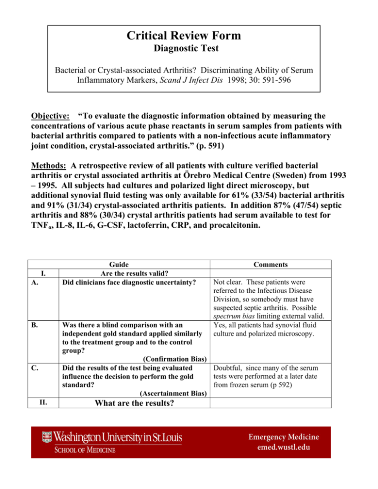
Critical Review Form Diagnostic Test Bacterial or Crystal-associated Arthritis? Discriminating Ability of Serum Inflammatory Markers, Scand J Infect Dis 1998; 30: 591-596 Objective: “To evaluate the diagnostic information obtained by measuring the concentrations of various acute phase reactants in serum samples from patients with bacterial arthritis compared to patients with a non-infectious acute inflammatory joint condition, crystal-associated arthritis.” (p. 591) Methods: A retrospective review of all patients with culture verified bacterial arthritis or crystal associated arthritis at Örebro Medical Centre (Sweden) from 1993 – 1995. All subjects had cultures and polarized light direct microscopy, but additional synovial fluid testing was only available for 61% (33/54) bacterial arthritis and 91% (31/34) crystal-associated arthritis patients. In addition 87% (47/54) septic arthritis and 88% (30/34) crystal arthritis patients had serum available to test for TNFα, IL-8, IL-6, G-CSF, lactoferrin, CRP, and procalcitonin. I. A. B. Guide Are the results valid? Did clinicians face diagnostic uncertainty? Comments Not clear. These patients were referred to the Infectious Disease Division, so somebody must have suspected septic arthritis. Possible spectrum bias limiting external valid. Yes, all patients had synovial fluid culture and polarized microscopy. Was there a blind comparison with an independent gold standard applied similarly to the treatment group and to the control group? (Confirmation Bias) Doubtful, since many of the serum Did the results of the test being evaluated tests were performed at a later date influence the decision to perform the gold from frozen serum (p 592) standard? (Ascertainment Bias) C. II. What are the results? A. What likelihood ratios were associated with the range of possible test results? • • • • • • • Septic arthritis patients were younger (median 72 vs. 78 years) with more rheumatoid arthritis (20% vs. 3%) than crystalloid arthritis. 15 septic arthritis cases involved prosthetic joints and arthroscopic surgery and three followed intra-articular injections. 36% of septic arthritis cases had positive blood cultures with the same organism. The predominant organisms were Staphylococcus aureus (48%), βhemolytic streptococci (20%), and coagnegative staph (11%) Gram staining revealed bacteria in only 42% of septic arthritis cases. 11% of bacterial arthritis cases died (vs. none of crystal arthritis). Half of crystal arthritis cases received antibiotics for mean of 2-days (versus mean 10-day antibiotic course for septic arthritis). Synovial WBC >100,000 + - Septic arthritis + 10 2 23 29 Synovial WBC >50,000 + - Septic arthritis + 19 8 14 23 • • • Sen = 30% Spec = 94% LR+ = 4.7 LR- = 0.75 Sen = 58% Spec =7 4% LR+ = 2.2 LR- = 0.57 Prevalence = 51.6% Medium jWBC in septic arthritis was 70,000 (range 4400-246,500) compared with crystal-associated arthritis 20,000 (range 140-104,000) which was significantly different (p = 0.009). A reduction in synovial glucose was seen in 64% septic arthritis vs. 15% crystal arthritis. ESR (81 vs. 54) and CRP (182 vs. 101) were both significantly higher inn septic arthritis as were TNFα (4.9 vs. 4.3), IL-8 (19.5 vs. 13.5), G-CSF (35 vs. 20), but significant overlap existed between each of these and optimal cut-points were not determined. • III. WBC and lactoferrin, IL-6, and procalcitonin levels did not differ between septic and crystalloid arthritis. How can I apply the results to patient care? A. Will the reproducibility of the test result and its interpretation be satisfactory in my clinical setting? B. Are the results applicable to the patients in my practice? C. Will the results change my management strategy? D. Will patients be better off as a result of the test? For serum WBC, ESR, and CRP likely yes. Since cytokines, lactoferrin procalcitonin are not readily available, perhaps not. Probably not since these were a highly select group already referred to ID, not undifferentiated ED patients. Probably not, since dissimilar patients are reported upon using a host of tests not readily available in 2007 and authors failing to report acceptable diagnostic performance measures such as ROC curve, AUC, optimal cut-points and likelihood ratios. Cannot deduce this from current paper. Limitations 1. Selection bias – recruited only subjects referred to ID with either positive crystals or bacterial growth on synovial fluid. These are different from ED patients with lower a prevalence of septic and crystal arthritis and therefore different diagnostic test characteristics. 2. Incomplete Gold standard. Given the limited sensitivity of culture, a composite Gold standard of positive culture or positive Gram stain or prevalent joint aspirate/operative drainage would have been superior. 3. Incomplete data reporting lacking ROC curve, AUC, optimal cut-points and LR’s. Additionally, failed to stratify data by co-morbidity (immunocompromised, rheumatoid arthritis, etc.) 4. Limited demographic reporting making assessment of external validity impossible. Bottom Line In a Swedish single center methodologically challenged retrospective review, patients referred to ID with septic arthritis or crystalloid arthritis might be distinguished by synovial WBC > 100,000 (LR+ = 4.70, 95% CI 1.1-20) (LR- = 0.75, 95% CI 0.58-0.95) or synovial WBC > 50,000 (LR+ = 2.2, 95% CI 1.2-4.3) (LR- = 0.57, 95% CI 0.37-0.90). ESR, CRF, TNFα, and G-CSF might be useful to distinguish the two arthropathies, but substantial overlap between septic and crystalloid arthritis exists for all of these. WBC, PCT, IL-6, and lactoferrin are clinically useless for this indication. Synovial fluid gram stain was only positive in 42% of septic arthritis cases and 11% of bacterial arthritis cases died.


