How its Made: Organisational Effects of Hormones on the
advertisement
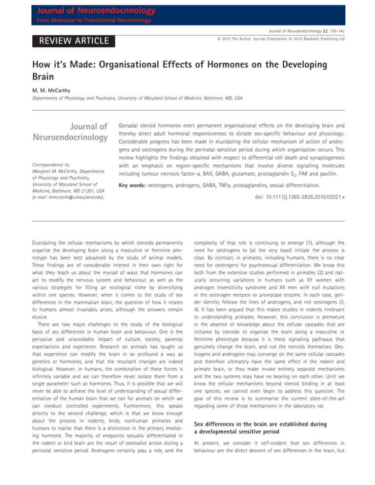
Journal of Neuroendocrinology From Molecular to Translational Neurobiology Journal of Neuroendocrinology 22, 736–742 ª 2010 The Author. Journal Compilation ª 2010 Blackwell Publishing Ltd REVIEW ARTICLE How it’s Made: Organisational Effects of Hormones on the Developing Brain M. M. McCarthy Departments of Physiology and Psychiatry, University of Maryland School of Medicine, Baltimore, MD, USA. Journal of Neuroendocrinology Correspondence to: Margaret M. McCarthy, Departments of Physiology and Psychiatry, University of Maryland School of Medicine, Baltimore, MD 21201, USA (e-mail: mmccarth@umaryland.edu). Gonadal steroid hormones exert permanent organisational effects on the developing brain and thereby direct adult hormonal responsiveness to dictate sex-specific behaviour and physiology. Considerable progress has been made in elucidating the cellular mechanism of action of androgens and oestrogens during the perinatal sensitive period during which organisation occurs. This review highlights the findings obtained with respect to differential cell death and synaptogenesis with an emphasis on region-specific mechanisms that involve diverse signalling molecules including tumour necrosis factor-a, BAX, GABA, glutamate, prostaglandin E2, FAK and paxillin. Key words: oestrogens, androgens, GABA, TNFa, prostaglandins, sexual differentiation. Elucidating the cellular mechanisms by which steroids permanently organise the developing brain along a masculine or feminine phenotype has been best advanced by the study of animal models. These findings are of considerable interest in their own right for what they teach us about the myriad of ways that hormones can act to modify the nervous system and behaviour, as well as the various strategies for filling an ecological niche by diversifying within one species. However, when it comes to the study of sex differences in the mammalian brain, the question of how it relates to humans almost invariably arises, although the answers remain elusive. There are two major challenges to the study of the biological basis of sex differences in human brain and behaviour. One is the pervasive and unavoidable impact of culture, society, parental expectations and experience. Research on animals has taught us that experience can modify the brain in as profound a way as genetics or hormones, and that the resultant changes are indeed biological. However, in humans, the combination of these forces is infinitely variable and we can therefore never isolate them from a single parameter such as hormones. Thus, it is possible that we will never be able to achieve the level of understanding of sexual differentiation of the human brain that we can for animals on which we can conduct controlled experiments. Furthermore, this speaks directly to the second challenge, which is that we know enough about the process in rodents, birds, nonhuman primates and humans to realise that there is a distinction in the primary mediating hormone. The majority of endpoints sexually differentiated in the rodent or bird brain are the result of oestradiol action during a perinatal sensitive period. Androgens certainly play a role, and the doi: 10.1111/j.1365-2826.2010.02021.x complexity of that role is continuing to emerge (1), although the need for oestrogens to (at the very least) initiate the process is clear. By contrast, in primates, including humans, there is no clear need for oestrogens for psychosexual differentiation. We know this both from the extensive studies performed in primates (2) and naturally occurring variations in humans such as XY women with androgen insensitivity syndrome and XX men with null mutations in the oestrogen receptor or aromatase enzyme. In each case, gender identity follows the lines of androgens, and not oestrogens (3, 4). It has been argued that this makes studies in rodents irrelevant to understanding primates. However, this conclusion is premature in the absence of knowledge about the cellular cascades that are initiated by steroids to organise the brain along a masculine or feminine phenotype because it is these signalling pathways that genuinely change the brain, and not the steroids themselves. Oestrogens and androgens may converge on the same cellular cascades and therefore ultimately have the same effect in the rodent and primate brain, or they make invoke entirely separate mechanisms and the two systems may have no bearing on each other. Until we know the cellular mechanisms beyond steroid binding in at least one species, we cannot even begin to address this question. The goal of this review is to summarise the current state-of-the-art regarding some of those mechanisms in the laboratory rat. Sex differences in the brain are established during a developmental sensitive period At present, we consider it self-evident that sex differences in behaviour are the direct descent of sex differences in the brain, but Organisational effects of hormones on the developing brain it was not always so. The beginning of the modern era is marked by a single publication in 1959 in the journal Endocrinology titled ‘Organising action of prenatally administered testosterone propionate on the tissues mediating mating behaviour in the female guinea pig,’ by Phoenix, Goy, Gerall and Young (5). There are two things about this paper that make it remarkable. The first is the clear articulation and testing of the hypothesis that developmental hormonal exposure determined adult behaviour. This has since been codified as the iconic organisational ⁄ activational hypothesis of sexual differentiation of the brain (Fig. 1). The second is the title; note the use of the word ‘tissue’ instead of brain. The concluding paragraph of the paper included the statement ‘An assumption seldom made explicit is that modification of behavior follows an alteration in the structure or function of the neural correlates of that behavior. We are assuming that testosterone or some metabolite acts on those central nervous tissues in which patterns of sexual behavior are organised.’ This was the authors planting the flag, their version of the Watson and Crick ‘It has not escaped our notice that the specific pairing we have postulated immediately suggests a possible copying mechanism for the genetic material’ at the end of their 1953 paper describing the structure of DNA. The claim of Phoenix et al. (5) that hormones permanently organised the developing brain to determine adult behaviour was equally heretical and transforming. A third no less remarkable aspect of this paper was that it also foreshadowed the discovery that, in many instances, it was the Sensitive period Male plasma testosterone levels Conception Late gestation E2 Birth Puberty T Organisation Activation Fig. 1. Hormonally-mediated sexual differentiation of the brain. The organisational ⁄ activational construct for sexual differentiation is based on the principle of an early sensitive period that is operationally defined as beginning with the onset of gonadal secretion of steroid hormones in the male, and as termination being that point at which the female becomes unresponsive to the impact of exogenously administered steroid hormone. The actions of gonadal steroids during this sensitive period are considered organisational, and generally permanent, and this differentiated neural substrate is then acted upon (activational) by the appropriate gonadal steroids in adulthood to produce stereotypic sexual behaviour or physiology. Different physiological and behavioural endpoints vary in the precise timing of the sensitive period, although most differentiation is complete by the end of the first postnatal week of life. E2, oestradiol. 737 metabolite of testosterone, oestradiol, and not testosterone itself that induces organisational effects on the developing brain. In the intervening 50 years, there have been major advances to support the dogma of the organisational ⁄ activational hypothesis of sexual differentiation of the brain, and many challenges as well. The goal of this review is to highlight some of the mechanistic advances and to discuss how understanding fundamental cellular processes can also speak to higher issues of the significance and impact of sex differences in the brain. Masculinisation of the preoptic area involves oestradiol-mediated changes in cell survival, cell death and synaptogenesis The preoptic area (POA) contains among the most robust sex differences in the brain and is critical to the normal expression of male sexual behaviour and pulsatile control of gonadotrophin secretion from the pituitary. High levels of oestrogen receptor (ER) and aromatase enzyme further implicate this as the key brain sight mediating oestradiol-induced masculinisation (6, 7). Three distinct cellular events occur here during the perinatal sensitive period; (i) cells in the female sexually dimorphic nucleus (SDN) die as a result of a lack of oestradiol; (ii) cells in the female anteroventral periventricular (AVPV) nucleus survive due to a lack of oestradiol; and (iii) neurones in the medial preoptic nucleus of males form approximately twice as many dendritic spines containing excitatory synapses. Each one of these events involves distinct non-overlapping cellular mechanisms. Perhaps the least understood is the SDN where oestradiol prevents apoptosis, resulting in males having three-fold more neurones than females (8). The cell death in females is classic BAX ⁄ caspase 3 apopotic cell death and higher levels of Bcl2 in males affords protection, but how BAX and Bcl2 are regulated is not yet clear. Several proteins of interest have been implicated including NMDA receptor glutamatergic signalling and neurotrophic proteins such as RNA binding motif protein 3 and alpha-tubulin (9, 10). A far greater understanding has been achieved for the basis of the sex difference in the volume of the AVPV. Tumour necrosis factor (TNF)-a is a proinflammatory cytokine that activates nuclear factor (NF)-kB receptors and promotes cell survival. This pathway is constitutively active in AVPV neurones of neonatal females but repressed in their male littermates. The suppression in males is the result of higher expression of an associated protein called TRIP (TNF receptor associated-associated factor 2-inhibiting protein) which inhibits both the TNFa-NF-kB survival pathway and the anti-apoptotic protein, bcl-2 (11). Whether the down-regulation of TRIP in males is directly a result of oestrogen action has not been established, although it has been determined that these events are exclusive to GABAergic neurones of the AVPV, and, equally importantly, TRIP is not expressed in the SDN-POA, which consists largely of GABAergic neurones (12). The selective involvement of GABA neurones of the AVPV is complemented by a parallel effect of oestrogen-induced activation of caspase-dependent cell death in the dopaminergic neurones of the AVPV, reducing the number of this class of neurones in males as well (13). The combination of oestra- ª 2010 The Author. Journal Compilation ª 2010 Blackwell Publishing Ltd, Journal of Neuroendocrinology, 22, 736–742 738 M. M. McCarthy fever (14). The cyclo-oxygenase enzymes, COX-1 and COX-2 limit the rate of prostaglandin synthesis, which includes up to eight distinct prostanoids determined by distinct synthetic enzymes from short-lived precursors (15). In the developing preoptic area, oestradiol induces a two-fold increase in COX-1 and -2 and a correspondingly greater increase in the prostaglandin, PGE2, but not other prostanoids (16). Making the connection from prostaglandins to changes in dendritic spine density involves the glutamate system of neurotransmission. AMPA ⁄ kainate and NMDA glutamate receptors are highly enriched on the surface of dendritic spines, the site at which an diol effects results in a male AVPV with significantly fewer dopaminergic and GABAergic neurones, comprising the two principle cell types in this nucleus, and appears to underlie at least in part the inability of males to respond to rising oestradiol in adulthood with positive feedback and a surge of luteinising hormone (LH) release. There has also been considerable progress made in elucidating the cellular pathway to greater dendritic spine density on male preoptic area neurones, which has direct implications for the control of male sexual behaviour. Prostaglandins are a class of lipid membrane-derived signalling molecules that subserve normal cellular functions relevant to reproduction, inflammation, hyperalgesia and (A) (B) Mean spine density (per microrn) 0.16 Male Female 0.12 Male + Indo Female + E2 0.08 Female + PGE2 0.04 0 Vehicle E2 PGE2 E2+PGE2 E2+ lndomethacin (C) Sex Prostaglandin synthesis POA Neurone Behaviour PGD2 PGF2alpha AA COX-2 PGE2 PGI2 TXA2 Feminisation PGD2 PGF2alpha AA COX-2 PGE2 PGI2 Masculinisation TXA2 Fig. 2. Prostaglandin E2 mediates masculinisation of preoptic area (POA) neurones and adult sexual behaviour of rats. The density of dendritic spines, and the excitatory synapses associated with these structures, is two- to three-fold higher on POA neurones in males than females. Oestradiol permanently organises this sex difference during the perinatal sensitive period. (A) Treatment of females with oestradiol (E2) or prostaglandin E2 (PGE2) significantly increases the density of dendritic spines, to the same level as seen in males. The effects of E2 and PGE2 are not additive, and the effects of E2 are completely blocked by the co-administration of the COX-2 inhibitor, indomethacin (indo). COX-2 is a key enzyme in prostaglandin synthesis (38). (B) Golgi impregnation of POA neurones further reveals the sex difference in dendritic spines and regulation by prostaglandins. These neurones were visualised in 20-day-old animals that had been treated with oestradiol, PGE2 or indomethacin on days 1 and 2 of life. The higher density of spines in males compared to females is evident, as is the induction of spines by E2 or PGE2 treatment of females. Treatment of males with indomethacin permanently down-regulated dendritic spines on POA neurones (16). (C) Schematic representation of the role of prostaglandins in masculinisation. Males have higher levels of the COX-2 enzyme, resulting in five-fold higher levels of the prostaglandin, PGE2, but not other prostanoids. Through a mechanism that at least partly involves the AMPA receptor, but that remains poorly understood, PGE2 permanently increases the density of dendritic spines on POA neurones. The resulting sex difference in neuronal morphology is correlated with (and presumably determines) adult sex differences in reproductive behaviour. However, the changes in brain regions outside the POA contributing to the change in behaviour cannot be ruled out. ª 2010 The Author. Journal Compilation ª 2010 Blackwell Publishing Ltd, Journal of Neuroendocrinology, 22, 736–742 Organisational effects of hormones on the developing brain estimated 95% of excitatory neurotransmission in the brain occurs. PGE2 signalling is propagated by G-protein coupled receptors named EP1–4 (17). Convergent evidence indicates EP2 and EP4 are required for PGE2-mediated masculinisation (18). Both receptors recruit protein kinase A (PKA) which helps to maintain AMPA ⁄ kainate receptor expression in the post-synaptic density of the dendritic spine (19). In the developing POA, activation of PKA increases the density of dendritic spines and inhibition of the AMPA ⁄ kainate glutamate receptors prevents this PKA-induced dendritic spine formation. (20). Taken together, these data allow us to construct a working model in which oestradiol increases the synthesis of COX-1 and -2 to promote PGE2 production in either neurones or glia (Fig. 2). PGE2 binds to EP2 and EP4 receptors, activating adenlyl cyclase, increasing cAMP and activating PKA, which assures the AMPA ⁄ kainate receptors are anchored in the dendritic spine. The induction and stabilisation of dendritic spines is aided by functional AMPA receptors. Disruption of any step in the above process, such as inhibiting the COX enzymes, blocking PKA activation or antagonising the AMPA glutamate receptors, will impair adult male rat sexual behaviour. More importantly, newborn female rats treated with PGE2 readily exhibit male sexual behaviour when given testosterone in adulthood, and the same effect can be achieved by activating the AMPA receptor during the critical period. Thus, both male sexual behaviour and its neurological underpinnings are organised by (A) oestradiol-induced prostaglandin synthesis, which alters glutamatergic neurotransmission during a perinatal sensitive period. Oestradiol mediates synaptogenesis in developing mediobasal hypothalamus by regulating glutamatergic neurotransmission The ventromedial nucleus (VMN) located in the mediobasal hypothalamus is a principle brain region controlling female sexual behaviour (21). The dendrites of neurones in the male VMN branch more frequently and therefore have more overall dendritic spine synapses than females (22, 23). Oestradiol-mediated dendritic spine formation begins with a rapid (approximately 1 h) ER-mediated activation of phosphoinositide 3-kinase (PI3) kinase and enhanced release of pre-synaptic glutamate (Fig. 3). The connection between PI3 kinase activation and glutamate release remains poorly characterised but is independent of protein synthesis. Increased synaptic glutamate leads to increased activation of post-synaptic NMDA receptors followed by dendritic branching and construction and stabilisation of dendritic spines (24). The latter process is dependent upon protein synthesis in the post-synaptic neurone, but does not require ER in the post-synaptic neurone. Activity dependent dendritic growth and synaptogenesis is a common theme throughout brain development but, in the case of sexual differentiation, it is (B) Paired-pulse stimulation Veh Males Females Females + E2 E2 50 pA 20 ms (C) 739 Sex Females + NMDA Glutamate release VMN neurone Behaviour Defeminised Feminised Fig. 3. Nongenomic steroid-induced organisation of the hypothalamus. Oestradiol rapidly induces glutamate release from hypothalamic neurones leading to activation of post-synaptic glutamate receptors and induction of dendritic spines. (A) Paired pulse stimulation ratios indicate enhanced release of glutamate on the second stimulation in neurones pretreated with oestradiol. The rate of destaining of FM4-64 visualised terminals confirms this, and the effect of oestradiol is blocked by the highly specific oestrogen receptor antagonist, ICI 182,780. The effect of oestradiol on FM4-64 destaining is apparent within 1 h of treatment and does not require protein synthesis but does require activation of phosphoinositide 3-kinase (PI3) kinase. (B) Golgi impregnated ventromedial nucleus (VMN) neurones reveal a higher number of dendritic spines on the neurones of males or females treated with oestradiol compared to control females (*P < 0.05 compared to all other groups, ANOVA). Treatment with the glutamate agonist, NMDA, mimics the effect of oestradiol, confirming an essential role for glutamate transmission in establishing sexually dimorphic dendritic morphology (25). (C) Schematic representation of the role of glutamate in defeminisation. Enhanced glutamate release in male neurones results in increased dendritic spines, which is correlated with a loss of expression of female sexual behaviour expression in adulthood. Treatment of neonatal females with NMDA successfully defeminises their adult behaviour, further confirming the role of glutamate in the organisational component of sexual differentiation. ª 2010 The Author. Journal Compilation ª 2010 Blackwell Publishing Ltd, Journal of Neuroendocrinology, 22, 736–742 740 M. M. McCarthy quite surprising that initiation begins with nongenomic effects of oestradiol. The effects of oestradiol on the pre-synaptic neurone are receptor mediated, and involve ERa. Thus, although the effects of oestradiol are nontraditional, the receptor mediating the effects are the classic ER. Moreover, these results illustrate the principle that oestradiol-mediated sexual differentiation of the mammalian brain is not a cell autonomous process in which only ER containing neurones change morphology in response to steroid exposure. Instead, a neurotransmitter serves as a signalling factor that elicits a morphological change in an entire population of cells, suggesting that all neuronal inputs to sexually differentiated brain regions such as the POA and VMN are considered and interpreted depending on the animal’s sex, regardless of whether the incoming signals are relevant to sex. The POA and VMN are two critical nodes in a larger network of brain regions controlling sex behaviour and they exert largely opposite influences. The POA is essential for expression of male sexual behaviour, and therefore a principle brain region subject to masculinisation, whereas the VMN is essential for female sexual behaviour and therefore presumably feminised. However, there is a third developmental process, defeminisation, in which the capacity for female sexual behaviour is suppressed. The observation that oestradiol increases the amount of excitatory input into both brain regions is consistent with the VMN being a principle brain region mediating defeminisation, and behavioural studies further support that view (25). Feminisation is an active process but difficult to study Understanding the cellular basis of feminisation is difficult on multiple levels. The definition of a feminised brain is one that supports the expression of female reproductive behaviour and oestradiol-initiated positive feedback control of LH release. Although the female brain is the default, it is not merely the absence of masculinisation. The fact that defeminisation is a distinct and active process negates the notion that the female brain is an undifferentiated brain. How do we gain an understanding of what cellular processes mediate feminisation? One approach is to identify factors that are inhibited or suppressed by the hormones mediating masculinisation, such as testosterone and oestradiol. A novel set of signalling molecules implicated as potential mediators of feminisation come from study of metastatic cancer, focal adhesion kinase (FAK) and its associated protein, paxillin. Both these proteins regulate cell adhesion and interaction with the extracellular matrix, and are therefore essential for dendritic and axonal growth and branching (26) as well as dendritic spine morphology (27). Newborn females have higher hypothalamic FAK and paxillin content than their male counterpoints, and treatment of females with oestradiol drives down levels to that of males (28). Higher FAK and paxillin is associated with reduced dendritic branching (26), and may be the basis for the fewer number of dendritic branches on the VMN neurones of females (Fig. 4). The above approach relies on the assumption that feminisation proceeds in the complete absence of gonadal steroids, although there has long been evidence that oestradiol plays an active but poorly understood role in feminisation (29). The advent of genetically modified mice has allowed for further exploration and support of this view (30). When considering the hormonal basis of sexual differentiation of the human brain, it is worth noting that an active role for oestradiol in feminisation has never been excluded. The organisational ⁄ activational hypothesis does not always strictly apply Behaviours directly associated with reproduction but not sexual behaviour per se are also often highly sexually dimorphic, with the best example being singing by male song birds, with females singing relatively little. Elaborate courtship displays, scent marking, territorial defence, mate guarding are all related behaviours shown by males to attract and then protect females. Conversely, next building, maternal behaviour, maternal aggression are more female typical behaviours, particularly in mammals. Despite the clear distinctions in motivation and division of labour between the sexes, a strict application of the organisational ⁄ activational hypothesis to explain the differences begins to break down and we instead observe a complex interplay of early hormone and experience combined with adult context and hormonal influences. As we move further away from behaviours directly relevant to reproduction and consider stress responding, pain perception, food preferences, learning and memory, drug addiction and so on, the applicability of the hypothesis becomes even less. This is not to say that there are no neural underpinnings that direct sex differences in behaviour, but that a strict adherence to early organisational hormonal effects activated by adult hormones does not satisfactorily explain the basis of the sex difference. Additional variables, including recent evidence for a genetic contribution (31), must be considered to create a more nuanced view of how and why males and females differ. Moreover, in some cases, what is taken for a sex difference is not really a sex difference at all but rather an example of hormonally-mediated plasticity in the adult (32). Oestradiol exerts profound effects on hippocampal morphology and function, inducing a 30% increase in the density of dendritic spines, increased long-term potentiation and enhanced spatial learning (33). These and similar data are often interpreted as evidence for a sex difference that is the result of sex differentiation, but, when males are considered, they fall somewhere in between the measures for females with and without oestradiol. Is this really a sex difference? The answer is ‘yes’ and ‘no’; there are some intrinsic sex differences in the hippocampus, and responses to extrinsic variables such as stress may impact the hippocampus differently in males versus females (34), but unambiguous evidence for classic sex differentiation is lacking. The hippocampus may prove a brain region that exemplifies a mix of hormonal, genetic and experiential influences to generate a sexspecific phenotype. Discerning the relative weight of each variable and its impact on function will provide a valuable integrative view of sex differences with relevance across a wide range of functions. The value of research on sex differences is wide ranging The motivation to incorporate sex differences into basic science research remains low, with over 50% of studies published in the field of neuroscience in 2009 being conducted either exclusively on ª 2010 The Author. Journal Compilation ª 2010 Blackwell Publishing Ltd, Journal of Neuroendocrinology, 22, 736–742 Organisational effects of hormones on the developing brain (B) 7 6 5 4 3 2 Male Female 1 0 2 (C) Sex FAK level FAK level (A) 8 6 8 10 4 Days postnatal FAK Hypothalamic neurone Expression FAK FAK M F M 741 F+E 100 80 60 40 20 0 Male Female+ Female E2 Sex behaviour Feminisation Masculinisation Fig. 4. Oestradiol down-regulates focal adhesion kinase (FAK) in the developing male brain. FAK is an important regulator of axonal growth and dendritic branching in the developing brain. Neurones of the developing hypothalamus have more dendritic branches in males than in females, an effect mediated by the higher levels of oestradiol in neonatal male brain, following its aromatisation from testicular androgens. (A) The level of FAK protein gradually increases in the female hypothalamus during the sensitive period of sexual differentiation, peaking on postnatal day 6, at which point it is significantly higher than in the male hypothalamus, where levels remain relatively constant. (B) Treatment of newborn females with oestradiol significantly reduces FAK levels within 24–48 h when assessed by quantitative western blot (28). (C) Schematic representation of FAK, which interacts with the integrins to control cell adhesion, thereby impacting on growth cone movement and branching. Increased FAK levels in females are speculated to decrease branching of hypothalamic neurones. The resulting sex difference in neuronal morphology is considered as a permanently organised change that is then activated by adult circulating gonadal steroids to regulate sexually dimorphic reproductive behaviour. males or with sex unspecified (35). Even in areas where there is a clear sex bias in the frequency or severity of a neurologic or mental health disorder, there remains a preponderance of research on only one sex and this is often not the sex that is dominantly afflicted by the disorder. Yet, the value of comparing physiology and behaviour of males and females has proven itself time and time again. Our discovery of a fundamental role for prostaglandins in masculinisation of the preoptic area is the first demonstration that this ubiquitous signalling molecule plays a role in normal brain development. Research in pain sensitivity identified a unique signalling pathway that is only active in females and could be the basis for novel therapeutic approaches (36). These are just two examples of the discovery potential inherent in comparing males versus females. This principle potentially extends to determining the sex bias in disorders such as autism, dyslexia, attention deficit disorder and early onset schizophrenia, all of which disproportionally afflict males (37). An additional pervading principle can also be extracted from our current knowledge of how steroid hormones act on the developing brain. There is a popular view that males have one kind of brain, whereas females have another, and never the twain shall meet. However, if we step back and consider what our data tells us, this uniform view of the male versus female brain conflicts with the discovery that the cellular mechanisms mediating steroid hormone action are extraordinarily diverse both within and between brain regions. Steroids are just the first step in highly complex cellular processes and variation at each node along the way could provide important variation to the final phenotypic outcome. For example, allelic variation in the expression or functionality of glutamate receptors, COX enzymes, PKA, TNFa, to name a few, would be predicted to impact on the effect that steroids have on the brain. As a result, the brain of one male is likely to be much different from that of another male because some regions or endpoints will be strongly masculinised, whereas others will be less male-like or even somewhat feminised. Likewise, specific females will have some brain regions that undergo a degree of masculinisation as a result of hormonal exposure from their male siblings in utero, or the natural processes of feminisation will vary depending on their genetic makeup. As a result, each brain is an individually specific mosaic of maleness and femaleness, and doesn’t that just fit perfectly with how we think of all the boys and girl, men and women we know and love? Received: 22 April 2010, revised 3 May 2010, accepted 3 May 2010 ª 2010 The Author. Journal Compilation ª 2010 Blackwell Publishing Ltd, Journal of Neuroendocrinology, 22, 736–742 742 M. M. McCarthy References 1 Morris JA, Jordan CL, Breedlove SM. Sexual differentiation of the vertebrate nervous system. Nat Neurosci 2004; 7: 1034–1039. 2 Wallen K. Hormonal influences on sexually differentiated behavior in nonhuman primates. Front Neuroendocrinol 2005; 26: 7–26. 3 Breedlove SM. Sexual differentiation of the human nervous system. Annu Rev Psychol 1994; 45: 389–418. 4 Hines M. Sexual differentiation of human brain and behavior. In: Pfaff D, ed. Hormones, Brain and Behavior. London: Academic Press, 2002: 425–462. 5 Phoenix CH, Goy RW, Gerall AA, Young WC. Organizing action of prenatally administered testosterone proprionate on the tissues mediating mating behavior in the female guinea pig. Endocrinology 1959; 65: 369–382. 6 DonCarlos LL, Handa RJ. Developmental profile of estrogen receptor mRNA in the preoptic area of male and female neonatal rats. Brain Res Dev Brain Res 1994; 79: 283–289. 7 Lephart ED. A review of brain aromatase cytochrome P450. Brain Res Rev 1996; 22: 1–26. 8 Davis EC, Popper P, Gorski RA. The role of apoptosis in sexual differentiation of the rat sexually dimorphic nucleus of the preoptic area. Brain Res 1996; 734: 10–18. 9 Tsukahara S et al. Estrogen modulates Bcl-2 family protein expression in the sexually dimorphic nucleus of the preoptic area of postnatal rats. Neurosci Lett 2008; 432: 58–63. 10 Tsukahara S, Kakeyama M, Toyofuku Y. Sex differences in the level of Bcl-2 family proteins and caspase-3 activation in the sexually dimorphic nuclei of the preoptic area in postnatal rats. J Neurobiol 2006; 66: 1411–1419. 11 Krishnan S et al. Central role of TRAF-interacting protein in a new model of brain sexual differentiation. Proc Natl Acad Sci USA 2009; 106: 16692–16697. 12 Searles RV et al. Sex differences in GABA turnover and glutamic acid decarboxylase (GAD(65) and GAD(67)) mRNA in the rat hypothalamus. Brain Res 2000; 2: 11–19. 13 Waters EM, Simerly RB. Estrogen induces caspase-dependent cell death during hypothalamic development. J Neurosci 2009; 29: 9714–9718. 14 Bos CL et al. Prostanoids and prostanoid receptors in signal transduction. Int J Biochem Cell Biol 2004; 36: 1187–1205. 15 Chandrasekharan NV, Simmons DL. The cyclooxygenases. Genome Biol 2004; 5: 241. 16 Amateau SK, McCarthy MM. Induction of PGE2 by estradiol mediates developmental masculinization of sex behavior. Nat Neurosci 2004; 7: 643–650. 17 Regan JW. EP2 and EP4 prostanoid receptor signaling. Life Sci 2003; 74: 143–153. 18 Wright CL, Burks SR, McCarthy MM. Identification of prostaglandin E2 receptors mediating perinatal masculinization of adult sex behavior and neuroanatomical correlates. Dev Neurobiol 2008; 68: 1406–1419. 19 Snyder EM et al. Role for A kinase-anchoring proteins (AKAPS) in glutamate receptor trafficking and long term synaptic depression. J Biol Chem 2005; 280: 16962–16968. 20 Wright CL, McCarthy MM. Prostaglandin E2-induced masculinization of brain and behavior requires protein kinase A, AMPA ⁄ kainate, and metabotropic glutamate receptor signaling. J Neurosci 2009; 29: 13274– 13282. 21 Pfaff DW et al. Cellular and molecular mechanisms of female reproductive behaviors. In: Knobil E, Neill JD, eds. Physiology of Reproduction. New York, NY: Raven Press, 1994: 107–220. 22 Mong JA, Glaser E, McCarthy MM. Gonadal steroids promote glial differentiation and alter neuronal morphology in the developing hypothalamus in a regionally specific manner. J Neurosci 1999; 19: 1464–1472. 23 Todd BJ, Schwarz JM, McCarthy MM. Prostaglandin-E2: a point of divergence in estradiol-mediated sexual differentiation. Horm Behav 2005; 48: 512–521. 24 Schwarz JM et al. Estradiol induces hypothalamic dendritic spines by enhancing glutamate release: a mechanism for organizational sex differences. Neuron 2008; 58: 584–598. 25 Schwarz JM, McCarthy MM. The role of neonatal NMDA receptor activation in defeminization and masculinization of sex behavior in the rat. Horm Behav 2008; 54: 662–668. 26 Ivankovic-Dikic I et al. Pyk2 and FAK regulate neurite outgrowth induced by growth factors and integrins. Nat Cell Biol 2000; 2: 574–581. 27 Girault JA et al. FAK and PYK2 ⁄ CAKbeta in the nervous system: a link between neuronal activity, plasticity and survival? Trends Neurosci 1999; 22: 257–263. 28 Speert DB et al. Focal adhesion kinase and paxillin: novel regulators of brain sexual differentiation? Endocrinology 2007; 148: 3391–3401. 29 Dohler KD, Hancke JL, Srivastava SS, Hofmann C, Shryne JE, Gorski RA. Participation of estrogens in female sexual differentiation of the brain; neuroanatomical, neuroendocrine and behavioral evidence. Prog Brain Res 1984; 61: 99117. 30 Bakker J, Baum MJ. Role for estradiol in female-typical brain and behavioral sexual differentiation. Front Neuroendocrinol 2008; 29: 1–16. 31 Arnold AP, Rissman EF, De Vries GJ. Two perspectives on the origin of sex differences in the brain. Ann NY Acad Sci 2003; 1007: 176–188. 32 McCarthy MM, Konkle AT. When is a sex difference not a sex difference? Front Neuroendocrinol 2005; 26: 85–102. 33 Woolley CS. Effects of estrogen in the CNS. Curr Opin Neurobiol 1999; 9: 349–354. 34 Shors TJ, Miesegaes G. Testosterone in utero and at birth dictates how stressful experience will affect learning in adulthood. Proc Natl Acad Sci USA 2002; 99: 13955–13960. 35 Wald C, Wu C. Biomedical research. Of mice and women: the bias in animal models. Science 2010; 327: 1571–1572. 36 Chanda ML, Mogil JS. Sex differences in the effects of amiloride on formalin test nociception in mice. Am J Physiol Regul Integr Comp Physiol 2006; 291: R335–R342. 37 McCarthy GM, de Vries MJ, Forger NG. Sexual differentiation of the brain: mode, mechanisms and meaning. In: Hormones, Brain and Behavior. Pfaff EW, Etgen AM, Fahrbach SE, Rubin RT, eds. San Diego, CA: Academic Press, 2009: 1707–1744. 38 Amateau SK, McCarthy MM. A novel mechanism of dendritic spine plasticity involving estradiol induction of prostglandin-E2. J Neurosci 2002; 22: 8586–8596. ª 2010 The Author. Journal Compilation ª 2010 Blackwell Publishing Ltd, Journal of Neuroendocrinology, 22, 736–742


