Association of Periorbital Edema and Fever in Acute Infectious
advertisement
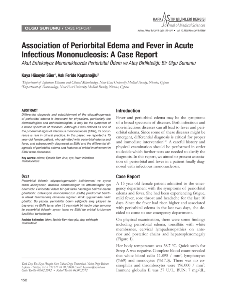
OLGU SUNUMU / CASE REPORT Kafkas J Med Sci 2013; 3(3):152–154 • doi: 10.5505/kjms.2013.02886 Association of Periorbital Edema and Fever in Acute Infectious Mononucleosis: A Case Report Akut Enfeksiyoz Mononukleozda Periorbital Ödem ve Ateș Birlikteliği: Bir Olgu Sunumu Kaya Hüseyin Süer1, Aslı Feride Kaptanoğlu2 Department of Infectious Diseases and Clinical Microbiology, Near East University Medical Faculty, Nicosia, Cyprus Department of Dermatology, Near East University Medical Faculty, Nicosia, Cyprus 1 2 ABSTRACT Differential diagnosis and establishment of the etiopathogenesis of periorbital edema is important for physicians, particularly the dermatologists and ophthalmologists. It may be the symptom of a broad spectrum of diseases. Although it was defined as one of the prodromal signs of infectious mononucleosis (EMN), its occurrence is rare in clinical practice. In this paper, we reported a 15 year-old female patient, who admitted with periorbital edema and fever, and subsequently diagnosed as EMN and the differential diagnosis of periorbital edema and features of orbital involvement in EMN were discussed. Key words: edema; Epstein-Barr virus; eye; fever; infectious mononucleosis Introduction Fever and periorbital edema may be the symptoms of a broad spectrum of diseases. Both infectious and non-infectious diseases can all lead to fever and periorbital edema. Since some of these diseases might be emergent, differential diagnosis is critical for proper and immediate intervention1,2. A careful history and physical examination should be performed in order to decide which further tests are needed to clarify the diagnosis. In this report, we aimed to present association of periorbital and fever in a patient finally diagnosed with infectious mononucleosis. ÖZET Case Report Periorbital ödemin etiyopatogenezinin belirlenmesi ve ayırıcı tanısı klinisyenler, özellikle dermatologlar ve oftalmologlar için önemlidir. Periorbital ödem bir çok farklı hastalığın belirtisi olarak görülebilir. Enfeksiyöz mononükleozun (EMN) prodromal belirtisi olarak tanımlanmıș olmasına rağmen klinik uygulamada nadir görülür. Bu yazıda, periorbital ödem eșliğinde ateș șikayeti ile bașvuran ve EMN tanısı alan 15 yașındaki bir kadın olgu sunumu ile periorbital ödemin ayırıcı tanısı ve EMN’de orbital tutulumun özellikleri tartıșılmıștır. A 15 year old female patient admitted to the emergency department with the symptoms of periorbital edema and fever. She had been experiencing fatigue, mild fever, sore throat and headache for the last 10 days. Since the fever had risen higher and associated with periorbital edema in the last two days, she decided to come to our emergency department. Anahtar kelimeler: ödem; Epstein-Barr virus; göz; ateș; enfeksiyöz mononükleoz Yard. Doç. Dr. Kaya Hüseyin Süer, Yakın Doğu Üniversitesi, Yakın Doğu Bulvarı Lefkoşa - Türkiye, Tel. 0 392 675 10 00 /2020 Email. kayasuer@mynet.com Geliş Tarihi: 09.02.2012 • Kabul Tarihi: 04.07.2012 152 On physical examination, there were some findings including periorbital edema, tonsillitis with white membranes, cervical lympadenopathies on anterior and posterior chains and hepatosplenomegaly (Figure 1). Her body temperature was 38.7 ºC. Quick swab for Strep A was negative. Complete blood count revealed that white blood cells: 11.890 / mm3, lymphocytes (%69) and monocytes (%17.3). There was no eosinophilia and thrombocytes were 196.000 / mm3. Immune globulin E was 37 U/L. BUN: 7 mg/dL, Kafkas J Med Sci nausea, vomiting, and coughing may help in diagnosis. In addition, the characteristics of the edema as being localized, diffuse, unilateral or bilateral may also be helpful2. Chronic forms of periorbital edema may originate from many reasons, however there are only a few diseases causing acute periorbital edema. In our patient, the onset of the periorbital edema was sudden in a few days, thus we excluded chronic reasons such as nephrotic syndrome or tumors. Bilateral periorbital edema in the absence of general edema is an important clue that is usualy seen in allergic reactions (angioedema), trichinosis, Kawasaki disease or bilateral periorbital cellulitis3-5. Figure 1. Bilateral periorbital edema in infectious mononucleasis. creatinin: 0.57 mg/dL. AST: 406 U/L, ALT: 377 U/L, LDH: 550 U/L, CRP: 1.87 mg/dL. Microscopic examination of the peripheral blood revealed Downey cells (%24). The initial laboratory results were compatible with lympho-monocytosis, thus we focused on Infectious mononucleosis (EMN). Serologically ELISA tests showed EBVEBNA Ig G (-), EBV VCA IgM: (+), EBV VCA– EA Ig G: (+). A cranial magnetic resonance imaging was performed to rule out other causes of dramatic periorbital edema. It was normal other than bilateral subcutaneous edema in palpebral areas. These results confirmed the diagnosis of EMN and the patient had received symptomatic treatment such as fluid replacement, paracetamol and anti-inflammatory mouthwash. She recovered clinically in 10 days. Discussion Periorbital edema may originate from a wide variety of diseases. Associated findings including fever, Allergic reactions like contact dermatitis and angioedema should be differentiated at first. In our patient, there were neither a suspicious exogen contact nor erythema or itching. Although angioedema in periorbital area is a very common cause, our case was not compatible with angioedema since its association with fever is not usual. Moreover, angioedema episodes of the periorbital region usually do not last more than 24 hours. However our patient was experiencing periorbital edema for the last two days. In addition, there was no history of drug use or suspicious food ingestion. Kawasaki disease may cause periorbital edema due to periorbital vasculitis, however can be differentiated by its other systemic features such as onset age and clinical presentation. Trichinosis also may present with periorbital edema, but generalized edema and, myalgia are dominant clinical presentations with an eosinofilic shift in peripheral blood count1. Bilateral cellulitis with its acute onset characteristic in association with fever rarely is seen bilaterally, however in our case it was excluded by MRI imaging which also helped to exclude a retro orbital tumor as well6, 7. EMN is an infectious disease caused by Epstein-Barr virüs (EBV). Major clinical symptoms are sore throat, fever, fatigue, anorexia, myalgia, headache and rarely nausea, coughing, vomiting and artralgia. Periorbital edema is also known to be a symptom of EMN8. However, in textbooks it is not classified as a major and classical symptom of EMN9. The association of periorbital edema with EMN was first described by Hoagland and is referred to as ‘’Hoagland sign’’ 10,11. Hoagland et al. reported 153 Kafkas J Med Sci periorbital edema in one third of EMN cases in 1952, whereas Mason et al. reported less frequencies in 19588,12. In 1991, Decker et al. reported a case of early EMN with periorbital edema. Similar to our patient, their patient was 18 years old and the edema was presented within the first few days of the disease13. In contrary with the above mentioned cases, in a study dealing with the clinical features of EMN in Turkish children, the authors did not observe periorbital edema in any of their patients14. Ophthalmologic involvement in EBV is unclear. Inflammation in the lacrimal gland with lymphocytic infiltrate was reported and regarded to cause ophthalmic involvement12. In addition, the mucosal edema in the sinuses was regarded to contribute in periorbital edema15. There are also reports of optic neuritis due to EBV3,16,17. In these reports, inflammation was present in optic nerve fibers. Anderson et al. pointed out that all cases of retrobulber neuritis associated with EBV were male patients with an age range of 6 and 29 years. He also noticed that neuritis developed 2-12 weeks following the acute infection and the finding suggested an immunologic mechanism3. However, the article did not provide information about the presence of periorbital edema or lacrimal gland involvement in those cases. Conclusion Periorbital edema may be an alerting symptom of EBV. Infectious mononucleosis should be kept in mind if the patients have association of periorbital edema and fever. Acknowledgement We would like to thank to our patient for providing the informed consent prior to publishing. 154 References 1. Rafailidis PI, Flags ME. Fever and periorbital edema: A review. Surv Ophthalmol 2007; 52(4): 422-33. 2. Scrivener Y, El Aboubi-Kühne S, Marquart-Elbaz C et al. Diagnosis of orbito-palpebral edema. Ann Dermatol Venereol 1999; 126: 844-8. 3. Anderson MD, Kennedy CA, Lewis A et al. Retrobulbar neuritis complicating acute Epstein-Barr virus infection. Clin Infect Dis 1994; 18: 799-801. 4. Kreel L, Poon WS, Nainby-Luxmoore JC. Trichinosis diagnosed by computed tomography. Postgrad Med J 1988; 64: 626-30. 5. Van Hasselt W, Schreuder R, Houwerzijl EV. Periorbital oedema. Neth J Med 2009; 67: 338-9. 6. Wald ER. Periorbital and orbital Infections. Infect Dis Clin North Am. 2007; 21: 393-408. 7. Aidan P, François M, Prunel M et al. Orbital cellulitis in children. Arch Pediatr 1994; 1; 879-85. 8. Mason WR Jr, Adams EK. Infectious mononucleosis; an analysis of 100 cases with particular attention to diagnosis, liver function tests and treatment of selected cases with prednisone. Am J Med Sci 1958; 236: 447-59. 9. Johannsen EC, Kaye MK. Epstein-Barr virus. In: Mandell GL, Bennett EJ, Dolin R, editors. Principles and Practice of Infectious Diseases. 7 th ed. Philadelphia; Churchill Livingstone; 2010: 1988-2010. 10. Burger J, Thurau S, Haritoqlou C. Bilateral lid swelling during infectious mononucleosis (Hoagland sign) Klin Monbl Augenheilkd 2005; 222: 1014-6. 11. Bass MH. Periorbital edema as the initial sign of infectious mononucleosis. J Pediatr 1954; 45(2): 204-5. 12. Hoagland RJ. Infectious mononucleosis. Am J Med 1952; 13: 158-71. 13. Decker GR, Berberian BJ, Sulica VI. Periorbital and eyelid edema: the initial manifestation of acute infectious mononucleosis. Cutis 1991; 47: 323-4. 14. Cengiz AB, Cultu-Kantaroğlu O, Secmeer G et al. Infectious mononucleosis in Turkish children. Turk J Pediatr 2010; 52: 245-54. 15. Demireller A. Rinosinuzit. Wilke Topçu A, Söyletir G, Doğanay M, editors. Enfeksiyon Hastalıkları ve Mikrobiyolojisi. 3.Baskı. İstanbul: Nobel Tıp Kitabevleri; 2008; 741-5. 16. Purvin V, Herr GJ, De Myer W. Chiasmal neuritis as a complication of Epstein-Barr virus infection. Arch Neurol 1988; 45: 458-60. 17. Demir T, Ulas F, Kibar Y. Bilateral optic neuritis associated with Epstein-Barr virus infection. T Klin Ophthalmol 2003; 12: 161-4.
