Carpal coalition : a rare coincidence with hand deficiencies
advertisement
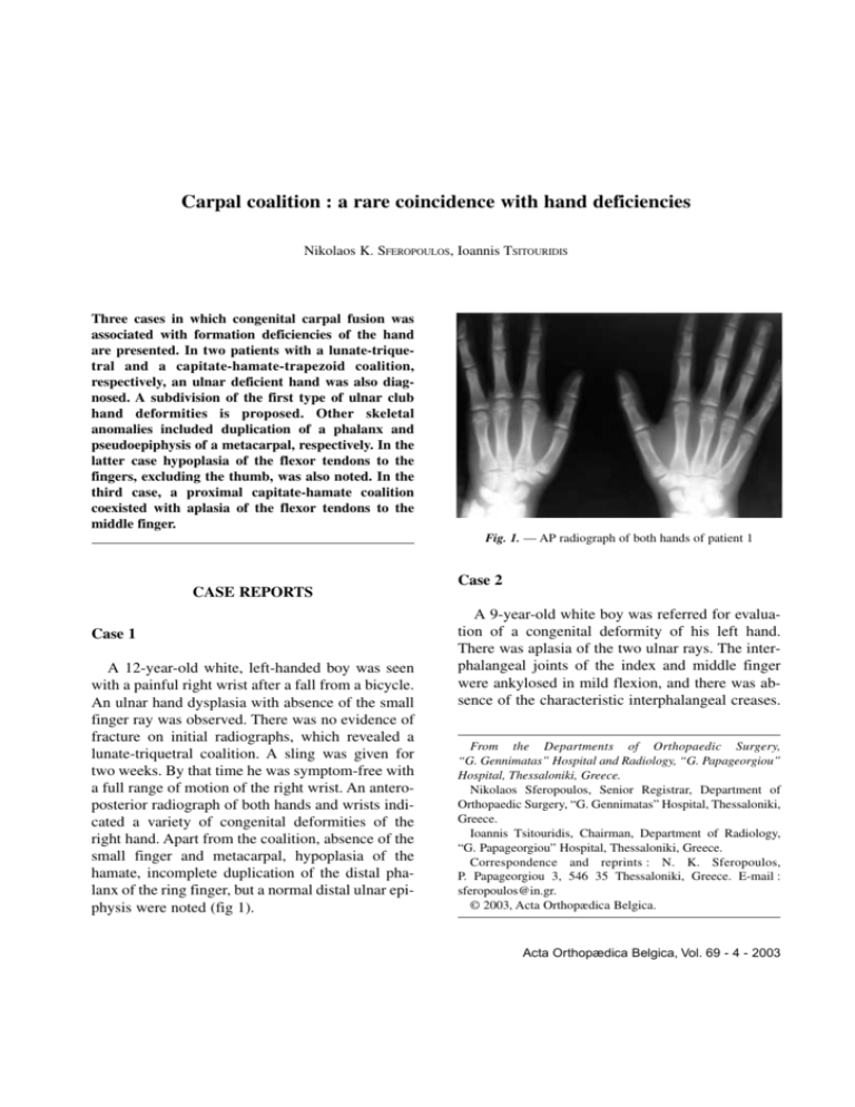
Carpal coalition : a rare coincidence with hand deficiencies Nikolaos K. SFEROPOULOS, Ioannis TSITOURIDIS Three cases in which congenital carpal fusion was associated with formation deficiencies of the hand are presented. In two patients with a lunate-triquetral and a capitate-hamate-trapezoid coalition, respectively, an ulnar deficient hand was also diagnosed. A subdivision of the first type of ulnar club hand deformities is proposed. Other skeletal anomalies included duplication of a phalanx and pseudoepiphysis of a metacarpal, respectively. In the latter case hypoplasia of the flexor tendons to the fingers, excluding the thumb, was also noted. In the third case, a proximal capitate-hamate coalition coexisted with aplasia of the flexor tendons to the middle finger. Fig. 1. — AP radiograph of both hands of patient 1 CASE REPORTS Case 1 A 12-year-old white, left-handed boy was seen with a painful right wrist after a fall from a bicycle. An ulnar hand dysplasia with absence of the small finger ray was observed. There was no evidence of fracture on initial radiographs, which revealed a lunate-triquetral coalition. A sling was given for two weeks. By that time he was symptom-free with a full range of motion of the right wrist. An anteroposterior radiograph of both hands and wrists indicated a variety of congenital deformities of the right hand. Apart from the coalition, absence of the small finger and metacarpal, hypoplasia of the hamate, incomplete duplication of the distal phalanx of the ring finger, but a normal distal ulnar epiphysis were noted (fig 1). Case 2 A 9-year-old white boy was referred for evaluation of a congenital deformity of his left hand. There was aplasia of the two ulnar rays. The interphalangeal joints of the index and middle finger were ankylosed in mild flexion, and there was absence of the characteristic interphalangeal creases. From the Departments of Orthopaedic Surgery, “G. Gennimatas” Hospital and Radiology, “G. Papageorgiou” Hospital, Thessaloniki, Greece. Nikolaos Sferopoulos, Senior Registrar, Department of Orthopaedic Surgery, “G. Gennimatas” Hospital, Thessaloniki, Greece. Ioannis Tsitouridis, Chairman, Department of Radiology, “G. Papageorgiou” Hospital, Thessaloniki, Greece. Correspondence and reprints : N. K. Sferopoulos, P. Papageorgiou 3, 546 35 Thessaloniki, Greece. E-mail : sferopoulos@in.gr. © 2003, Acta Orthopædica Belgica. Acta Orthopædica Belgica, Vol. 69 - 4 - 2003 318 N. K. SFEROPOULOS, I. TSITOURIDIS ankylosed and there was absence of the interphalangeal creases. There was neither instability of the joints nor any significant contracture of the skin. No other abnormality was detected to the other fingers or to the adjacent wrist. Plain radiographs of both hands and wrists and a magnetic resonance imaging (MRI) that followed revealed a proximal osseous bridge with distal notch between capitate and hamate on the right hand (fig 3a). The latter also indicated aplasia of the flexor tendons to the middle finger (fig 3b). DISCUSSION Fig. 2. — AP radiographic appearance of patient 2 It was reported that a congenital absence of the flexor tendons to both fingers was diagnosed and treated, five years previously, with transfers from the toe extensor tendons, but resulted in no significant improvement. No functional abnormality was detected to the thumb, wrist and elbow joint. Anteroposterior and lateral radiographs revealed absence of both small and ring fingers and metacarpals, a pseudoepiphysis of the second metacarpal, a coalition between trapezoid-capitatehamate and a delayed skeletal age of the wrist and forearm bones. No osseous abnormalities to the forearm and elbow were detected (fig 2). Case 3 A 3-year-old white girl presented with absence of movement of the middle finger of the right hand. On examination both interphalangeal joints were Acta Orthopædica Belgica, Vol. 69 - 4 - 2003 Failure of differentiation of the individual carpal bones results in congenital fusions. Every combination of congenital coalitions has been described. The most common synostosis is between the lunate and the triquetrum, followed by capitate-hamate, pisiform-triquetral and trapezium-trapezoid. The incidence in the white population is about 0.095% but it can be 100 times higher in blacks, and two times higher in females than in males. It is often bilateral and may be familial in some cases. It is not classically sex-linked but multifactorial in nature (8). Although carpal fusions may occur as part of a syndrome of congenital malformations, the cases presented in this report were associated only with other formation deficiencies involving the ipsilateral hand. As has been noted with most of the reported carpal coalitions, and unlike the situation in tarsal coalitions, no clinical symptoms related to the fusion could be noted in our patients. Synostosis of the carpal bones is not always radiologically complete. The lunate-triquetral coalitions are divided into four categories according to the degree of fusion and the associated anomalies (7). The same classification could be applied to any other carpal coalition. Therefore, patient 1 has been classified as type IV (fusion with other associated carpal anomalies), patient 2 as type III (complete fusion) and patient 3 as type II (proximal osseous bridge with distal notch). An incomplete fusion resembling a pseudarthrosis (type I coalitions) is not easily diagnosed and is likely to be missed by plain radiological examination. 319 CARPAL COALITION ulnar deficiency by the presenting deficit in the thumb and first web, while Ogino and Kato (6) have indicated a close relationship between the severity of ulnar deficiency and the degree of deficiency of fingers. However, none of the classification systems includes the ulnar-deficient hand with a normal ulna (4). It would appear reasonable to subdivide the first type of ulnar club deformities in two categories : Type 1a including the ulnar-deficient hand (a hand without the fourth and/or fifth ray) with a normal ulna, and type 1b indicating a hypoplastic ulna. The formation of a pseudoepiphysis at the distal end of the first metacarpal (3) or in the nonepiphyseal end of bones of the hands and feet (5) is very rare. It was noted at the proximal end of the second metacarpal in one case (patient 2). Finally, in two patients (case 2 and 3) congenital absence of flexor tendons to the fingers (excluding the thumb) was noted. This is a rare anomaly (9) which has not been included even in the most recent literature. Division of this deformity into two major types is suggested : (1) absence of flexor tendons to a single finger (patient 3) and (2) absence of the flexors of all the fingers –excluding the thumb (patient 2). In conclusion, carpal fusions usually present without any obvious clinical symptoms and may occasionally be associated with any skeletal or functional hand deficiency. The evaluation of this rare entity is very easy with conventional radiographs as well as with MRI axial and coronal T1weighted images. REFERENCES Fig. 3. — MRI examination indicating osseous (a) and tendon anomalies (b) of patient 3. Two cases (patients 1 and 2) were associated with an ulnar or postaxial deficiency. Bayne (1) has classified ulnar club hand into four groups based on the muscluloskeletal abnormalities of the elbow and forearm. Cole and Manske (2) further classified 1. Bayne LG. Ulnar club hand (ulnar deficiencies). In Green DP (Ed) Operative Hand Surgery, Churchill Livingstone, New York, 1993, pp 288-303. 2. Cole RJ, Manske PR. Classification of ulnar deficiency according to the thumb and first web. J Hand Surg 1997 ; 22-A : 479-488. 3. Haines RW. The pseudoepiphysis of the first metacarpal of man. J Anat 1974 ; 117 : 145-158. 4. Manske PR. Longitudinal failure of upper-limb formation. Instr Course Lect 1997 ; 46 : 83-110. 5. Ogden JA, Ganey TM, Light TR, Belsole RJ, Greene TL. Ossification and pseudoepiphysis formation in Acta Orthopædica Belgica, Vol. 69 - 4 - 2003 320 N. K. SFEROPOULOS, I. TSITOURIDIS the “nonepiphyseal” end of bones of the hands and feet. Skeletal Radiol 1994 ; 23 : 3-13 . 6. Ogino T, Kato H. Clinical and experimental studies on ulnar ray deficiency. Handchir Mikrochir Plast Chir 1988 ; 20 : 330-337. 7. Simmons BP, McKenzie WD. Symptomatic carpal coalition. J Hand Surg 1985 ; 10-A : 190-193. Acta Orthopædica Belgica, Vol. 69 - 4 - 2003 8. Simmons BP. Injuries to and developmental deformities of the wrist and carpus. In : The Pediatric Upper Extremity. Diagnosis and Management. F. William Bora, Jr (Ed). W.B. Saunders, Philadelphia, 1986, pp 176-178. 9. Verdan C. Anomalies of muscles and tendons in hand and wrist. Rev Chir Orthop 1981 : 67 : 221-30.
