optomap plus
®
Case Study: Preeclampsia-Eclampsia
Hypertensive Retinopathy
Eugene W. Ng, MD, MBA | Karl Waite, MD | Michael Bennett, MD
Retina Institute of Hawaii
Background
History
Preeclampsia-Eclampsia Syndrome is a pregnancy-specific
hypertensive disorder affecting multiple systems with a maternal
mortality rate of 1.8%.1 Risk factors for preeclampsia include very
young or advanced maternal age, multi-fetal pregnancy, hemolytic
disease of the newborn, diabetes mellitus, chronic systemic
hypertension, and renal disease. In ophthalmology, visual disturbances
such as scotoma, diplopia, dimness of vision, and photopsias were
documented in 25% of patients with severe preeclampsia and up to
50% of patients with eclampsia.2 The ocular findings of preeclampsia-eclampsia are associated with severe retinopathy, serous retinal
detachments, yellow, opaque retinal pigment epithelial lesions, and
cortical blindness.3 Preeclampisa typically occurs after 20 weeks
gestation with an incidence of 5%.4 Eclampsia is the next phase that
progresses due to the onset of seizures.4
A 43 year-old pregnant woman went to her optometrist complaining
of having blurry vision for the prior 3 days. The optometrist did not
perform a retinal exam, but noted visual field loss and a visual acuity
of 20/400. Based on those tests, the optometrist thought she may
have a retinal detachment and immediately referred her to the Retina
Institute of Hawaii. The patient stated she was 20 weeks pregnant
and had not received any prenatal care. Her best corrected visual
acuity was 20/80 in her right eye and 20/400 in her left eye. The patient’s blood pressure was 261/140.
Examination
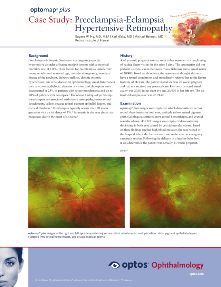
optomap® plus images were captured, which demonstrated serous
retinal detachments in both eyes, multiple yellow retinal pigment
epithelial plaques, scattered intra-retinal hemorrhages, and cystoid
macular edema. SD-OCT images were captured demonstrating
thickening in both eyes caused by cystoid macular edema. Based
on these findings and her high blood pressure, she was rushed to
the hospital where she had a seizure and underwent an emergency
caesarean section. Following the delivery of a healthy baby boy,
it was determined the patient was actually 33 weeks pregnant.
(over)
optomap® plus images of the right and left eyes demonstrating serous retinal detachments, multiple yellow retinal pigment epithelial plaques,
scattered intra-retinal hemorrhages, and cystoid macular edema
optos.com
©2011 Optos. All rights reserved. Optos® and optos® are registered trademarks of Optos plc. P/N 464001
optomap plus
®
Case Study: Preeclampsia-Eclampsia
Hypertensive Retinopathy
Eugene W. Ng, MD, MBA | Karl Waite, MD | Michael Bennett, MD
Retina Institute of Hawaii
Discussion
Conclusion
Optomap images are a useful tool for documenting signs of
preeclampsia-eclampsia in pregnant women. Findings from the ultrawidefield images demonstrate the need to image outside the posterior
pole for this disease as pathological symptoms can often manifest in
the mid to far periphery. In addition, viewing the image in the green
channel in the V2 Vantage Pro Review software can make
hemorrhages and serous detachment more evident.
The patient’s final diagnosis was eclampsia due to the seizure episode.
Follow-up optomap® plus images were captured 3 weeks later
demonstrating elshnig spots, which is a typical finding after recovering
from eclampsia. The treatment was the delivery of the baby via a
caesarean section which halted the progression of this disease and the
patient’s vision returned to 20/20. The prompt response may have
saved the lives of the patient and her baby boy.
You can find the complete case study published in Retina Today,
Retinal Manifestations of Preeclampsia. September 2010 p. 32-34.
References:
1. Douglas, K. A., Redman, C. W. Eclampsia in the United Kingdom. BMJ. 1994; 309:13951400. 2. Dieckmann WJ. The toxemias of pregnancy, 2nd edn. St Louis: Mosby, 1952. 3.
Cunningham, F.G., Fernandez, C.O., Hernandez, C. Blindness Associated with Preeclampsia
and Eclampsia. Am J Obstet Gynecol. 1995;172(4):1291-1298. 4.Wagner, L.K. Diagnosis
and Management of Preeclampsia. Am Fam Physician. 2004;70(12):2317-2324.
optomap® plus of the right eye viewed in the green channel
optomap® plus 3 weeks post delivery demonstrating elshnig spots
optos.com
©2011 Optos. All rights reserved. Optos® and optos® are registered trademarks of Optos plc. P/N 478001


