Skeletal System worksheet
advertisement

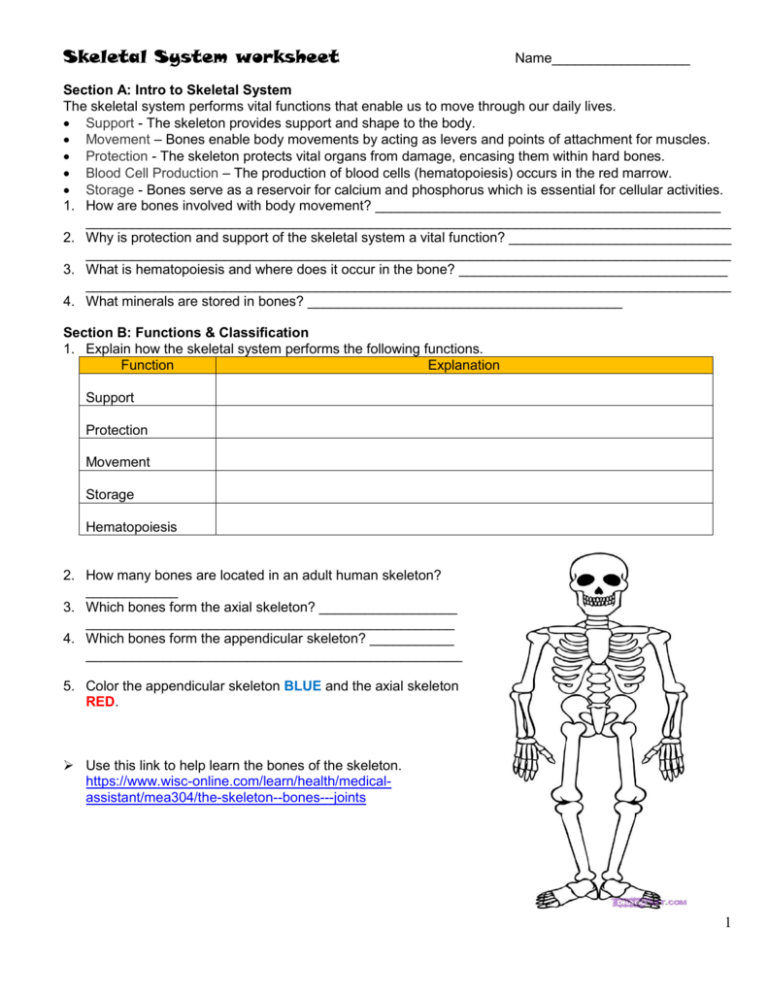
Skeletal System worksheet Name__________________ Section A: Intro to Skeletal System The skeletal system performs vital functions that enable us to move through our daily lives. Support - The skeleton provides support and shape to the body. Movement – Bones enable body movements by acting as levers and points of attachment for muscles. Protection - The skeleton protects vital organs from damage, encasing them within hard bones. Blood Cell Production – The production of blood cells (hematopoiesis) occurs in the red marrow. Storage - Bones serve as a reservoir for calcium and phosphorus which is essential for cellular activities. 1. How are bones involved with body movement? _____________________________________________ ____________________________________________________________________________________ 2. Why is protection and support of the skeletal system a vital function? _____________________________ ____________________________________________________________________________________ 3. What is hematopoiesis and where does it occur in the bone? ___________________________________ ____________________________________________________________________________________ 4. What minerals are stored in bones? _________________________________________ Section B: Functions & Classification 1. Explain how the skeletal system performs the following functions. Function Explanation Support Protection Movement Storage Hematopoiesis 2. How many bones are located in an adult human skeleton? ____________ 3. Which bones form the axial skeleton? __________________ ________________________________________________ 4. Which bones form the appendicular skeleton? ___________ _________________________________________________ 5. Color the appendicular skeleton BLUE and the axial skeleton RED. Use this link to help learn the bones of the skeleton. https://www.wisc-online.com/learn/health/medicalassistant/mea304/the-skeleton--bones---joints 1 6. Label the bones on the skeleton. sacrum, coccyx, pelvis, cervical vertebrae, thoracic vertebrae, lumbar vertebrae, cranium, mandible, humerus, ribs, sternum, clavicle, scapula, radius, ulna, carpals, phalanges, metacarpals, femur, tibia, fibula, patella, tarsals, metatarsals, calcaneus, phalanges 7. Fill in the missing information on the chart. Type of bone: Bone Long, Short, Flat, Irregular Carpals Skull Humerus Pelvis Scapula Ulna Vertebrae Ribs Metatarsals Skeletal Division: Appendicular or Axial 8. What are the functions of bone markings? _______________________________________________ _________________________________________________________________________________ 2 Section C: Bones Tissue Compact and spongy bones are considered the two basic bone types. In a long bone, spongy tissue is found at the ends while compact can be found at the outer layer and shaft of a long bone. In other type of bones, spongy fills the inside and compact forms the outer layers. Compact bone is made of osteons (Haversian systems) which form longitudinally within the bone. These osteons form the structure of compact bone. Osteons have a central canal which carries vessels and nerves. Compact bone is heavy, extremely tough and forms in layers. It accounts for more than 75-80% of the entire skeleton. Spongy (calcellous) bone consists of a lattice of thin threads of bone called trabeculae. It is less dense than compact bone. The orientation of trabeculae is affected by exposure to mechanical stress. Trabeculae bone gives supporting strength to the ends of the weight-bearing bone. 1. Compare and contrast the two types of bone tissue – spongy and compact. Include structure, location and arrangement of cells, functions and any other characteristic. COMPACT SPONGY Section D: Macroscopic Anatomy 1. Using the key choices, identify the following statements relating to long bones. Diaphysis Medullary cavity Epiphyseal plate Red marrow Osteoblast Epiphysis Articular cartilage Periosteum Endosteum Osteoclast a. _____________________ Location of spongy bone in a long bone b. _____________________ Location of only compact bone in a long bone c. _____________________ Site of hematopoiesis in the adult d. _____________________ Cells that form bone e. _____________________ Cells that destroy bone f. _____________________ Bone shaft g. _____________________ Membrane lining trabeculae and internal compact bone h. _____________________ Outer membrane covering compact bone i. _____________________ Site of fat storage in the adults j. _____________________ Site of longitudinal growth in a child k. _____________________ Ends of a long bone l. _____________________ Covers the epiphysis to provide cushion between bones m. _____________________ Provides anchoring points for ligaments and tendons n. _____________________ Contains osteogenic layers consisting of osteoblasts and osteoclasts _____________________ 3 2. Anatomy of a Long Bone – color the diagram. EPIPHYSIS (end) (a), EPIPHYSIAL LINE (a) - purple The epiphysis is the end of a long bone. Externally it has a thin layer of compact bone, while internally the bone is cancellous. The Epiphysis is capped with articular cartilage. DIAPHYSIS (shaft) (b) The diaphysis is the shaft of the long bone. It has compact bone with a central cavity. ARTICULAR CARTILAGE (c) - green The articular cartilage is found on the ends of long bones. It is smooth, slippery, and bloodless. PERIOSTEUM (d) – dark blue Periosteum is a fibrous, vascular, sensitive life support covering for bone. It provides nutrient-rich blood for bone cells and is a source of bone-developing cells during growth or after a fracture. CANCELLOUS (spongy) BONE (e) and MARROW (e) – light blue The cancellous bone appears as tiny beams of bone arranged like a lattice. Red marrow packs the spaces between beams. COMPACT BONE (f) - pink The compact bone is a dense bone found in the diaphysis. Its repeated pattern is arranged in concentric layers of solid bone tissue. MEDULLARY CAVITY (g), YELLOW MARROW (g) - yellow The medullar cavity of the diaphysis serves to lighten bone weight and provide space for its marrow. NUTRIENT ARTERY (h) - red Each long bone contains a tunnel in its shaft for the passage of a nutrient artery, which supplies the shaft. Section E: Gross Anatomy Concept Check 1. Explain how the function of the endosteum and the periosteum. ___________________________________________________________________________________ ___________________________________________________________________________________ 2. Which type of bone contains osteons? ______________________ 3. Which type of bone contains trabeculae? ____________________ 4. What’s the membrane lining the medullary cavity called? ______________________ 5. What’s the membrane lining the diaphysis called? _____________________ 6. Describe the location of spongy and compact bone in a flat bone. _______________________________ ____________________________________________________________________________________ 7. Where are the osteogenic layers of osteoblast and osteoclast found? ____________________________ ___________________________________________________________________________________ 8. What is the function of the epiphyseal plate? _______________________________________________ ___________________________________________________________________________________ 9. In an adult long bone, where are the location of yellow marrow and the location of red marrow? ___________________________________________________________________________________ 4 10. Label the long bone diagram. a. ____________________________ b. ____________________________ c. ____________________________ d. ____________________________ e. ____________________________ f. ____________________________ g. ____________________________ h. ____________________________ Long Bone Structure: http://media.pearsoncmg.com/bc/bc_marieb_happlace_7/labeling/fig_0603.html Section F: Microscopic Anatomy 1. Mature bone cells, called __________________________ maintain bone in a viable state. 2. ________________________ are stem cells that divide by mitosis & are located under the membranes. 3. __________________________ causes calcium to be deposited in bones as calcium salts. 4. Bone cells that liquefy matrix and release Ca+ to the blood are called _________________________. 5. What materials form the matrix that surrounds bone cells? _____________________________________ 6. What type of cells divide and differentiate into osteoblast or osteoclast? _________________________ 7. Lacunae arrange themselves in concentric rings called ____________________________. 8. What is the structural unit of compact bone? _______________________ 9. What are the small cavities that contain osteocytes called? ________________________ 10. The _________________________ is the central canal running vertically in an osteon. 11. What canal connects the periosteum to the Haversian canal? _________________________ 12. What canal connects lacunae to each other and to the Haversian canal? ______________________ 13. Label the diagram. a. ________________________ b. ________________________ c. ________________________ d. ________________________ e. ________________________ f. ________________________ Micro of a long bone: http://media.pearsoncmg.com/bc/bc_marieb_happlace_7/labeling/fig_0606.html 5 Section G: Bone Growth & Remodeling Growth in length: A sequence of steps occurs for bone to grow longer at the epiphyseal plate. At the plate, chondrocytes first produce hyaline cartilage. The cartilage then becomes calcified or ossified to form hard bone tissue (involves addition of Ca+ and Phosphorous ions). The chondrocytes produce cartilage on one side of the plate and push the end of the bone up. The other side of the epiphyseal plate gradually becomes calcified. Once a person reached adulthood and the bones have reached maximum length, and the whole plate gets calcified. It forms a visible line called the epiphyseal line. Growth in diameter: Making a bone grow in diameter is a more straightforward process. To make a bone thicker, just add new bone tissue to the outside. Osteoblasts on the periosteal side add bone to increase diameter and osteoclasts on the endosteal side remove some bone tissue resulting in a wider medullary cavity. It’s important to take excess bone tissue away from the inside otherwise bone would get too thick and fill completely with bone tissue. Bone Remodeling: There is a tremendous amount of activity by bone cells throughout life to maintain healthy bone and keep it from getting brittle. As old bone tissue gets hard and brittle so it must be removed and new tissue replaces the old. This process is stimulated in response to stress (like any exercise). Thus bone is reshaped when exposed to gravity by placing weight on it. 1. Where does bone growth to increase length occur? __________________________________ 2. What do chondrocytes produce? ____________________________ 3. When bones stop growing in length, what is left? ____________________________________ 4. How do bones grow wider? ____________________________________________________________ 5. What prevents bone from becoming too thick and completely filled with bone tissue due osteoblasts on the periosteal side? ___________________________________________________________________ 6. Do bones activity stop when you finishing growing? Explain your answer. _________________________ ____________________________________________________________________________________ 7. What factors stimulate bone remodeling? __________________________________________________ Section H: Bone Development, Growth & Remodeling 1. When does osteogenesis occur in an embryo? ________________________________ 2. What is intramembranous ossification? ____________________________________________________ ___________________________________________________________________________________ 3. What is endochondral ossification and where does it occur? ____________________________________ ____________________________________________________________________________________ 4. When babies are born ossification is complete for the most part except for the hyaline cartilage founding the articular cartilage and epiphyseal plate. Why is it vital that the hyaline cartilage in these two areas NOT prematurely ossify? _______________________________________________________________ 5. In your own words, explain how bones grow in width. _________________________________________ ____________________________________________________________________________________ 6. During remodeling, what cells cause bone deposition? _______________________ 7. During remodeling, what cells cause bone reabsorption? ____________________________ 8. During remodeling, most bone reabsorption occurs under the ____________________________. 9. During remodeling, most bone deposition occurs under the ____________________________. 10. New cartilage continuously divides, matures and is ossified. What replaces this ossified cartilage cells? __________________________________________________________________________________ Section I: Factors Affecting Bone Growth/Remodeling 1. Briefly explain how mineral balance is important for bone growth. _______________________________ ___________________________________________________________________________________ 2. Explain how osteoblastic activity is stimulated by hormones. Rising blood Ca+ levels Stimulates _____________________ gland _____________________ hormone To produce ______________________ (cells) deposit Ca+ salts in bone 6 3. Explain how osteoclastic activity is stimulated by hormones. Falling blood Ca+ levels Stimulates _____________________ gland _____________________ hormone To produce ______________________ (cells) to break down bone matrix and release Ca+ salts into blood 4. Besides PTH and calcitonin, what other hormones influence bone growth? ___________________________________________________________________________________ 5. How is the bone tissue of a person that is active and working out different than that of an individual that is sedentary “couch potato” or bedridden? ____________________________________________________ ____________________________________________________________________________________ 6. What happens to your bones as you age? __________________________________________________ ____________________________________________________________________________________ Section J: Calcium Metabolism -- Alcohol Disrupts the Balance in Your Body What does alcohol do to bone metabolism? Even one night of drinking causes temporary PTH deficiency and increased urinary calcium excretion. This means you lose calcium from your body. Furthermore, if you choose to drink chronically, vitamin D metabolism will be obstructed, and thus you will not be able to absorb calcium that you get from your diet. Several studies indicate that alcohol is directly damages the cells that form your bones and indirectly contributes to nutritional deficiencies of calcium or vitamin D. Alcohol also indirectly causes inefficient bone metabolism through liver disease and altered levels of reproductive hormones. Why are alcoholics more likely to suffer from bone disease? Alcohol makes it difficult for calcium to be absorbed from the gastrointestinal tract. This causes serum calcium levels to fall which feeds back to parathyroid glands resulting in an increase secretion of parathyroid hormone (PTH). This increase in PTH then leads to calcium resorption from bone causing demineralization of bone and osteoporosis bone disease. PTH could also directly inhibit bone-forming cells called osteoblasts. As you may recall from the effects of alcohol on the reproductive organs, alcohol reduces testosterone levels. Reduced testosterone levels cause bone demineralization; many studies have indicated that androgens in the male are necessary for preserving bone mass. Low testosterone levels that are induced by alcohol use may cause osteoporosis which then leads to the greater possibility of breaking your bones. http://www.montana.edu/wwwai/imsd/alcohol/Vanessa/vwcalcium.htm 1. How does alcohol affect bone metabolism? ________________________________________________ ___________________________________________________________________________________ ___________________________________________________________________________________ 2. Why are alcoholics more likely to suffer from bone disease? ___________________________________ ___________________________________________________________________________________ ___________________________________________________________________________________ Section K: At the Clinic 1. A doctor has just viewed the x-rays of a 12 year old male and has found that where the growth plate should be is instead filled with what seems to be newly formed compact bone. The doctor has diagnosed the boy with premature ossification of the epiphyseal plate. What effects if any will the doctor now need to share with the parents related to their son’s future growth? _________________________________________________________________ _________________________________________________________________ 2. Jessie fractured her tibia. The orthopedic surgeon tells her the fracture is nondisplaced, open and complete. Describe the fracture based on the surgeon’s diagnosis. _________________________________________________________________________________ _________________________________________________________________________________ 7 3. A 75 year-old woman and her 9 year old- granddaughter were in a train crash in which both sustained trauma to the chest while seated next to each other. X ray showed that the grandmother had several fractured ribs, while her granddaughter had none. Explain these surprisingly different findings. _________________________________________________________________________________ 4. Bernice, a 75- year old woman, stumbled slightly while walking then felt a terrible pain in her left hip. At the hospital, X rays revealed that the hip was broken. Also, the compact and spongy bone throughout her spine was very thin. What was her probable condition? _______________________________ 5. A middle-aged woman comes to the clinic complaining of stiff, painful joints. A glance at her hands reveals knobby, deformed knuckles. What condition will she be tested for? ________________________ 6. Patient X has a tumor of the thyroid gland that causes a hypersecretion from this gland. Predict the effect on the skeletal system and on the secretion of calcitonin. ______________________________________ ____________________________________________________________________________________ ____________________________________________________________________________________ 7. After being picked up from the Atlantic Ocean after ‘splashdown’, the American astronaut team was brought to the Naval Hospital for checkups. X rays revealed decreased bone mass in all of them. Isn’t that surprising in view of the fact that they do exercises in the capsule while in space? Why or why not? ____________________________________________________________________________________ ____________________________________________________________________________________ 8. Where would you find these 3 types of joints? a. Synarthrotic - ________________________________________________________________ b. Amphiarthrotic - ______________________________________________________________ c. Diarthrotic - _________________________________________________________________ Section L: Learning from Skeletons http://www.pbs.org/opb/historydetectives/technique/learning-from-skeletons/ Skull - Look for the sagittal suture – the squiggly line that runs the length of the skull – and note whether is it's completely fused. If it is, the remains are likely to be of someone older than 35. Look for a second line at the front of the skull -- the coronal suture – which fully fuses by age 40. Teeth - They can determine how old a person was at death, what kind of health they were in and what kind of diet they had. Pelvis - Look for the pubic symphysis, which is the joint located in the pelvis. The older the person at death, the more pitted and craggy these bones will be. Check if there are any soft marks on the cartilage which are left by childbirth as the bones soften to allow easier birth. To identify gender, assess the pelvis shape; men have a narrow, deep pelvis and women a wider, shallower pelvis, better-suited to carrying a baby. For a quick identification in the field, a forensic anthropologist will find the notch in the fan-shaped bone of the pelvis and stick their thumb into it. If there's room to wiggle the thumb, then it's a female; if it's a tight fit, it's the skeleton of a man Wrist - Examine the wrists, as bones often hold clues to the primary work of the decedent. Bony ridges form where the muscles were attached and pulled over the years. A forensic anthropologist might find a bony ridge on the wrist and decide the dead person may have been someone who used their hands for a living. 1. How can the skull be used to determine age? ______________________________________________ ___________________________________________________________________________________ ___________________________________________________________________________________ 2. What can teeth tell you about the deceased? _______________________________________________ ___________________________________________________________________________________ 3. How is the pelvis of a female different from a male? __________________________________________ ____________________________________________________________________________________ ____________________________________________________________________________________ 4. What information about the deceased can you derive from the bones in the hand? __________________ ____________________________________________________________________________________ 8


