Chapter 3
advertisement

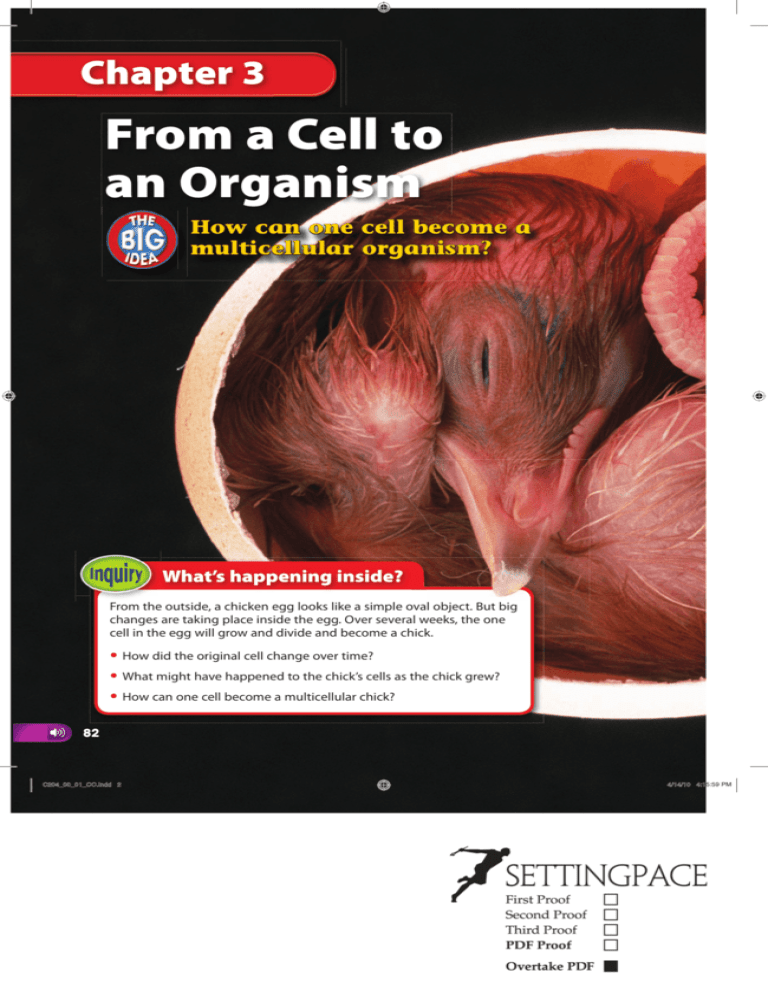
Chapter 3 From a Cell to an Organism How can one cell become a multicellular organism? What’s happening inside? From the outside, a chicken egg looks like a simple oval object. But big changes are taking place inside the egg. Over several weeks, the one cell in the egg will grow and divide and become a chick. • How did the original cell change over time? • What might have happened to the chick’s cells as the chick grew? • How can one cell become a multicellular chick? 82 C204_00_01_CO.indd 2 4/14/10 4:15:59 PM Get Ready to Read What do you think? Before you read, decide if you agree or disagree with each of these statements. As you read this chapter, see if you change your mind about any of the statements. 1 Cell division produces two identical cells. 2 Cell division is important for growth. 3 At the end of the cell cycle, the original cell no longer exists. 4 Unicellular organisms do not have all the characteristics of life. 5 All the cells in a multicellular organism are the same. 6 Some organs work together as part of an organ system. Your one-stop online resource connectED.mcgraw-hill.com ? Video WebQuest Audio Assessment Review Concepts in Motion Inquiry g Multilingual eGlossary 83 C204_00_01_CO.indd 1 12/17/09 10:18:15 AM Lesson 1 Reading Guide Key Concepts ESSENTIAL QUESTIONS • What are the phases of the cell cycle? • Why is the result of the cell cycle important? Vocabulary The Cell Cycle and Cell Division cell cycle p. 85 interphase p. 86 sister chromatid p. 88 centromere p. 88 mitosis p. 89 cytokinesis p. 89 daughter cell p. 89 g Multilingual eGlossary Video BrainPOP® Time to Split? Unicellular organisms such as these reproduce when one cell divides into two new cells. The two cells are identical to each other. What do you think happened to the contents of the original cell before it divided? 84 • C204_02_12_L1.indd 2 Chapter 3 ENGAGE 12/28/09 9:26:02 AM Launch Lab 15 minutes Why isn’t your cell like mine? All living things are made of cells. Some are made of only one cell, while others are made of trillions of cells. Where do all those cells come from? 1 Read and complete a lab safety form. 2 Ask your team members to face away from you. Draw 3 4 5 6 an animal cell on a sheet of paper. Include as many organelles as you can. Use scissors to cut the cell drawing into equal halves. Fold each sheet of paper in half so the drawing cannot be seen. Ask your team members to face you. Give each team member half of the cell drawing. Have team members sit facing away from each other. Each person should use a glue stick to attach the cell half to one side of a sheet of paper. Then, each person should draw the missing cell half. Compare the two new cells to your original cell. Think About This 1. How did the new cells compare to the original cell? 2. Key Concept What are some things that might be done in the early steps to produce two new cells that are more like the original cell? The Cell Cycle No matter where you live, you have probably noticed that the weather changes in a regular pattern each year. Some areas experience four seasons—winter, spring, summer, and fall. In other parts of the world, there are only two seasons—rainy and dry. As seasons change, temperature, precipitation, and the number of hours of sunlight vary in a regular cycle. These changes can affect the life cycles of organisms such as trees. Notice how the tree in Figure 1 changes with the seasons. Like changing seasons or the growth of trees, cells go through cycles. Most cells in an organism go through a cycle of growth, development, and division called the cell cycle. Through the cell cycle, organisms grow, develop, replace old or damaged cells, and produce new cells. Figure 1 This maple tree changes in response to a seasonal cycle. Visual Check List the seasonal changes of this maple tree. Lesson 1 EXPLORE C204_02_12_L1.indd 3 • 85 6/7/11 11:17:55 PM Phases of the Cell Cycle Make a folded book from a sheet of paper. Label the front The Cell Cycle, and label the inside of the book as shown. Open the book completely and use the full sheet to illustrate the cell cycle. Interphase Mitotic Phase There are two main phases in the cell cycle—interphase and the mitotic (mi TAH tihk) phase. Interphase is the period during the cell cycle of a cell’s growth and development. A cell spends most of its life in interphase, as shown in Figure 2. During interphase, most cells go through three stages: • rapid growth and replication, or copying, of the membranebound structures called organelles; • copying of DNA, the genetic information in a cell; and • preparation for cell division. Interphase is followed by a shorter period of the cell cycle known as the mitotic phase. A cell reproduces during this phase. The mitotic phase has two stages, as illustrated in Figure 2. The nucleus divides in the first stage, and the cell’s fluid, called the cytoplasm, divides in the second stage. The mitotic phase creates two new identical cells. At the end of this phase, the original cell no longer exists. Key Concept Check What are the two main phases of the cell cycle? The Cell Cycle Figure 2 A cell spends most of its life growing and developing during interphase. Interphase S DNA replication G2 Preparation for cell division Mitotic phase Mitosis Cytokinesis G1 Rapid growth and replication of organelles Visual Check Which stage of interphase is the longest? 86 • C204_02_12_L1.indd 4 Chapter 3 EXPLAIN 6/6/10 9:31:55 AM 2-cell stage 4-cell stage 32-cell stage 256-cell stage SEM Magnification: 160× SEM Magnification: 155× SEM Magnification: 150× SEM Magnification: 130× Length of a Cell Cycle The time it takes a cell to complete the cell cycle depends on the type of cell that is dividing. Recall that a eukaryotic cell has membrane-bound organelles, including a nucleus. For some eukaryotic cells, the cell cycle might last only eight minutes. For other cells, the cycle might take as long as one year. Most dividing human cells normally complete the cell cycle in about 24 hours. As illustrated in Figure 3, the cells of some organisms divide very quickly. ▲ Figure 3 The fertilized egg of a zebra fish divides into 256 cells in 2.5 hours. REVIEW VOCABULARY eukaryotic a cell with membrane-bound structures Interphase As you have read, interphase makes up most of the cell cycle. Newly produced cells begin interphase with a period of rapid growth—the cell gets bigger. This is followed by cellular activities such as making proteins. Next, actively dividing cells make copies of their DNA and prepare for cell division. During interphase, the DNA is called chromatin (KROH muh tun). Chromatin is long, thin strands of DNA, as shown in Figure 4. When scientists dye a cell in interphase, the nucleus looks like a plate of spaghetti. This is because the nucleus contains many strands of chromatin tangled together. Figure 4 During interphase, the nuclei of an animal cell and a plant cell contain long, thin strands of DNA called chromatin. ▼ LM Magnification: 250× Color-Enhanced TEM Magnification: 10,900× Interphase Chromatin Plant Cell Animal Cell Lesson 1 EXPLAIN • 87 (l)Dr. Richard Kessel & Dr. Gene Shih/Visuals Unlimited. C204_02_12_L1.indd 5 12/21/09 9:15:38 AM Table 1 Phases of the Cell Cycle Phase Concepts in Motion Stage Interphase Interactive Table Description G1 growth and cellular functions; organelle replication S growth and chromosome replication; organelle replication G2 growth and cellular functions; organelle replication mitosis division of nucleus Mitotic phase cytokinesis ▲ Table 1 The two phases of the cell cycle can each be divided into different stages. Figure 5 The coiled DNA forms a duplicated chromosome made of two sister chromatids connected at the centromere. ▼ Sister Chromatid Sister Chromatid Centromere division of cytoplasm Phases of Interphase Scientists divide interphase into three stages, as shown in Table 1. Interphase begins with a period of rapid growth—the G1 stage. This stage lasts longer than other stages of the cell cycle. During G1, a cell grows and carries out its normal cell functions. For example, during G1, cells that line your stomach make enzymes that help digest your food. Although most cells continue the cell cycle, some cells stop the cell cycle at this point. For example, mature nerve cells in your brain remain in G1 and do not divide again. During the second stage of interphase—the S stage—a cell continues to grow and copies its DNA. There are now identical strands of DNA. These identical strands of DNA ensure that each new cell gets a copy of the original cell’s genetic information. Each strand of DNA coils up and forms a chromosome. Identical chromosomes join together. The cell’s DNA is now arranged as pairs of identical chromosomes. Each pair is called a duplicated chromosome. Two identical chromosomes, called sister chromatids, make up a duplicated chromosome, as shown in Figure 5. Notice that the sister chromatids are held together by a structure called the centromere. The final stage of interphase—the G2 stage—is another period of growth and the final preparation for the mitotic phase. A cell uses energy copying DNA during the S stage. During G2, the cell stores energy that will be used during the mitotic phase of the cell cycle. Duplicated chromosome 88 • C204_02_12_L1.indd Reading Check Describe what happens in the G2 phase. Chapter 3 EXPLAIN 6 12/13/10 5:17:58 PM TEM Magnification: Unavailable Organelle Replication During cell division, the organelles in a cell are distributed between the two new cells. Before a cell divides, it makes a copy of each organelle. This enables the two new cells to function properly. Some organelles, such as the energy-processing mitochondria and chloroplasts, have their own DNA. These organelles can make copies of themselves on their own, as shown in Figure 6. A cell produces other organelles from materials such as proteins and lipids. A cell makes these materials using the information contained in the DNA inside the nucleus. Organelles are copied during all stages of interphase. Figure 6 This mitochondrion is in the final stage of dividing. The Mitotic Phase The mitotic phase of the cell cycle follows interphase. It consists of two stages: mitosis (mi TOH sus) and cytokinesis (si toh kuh NEE sus). In mitosis, the nucleus and its contents divide. In cytokinesis, the cytoplasm and its contents divide. Daughter cells are the two new cells that result from mitosis and cytokinesis. WORD ORIGIN mitosis from Greek mitos, means “warp thread”; and Latin –osis, means “process” During mitosis, the contents of the nucleus divide, forming two identical nuclei. The sister chromatids of the duplicated chromosomes separate from each other. This gives each daughter cell the same genetic information. For example, a cell that has ten duplicated chromosomes actually has 20 chromatids. When the cell divides, each daughter cell will have ten different chromatids. Chromatids are now called chromosomes. In cytokinesis, the cytoplasm divides and forms the two new daughter cells. Organelles that were made during interphase are divided between the daughter cells. Lesson 1 EXPLAIN C204_02_12_L1.indd 7 • 89 12/17/09 10:46:35 AM Phases of Mitosis Like interphase, mitosis is a continuous process that scientists divide into different phases, as shown in Figure 7. Prophase During the first phase of mitosis, called prophase, the copied chromatin coils together tightly. The coils form visible duplicated chromosomes. The nucleolus disappears, and the nuclear membrane breaks down. Structures called spindle fibers form in the cytoplasm. Metaphase During metaphase, the spindle fibers pull and push the duplicated chromosomes to the middle of the cell. Notice in Figure 7 that the chromosomes line up along the middle of the cell. This arrangement ensures that each new cell will receive one copy of each chromosome. Metaphase is the shortest phase in mitosis, but it must be completed successfully for the new cells to be identical. Concepts in Motion Phases of Mitosis Animation Prophase Copied DNA condenses into chromosomes. The nucleolus disappears, and the nuclear membrane breaks down. Spindle fibers begin to form. LM Magnification: 250× Metaphase Chromosomes line up in single file at the middle of the cell. Figure 7 Mitosis begins when replicated chromatin coils together and ends when two identical nuclei are formed. 90 • C204_02_12_L1.indd 8 LM Magnification: 250× Chapter 3 EXPLAIN 12/17/09 10:46:38 AM Anaphase In anaphase, the third stage of mitosis, the two sister chromatids in each chromosome separate from each other. The spindle fibers pull them in opposite directions. Once separated, the chromatids are now two identical single-stranded chromosomes. As they move to opposite sides of a cell, the cell begins to get longer. Anaphase is complete when the two identical sets of chromosomes are at opposite ends of a cell. Telophase During telophase, the spindle fibers begin to disappear. Also, the chromosomes begin to uncoil. A nuclear membrane forms around each set of chromosomes at either end of the cell. This forms two new identical nuclei. Telophase is the final stage of mitosis. It is often described as the reverse of prophase because many of the processes that occur during prophase are reversed during telophase. Reading Check What are the phases of mitosis? LM Magnification: 250× Telophase A nuclear membrane forms around the chromatin. Chromosomes begin to unwind. Spindle fibers begin to break down. Two identical nuclei form. Anaphase Sister chromatids separate. Spindle fibers begin to shorten, pulling chromatids toward opposite sides of the cell. The cell begins to lengthen. LM Magnification: 250× Lesson 1 EXPLAIN C204_02_12_L1.indd 9 • 91 12/17/09 10:46:40 AM Cytokinesis Furrow Cell plate Animal Cell Figure 8 Cytokinesis differs in animal cells and plant cells. Math Skills Use Percentages A percentage is a ratio that compares a number to 100. If the length of the entire cell cycle is 24 hours, 24 hours equals 100%. If part of the cycle takes 6.0 hours, it can be expressed as 6.0 hours/ 24 hours. To calculate percentage, divide and multiply by 100. Add a percent sign. 6.0 _ = 0.25 × 100 = 25% 24 Practice Interphase in human cells takes about 23 hours. If the cell cycle is 24 hours, what percentage is interphase? Review • Math Practice • Personal Tutor 92 • C204_02_12_L1.indd 10 Color-Enhanced SEM Magnification: 1500× Plant Cell LM Magnification: 400× Dividing the Cell’s Components Following the last phase of mitosis, a cell’s cytoplasm divides in a process called cytokinesis. The specific steps of cytokinesis differ depending on the type of cell that is dividing. In animal cells, the cell membrane contracts, or squeezes together, around the middle of the cell. Fibers around the center of the cell pull together. This forms a crease, called a furrow, in the middle of the cell. The furrow gets deeper and deeper until the cell membrane comes together and divides the cell. An animal cell undergoing cytokinesis is shown in Figure 8. Cytokinesis in plants happens in a different way. As shown in Figure 8, a new cell wall forms in the middle of a plant cell. First, organelles called vesicles join together to form a membrane-bound disk called a cell plate. Then the cell plate grows outward toward the cell wall until two new cells form. Reading Check Compare cytokinesis in plant and animal cells. Results of Cell Division Recall that the cell cycle results in two new cells. These daughter cells are genetically identical to each other and to the original cell that no longer exists. For example, a human cell has 46 chromosomes. When that cell divides, it will produce two new cells with 46 chromosomes each. The cell cycle is important for reproduction in some organisms, growth in multicellular organisms, replacement of worn out or damaged cells, and repair of damaged tissues. Chapter 3 EXPLAIN 12/17/09 10:46:43 AM Reproduction In some unicellular organisms, cell division is a form of reproduction. For example, an organism called a paramecium often reproduces by dividing into two new daughter cells or two new paramecia. Cell division is also important in other methods of reproduction in which the offspring are identical to the parent organism. Growth Cell division allows multicellular organisms, such as humans, to grow and develop from one cell (a fertilized egg). In humans, cell division begins about 24 hours after fertilization and continues rapidly during the first few years of life. It is likely that during the next few years you will go through another period of rapid growth and development. This happens because cells divide and increase in number as you grow and develop. MiniLab 20 minutes How does mitosis work? The dolix is a mythical animal whose cells contain just two chromosomes. What happens to a dolix cell nucleus during mitosis? 1 Read and complete a lab safety form. 2 Form four 60-cm lengths of yarn into large circles on four separate sheets of paper. Each piece of paper represents one phase of mitosis, and the yarn represents the cell membrane. 3 On each sheet of paper, model one phase of mitosis using different colors of yarn to represent the nuclear membrane, the spindles, and the chromosomes. Use twist ties to represent centromeres. Tape the yarn in place. 4 Label your models, or develop a key to indicate which color is used for which part. Replacement Even after an organism is fully grown, cell division continues. It replaces cells that wear out or are damaged. The outermost layer of your skin is always rubbing or flaking off. A layer of cells below the skin’s surface is constantly dividing. This produces millions of new cells daily to replace the ones that are rubbed off. Repair Cell division is also critical for repairing damage. When a bone breaks, cell division produces new bone cells that patch the broken pieces back together. Not all damage can be repaired, however, because not all cells continue to divide. Recall that mature nerve cells stop the cell cycle in interphase. For this reason, injuries to nerve cells often cause permanent damage. Analyze and Conclude 1. Identify If you were to model a dolix cell’s nucleus before mitosis began, what would your model look like? Would you be able to see the individual chromosomes? 2. Integrate What would a model of your cell look like during the stage immediately following mitosis? What is this stage? 3. Key Concept During mitosis, a cell forms two new, identical nuclei. Use your models to explain why, in order to do this, mitosis must occur after events in interphase. Key Concept Check Why is the result of the cell cycle important? Lesson 1 EXPLAIN C204_02_12_L1.indd 11 • 93 12/17/09 10:46:48 AM Lesson 1 Review Visual Summary Online Quiz Assessment Use Vocabulary During interphase, most cells go through periods of rapid growth and replication of organelles, copying DNA, and preparation for cell division. The nucleus and its contents divide during mitosis. 1 Distinguish between mitosis and cytokinesis. 2 A duplicated chromosome is made of two . 3 Use the term interphase in a sentence. Understand Key Concepts 4 Which is NOT part of mitosis? A. anaphase C. prophase B. interphase D. telophase 5 Construct a table to show the different phases of mitosis and what happens during each. 6 Give three examples of why the result of the cell cycle is important. Interpret Graphics The cytoplasm and its contents divide during cytokinesis. Use your lesson Foldable to review the lesson. Save your Foldable for the project at the end of the chapter. 7 Identify The animal cell on the right is in what phase of mitosis? Explain your answer. 8 Organize Copy and fill in the graphic organizer below to show the results of cell division. Results of cell division What do you think You first read the statements below at the beginning of the chapter. 1. Cell division produces two identical cells. 2. Cell division is important for growth. 3. At the end of the cell cycle, the original cell no longer exists. Did you change your mind about whether you agree or disagree with the statements? Rewrite any false statements to make them true. 94 • C204_02_12_L1.indd 12 Critical Thinking 9 Predict what might happen to a cell if it were unable to divide by mitosis. Math Skills Review Math Practice 10 The mitotic phase of the human cell cycle takes approximately 1 hour. What percentage of the 24-hour cell cycle is the mitotic phase? Chapter 3 EVALUATE 12/28/09 10:32:19 AM DNA Fingerprinting DNA Solving Crimes One Strand at a Time E very cell in your body has the same DNA in its nucleus. Unless you are an identical twin, your DNA is entirely unique. Identical twins have identical DNA because they begin as one cell that divides and separates. When your cells begin mitosis, they copy their DNA. Every new cell has the same DNA as the original cells. That is why DNA can be used to identify people. Just as no two people have the same fingerprints, your DNA belongs to you alone. Using scientific methods to solve crimes is called forensics. DNA fingerprinting is now a basic tool in forensics. Samples collected from a crime scene can be compared to millions of samples previously collected and indexed in a computer. Every day, everywhere you go, you leave a trail of DNA. It might be in skin cells. It might be in hair or in the saliva you used to lick an envelope. If you commit a crime, you will most likely leave DNA behind. An expert crime scene investigator will know how to collect that DNA. DNA evidence can prove innocence as well. Investigators have reexamined DNA found at old crime scenes. Imprisoned persons have been proven not guilty through DNA fingerprinting methods that were not yet available when a crime was committed. DNA fingerprinting can also be used to identify bodies that had previously been known only as a John or Jane Doe. The Federal Bureau of Investigation (FBI) has a nationwide index of DNA samples called CODIS (Combined DNA Index System). ondria. alled mitoch c s lle e n a rg ontain o . Your Your cells c ondrial DNA DISCOVER own DNA, called mitoch er’s mitochondrial th eir l to your mo They have th A is identica N D d. l a se ri u d is n o n h mitoc formatio in is th w o h ut DNA. Find o Lesson 1 EXTEND C204_13_13_CMyK.indd 13 • 95 12/28/09 9:56:08 AM Lesson 2 Reading Guide Key Concepts ESSENTIAL QUESTIONS • How do unicellular and multicellular organisms differ? Levels of Organization • How does cell differentiation lead to the organization within a multicellular organism? Vocabulary cell differentiation p. 99 stem cell p. 100 tissue p. 101 organ p. 102 organ system p. 103 g Multilingual eGlossary Video BrainPOP® Scales on Wings? This butterfly has a distinctive pattern of colors on its wings. The pattern is formed by clusters of tiny scales. In a similar way, multicellular organisms are made of many small parts working together. 96 • C204_14_25_L2.indd 14 Chapter 3 ENGAGE 12/17/09 1:29:57 PM Launch Lab 15 minutes How is a system organized? The places people live are organized in a system. Do you live in or near a city? Cities contain things such as schools and stores that enable them to function on their own. Many cities together make up another level of organization. 1 Read and complete a lab safety form. 2 Using a metric ruler and scissors, measure and cut squares of construction paper that are 4 cm, 8 cm, 12 cm, 16 cm, and 20 cm on each side. Use a different color for each square. 3 Stack the squares from largest to smallest, and glue them together. 4 Cut apart the City, Continent, Country, County, and State labels your teacher gives you. 5 Use a glue stick to attach the City label to the smallest square. Sort the remaining labels from smallest to largest, and glue to the corresponding square. Think About This 1. What is the largest level of organization a city belongs to? 2. Can any part of the system function without the others? Explain. 3. Key Concept How do you think the system used to organize where people live is similar to how your body is organized? Color-Enhanced SEM Magnification: 12× Life’s Organization You might recall that all matter is made of atoms and that atoms combine and form molecules. Molecules make up cells. A large animal, such as a Komodo dragon, is not made of one cell. Instead, it is composed of trillions of cells working together. Its skin, shown in Figure 9, is made of many cells that are specialized for protection. The Komodo dragon has other types of cells, such as blood cells and nerve cells, that perform other functions. Cells work together in the Komodo dragon and enable it to function. In the same way, cells work together in you and in other multicellular organisms. Recall that some organisms are made of only one cell. These unicellular organisms carry out all the activities necessary to survive, such as absorbing nutrients and getting rid of wastes. But no matter their sizes, all organisms are made of cells. Figure 9 Skin cells are only one of the many kinds of cells that make up a Komodo dragon. Lesson 2 EXPLORE C204_14_25_L2.indd 15 • 97 12/17/09 1:30:36 PM Unicellular Organisms Figure 10 Unicellular organisms carry out life processes within one cell. Contractile vacuole LM Magnification: 16× This unicellular amoeba captures a desmid for food. Unicellular Organisms As you read on the previous page, some organisms have only one cell. Unicellular organisms do all the things needed for their survival within that one cell. For example, the amoeba in Figure 10 is ingesting another unicellular organism, a type of green algae called a desmid, for food. Unicellular organisms also respond to their environment, get rid of waste, grow, and even reproduce on their own. Unicellular organisms include both prokaryotes and some eukaryotes. Prokaryotes Recall that a cell without a membranebound nucleus is a prokaryotic cell. In general, prokaryotic cells are smaller than eukaryotic cells and have fewer cell structures. A unicellular organism made of one prokaryotic cell is called a prokaryote. Some prokaryotes live in groups called colonies. Some can also live in extreme environments, as shown in Figure 10. Eukaryotes Color-Enhanced TEM Magnification: 6000× You might recall that a eukaryotic cell has a nucleus surrounded by a membrane and many other specialized organelles. For example, the amoeba shown in Figure 10 has an organelle called a contractile vacuole. It functions like a bucket that is used to bail water out of a boat. A contractile vacuole collects excess water from the amoeba’s cytoplasm. Then it pumps the water out of the amoeba. This prevents the amoeba from swelling and bursting. These heat-loving bacteria are often found in hot springs as shown here. They get their energy to produce food from sulfur instead of from light like plants. A unicellular organism that is made of one eukaryotic cell is called a eukaryote. There are thousands of different unicellular eukaryotes, such as algae that grow on the inside of an aquarium and the fungus that causes athlete’s foot. Reading Check Give an example of a unicellular eukaryotic organism. 98 • C204_14_25_L2.indd 16 Chapter 3 EXPLAIN 12/17/09 1:30:44 PM Multicellular Organisms Multicellular organisms are made of many eukaryotic cells working together, like the crew on an airplane. Each member of the crew, from the pilot to the mechanic, has a specific job that is important for the plane’s operation. Similarly, each type of cell in a multicellular organism has a specific job that is important to the survival of the organism. Key Concept Check How do unicellular and multicellular organisms differ? Cell Differentiation As you read in the last lesson, all cells in a multicellular organism come from one cell—a fertilized egg. Cell division starts quickly after fertilization. The first cells made can become any type of cell, such as a muscle cell, a nerve cell, or a blood cell. The process by which cells become different types of cells is called cell differentiation (dihf uh ren shee AY shun). You might recall that a cell’s instructions are contained in its chromosomes. Also, nearly all the cells of an organism have identical sets of chromosomes. If an organism’s cells have identical sets of instructions, how can cells be different? Different cell types use different parts of the instructions on the chromosomes. A few of the many different types of cells that can result from human cell differentiation are shown in Figure 11. Cell Differentiation in Eukaryotes Make a layered book from three sheets of notebook paper. Label it as shown. Use your book to describe the levels of organization that make up organisms. Levels of Organization Cell Tissue Organ Organ System Organism Figure 11 A fertilized egg produces cells that can differentiate into a variety of cell types. Review Personal Tutor Nerve cell Egg Red blood cell Fertilized egg Bone cell Sperm Muscle cell Lesson 2 EXPLAIN C204_14_25_L2.indd 17 • 99 12/17/09 1:30:50 PM SCIENCE USE V. COMMON USE fiber Animal Stem Cells Not all cells in a developing animal differentiate. Stem cells are unspecialized cells that are able to develop into many different cell types. There are many stem cells in embryos but fewer in adult organisms. Adult stem cells are important for the cell repair and replacement you read about in Lesson 1. For example, stem cells in your bone marrow can produce more than a dozen different types of blood cells. These replace ones that are damaged or worn out. Stem cells have also been discovered in skeletal muscles. These stem cells can produce new muscle cells when the fibers that make up the muscle are torn. Science Use a long muscle cell Plant Cells Plants also have unspecialized cells similar to ani- Common Use a thread mal stem cells. These cells are grouped in areas of a plant called meristems (MER uh stemz). Meristems are in different areas of a plant, including the tips of roots and stems, as shown in Figure 12. Cell division in meristems produces different types of plant cells with specialized structures and functions, such as transporting materials, making food, storing food, or protecting the plant. These cells might become parts of stems, leaves, flowers, or roots. Figure 12 Plant meristems produce cells that can become part of stems, leaves, flowers, or roots. Stem meristem Root meristem 100 • C204_14_25_L2.indd 18 Chapter 3 EXPLAIN 12/17/09 1:31:01 PM Tissues Color-Enhanced SEM Magnification: 113× Plant vascular tissue Animal muscle tissue LM Magnification: 100× Figure 13 Similar cells work together and form tissues such as this animal muscle tissue that contracts the stomach to help digestion. Plant vascular tissue, indicated by red arrows, moves water and nutrients throughout a plant. Tissues In multicellular organisms, similar types of cells are organized into groups. Tissues are groups of similar types of cells that work together to carry out specific tasks. Humans, like most other animals, have four main types of tissue—muscle, connective, nervous, and epithelial (eh puh THEE lee ul). For example, the animal tissue shown in Figure 13 is smooth muscle tissue that is part of the stomach. Muscle tissue causes movement. Connective tissue provides structure and support and often connects other types of tissue together. Nervous tissue carries messages to and from the brain. Epithelial tissue forms the protective outer layer of the skin and the lining of major organs and internal body cavities. WORD ORIGIN tissue from Latin texere, means “weave” Plants also have different types of tissues. The three main types of plant tissue are dermal, vascular (VAS kyuh lur), and ground tissue. Dermal tissue provides protection and helps reduce water loss. Vascular tissue, shown in Figure 13, transports water and nutrients from one part of a plant to another. Ground tissue provides storage and support and is where photosynthesis takes place. Reading Check Compare animal and plant tissues. Lesson 2 EXPLAIN C204_14_25_L2.indd 19 • 101 12/17/09 1:31:07 PM Organs ACADEMIC VOCABULARY complex (adjective) made of two or more parts Figure 14 A plant leaf is an organ made of several different tissues. Visual Check Which plant tissue makes up the thinnest layer? Complex jobs in organisms require more than one type of tissue. Organs are groups of different tissues working together to perform a particular job. For example, your stomach is an organ specialized for breaking down food. It is made of all four types of tissue: muscle, epithelial, nervous, and connective. Each type of tissue performs a specific function necessary for the stomach to work properly. Layers of muscle tissue contract and break up pieces of food, epithelial tissue lines the stomach, nervous tissue sends signals to indicate the stomach is full, and connective tissue supports the stomach wall. Plants also have organs. The leaves shown in Figure 14 are organs specialized for photosynthesis. Each leaf is made of dermal, ground, and vascular tissues. Dermal tissue covers the outer surface of a leaf. The leaf is a vital organ because it contains ground tissue that produces food for the rest of the plant. Ground tissue is where photosynthesis takes place. The ground tissue is tightly packed on the top half of a leaf. The vascular tissue moves both the food produced by photosynthesis and water throughout the leaf and the rest of the plant. Reading Check List the tissues in a leaf organ. LM Magnification: 50× Dermal tissue Ground tissue Vascular tissue 102 • C204_14_25_L2.indd 20 Chapter 3 EXPLAIN 12/17/09 1:31:10 PM Organ Systems Usually organs do not function alone. Instead, organ systems are groups of different organs that work together to complete a series of tasks. Human organ systems can be made of many different organs working together. For example, the human digestive system is made of many organs, including the stomach, the small intestine, the liver, and the large intestine. These organs and others all work together to break down food and take it into the body. Blood absorbs and transports nutrients from broken down food to cells throughout the body. Plants have two major organ systems—the shoot system and the root system. The shoot system includes leaves, stems, and flowers. Food and water are transported throughout the plant by the shoot system. The root system anchors the plant and takes in water and nutrients. Reading Check What are the major organ systems in plants? MiniLab 25 minutes How do cells work together to make an organism? In a multicellular organism, similar cells work together and make a tissue. A tissue can perform functions that individual cells cannot. Tissues are organized into organs, then organ systems, then organisms. How can you model the levels of organization in an organism? 1 Read and complete a lab safety form. 2 Your teacher will give you a cardboard 3 4 5 6 shape, macaroni, and a permanent marker. The macaroni represent cells. Use the marker to draw a small circle on each piece of macaroni. This represents the nucleus. Arrange and glue enough macaroni on the blank side of the cardboard shape to cover it. Your group of similar cells represents a tissue. One of the squares on the back of your shape is labeled A, B, C, or D. Find the group with a matching letter. Line up these squares, and use tape to connect the two tissues. This represents an organ. Repeat step 4 with the squares labeled E or F. This represents an organ system. 7 Connect the organ systems by aligning the squares labeled G to represent an organism. Analyze and Conclude 1. Each group had to work with other groups to make a model of an organism. Do cells, tissues, and organs need to work together in organisms? Explain. 2. Key Concept How does your model show the levels of organization in living things? Lesson 2 EXPLAIN C204_14_25_L2.indd 21 • 103 12/17/09 1:31:18 PM Organisms Multicellular organisms usually have many organ systems. These systems work together to carry out all the jobs needed for the survival of the organisms. For example, the cells in the leaves and the stems of a plant need water to live. They cannot absorb water directly. Water diffuses into the roots and is transported through the stem to the leaves by the transport system. Bone cell Bone tissue In the human body, there are many major organ systems. Each organ system depends on the others and cannot work alone. For example, the cells in the muscle tissue of the stomach cannot survive without oxygen. The stomach cannot get oxygen without working together with the respiratory and circulatory systems. Figure 15 will help you review how organisms are organized. Key Concept Check How does cell differentiation lead to the organization within a multicellular organism? Concepts in Motion Animation Bone (organ) Respiratory system Nervous system Skeletal system Figure 15 An organism is made of organ systems, organs, tissues, and cells that all function together and enable the organism’s survival. 104 • C204_14_25_L2.indd 22 Digestive system Person (organism) Circulatory system Muscular system Chapter 3 EXPLAIN 12/17/09 1:31:25 PM Lesson 2 Review Visual Summary Assessment ? Inquiry Online Quiz Virtual Lab Use Vocabulary 1 Define cell differentiation in your own A unicellular organism carries out all the activities necessary for survival within one cell. words. 2 Distinguish between an organ and an organ system. Understand Key Concepts 3 Explain the difference between a Cells become specialized in structure and function during cell differentiation. unicellular organism and a multicellular organism. 4 Describe how cell differentiation produces different types of cells in animals. 5 Which is the correct sequence of the levels Organs are groups of different tissues that work together to perform a job. of organization? A. cell, organ, tissue, organ system, organism B. organism, organ, organ system, tissue, cell C. cell, tissue, organ, organ system, organism D. tissue, organ, organism, organ system, cell Interpret Graphics Use your lesson Foldable to review the lesson. Save your Foldable for the project at the end of the chapter. 6 Organize Copy and fill in the table below to summarize the characteristics of unicellular and multicellular organisms. Organism Characteristics Unicellular Multicellular What do you think What do you think You first read the statements below at the beginning of the chapter. 4. Unicellular organisms do not have all the characteristics of life. 5. All the cells in a multicellular organism are the same. 6. Some organs work together as part of an organ system. Did you change your mind about whether you agree or disagree with the statements? Rewrite any false statements to make them true. Critical Thinking 7 Predict A mistake occurs during mitosis of a muscle stem cell. How might this affect muscle tissue? 8 Compare the functions of a cell to the functions of an organism, such as getting rid of wastes. Lesson 2 EVALUATE C204_14_25_L2.indd 23 • 105 12/17/09 1:31:31 PM Lab 90 minutes Cell Differentiation Materials cooked eggs boiled chicken leg forceps dissecting scissors plastic knife It’s pretty amazing that a whole chicken with wings, feet, beak, feathers, and internal organs can come from one cell, a fertilized egg. Shortly after fertilization, the cell begins to divide. The new cells in the developing embryo become specialized both in structure and function. The process by which cells become specialized is called cellular differentiation. Question How does a single cell become a multicellular organism? Procedure 1 Read and complete a lab safety 2 3 paper towels Safety 4 5 6 106 • C204_14_25_L2.indd 24 form. Carefully examine the outside of your egg. Remove the shell. Dissect the egg on a paper towel, cutting it in half from tip to rounded end. Examine the inside. Record your observations in your Science Journal. Include a labeled drawing. Infer the function of each part. Discard all your trash in the container provided. Examine the outside of the chicken leg. Describe the skin and its functions. 3 Chapter 3 EXTEND 12/17/09 1:31:44 PM 7 Carefully remove the skin using forceps and 7 dissecting scissors. Put the skin in your discard container. Now you should see evidence of fat and muscles. You may also be able to see some blood vessels and tendons, but these are not always visible after cooking. Describe each part that you see and explain its function. 8 Peel back the muscles to reveal the bones. Tendons, ligaments, and cartilage holding the bones in place may also be evident. 9 Put all your trash in the discard container. Your teacher will give you instructions about cleaning up. Analyze and Conclude 10 The Big Idea A single cell can become a multicellular organism through the process of cell differentiation. How do the organization of the egg and the chicken leg compare? 11 Summarize How many different types of cell differentiation did you observe in the chicken leg? Lab 4HOR Work slowly and carefully on your dissections so as not to destroy any structures. Report any accidents to your teacher immediately. Cleaning up is important! Communicate Your Results Make a poster about how an egg transforms into a chicken through the process of cell differentiation. Extension Examine a whole raw chicken or a raw chicken leg that is still attached to a thigh. You might be able to move the muscles in the legs or wings and see parts that were not visible in this lab. Be sure to wear gloves and to wash well with soap and water after touching the raw chicken. Remember to use scientific methods. BV`ZDWhZgkVi^dch 6h`VFjZhi^dc ;dgbV=nedi]Zh^h IZhindjg=nedi]Zh^h ana 6cVanoZVcY8dcXajYZ 8dbbjc^XViZGZhjaih Lesson 2 EXTEND C204_14_25_L2.indd 25 • 107 12/17/09 1:31:53 PM Chapter 3 Study Guide WebQuest Through cell division, one cell can produce new cells to grow and develop into a multicellular organism. Key Concepts Summary Vocabulary Lesson 1: The Cell Cycle and Cell Division cell cycle p. 85 • The cell cycle consists of two phases. During interphase, a cell grows and its chromosomes and organelles replicate. During the mitotic phase of the cell cycle, the nucleus divides during mitosis, and the cytoplasm divides during cytokinesis. • The cell cycle results in two genetically identical daughter cells. The original parent cell no longer exists. • The cell cycle is important for growth in multicellular organisms, reproduction in some organisms, replacement of worn-out cells, and repair of damaged cells. interphase p. 86 Lesson 2: Levels of Organization cell differentiation p. 99 • The one cell of a unicellular organism is able to obtain all the materials that it needs to survive. • In a multicellular organism, cells cannot survive alone and must work together to provide the organism’s needs. • Through cell differentiation, cells become different types of cells with specific functions. Cell differentiation leads to the formation of tissues, organs, and organ systems. stem cell p. 100 sister chromatid p. 88 centromere p. 88 mitosis p. 89 cytokinesis p. 89 daughter cell p. 89 tissue p. 101 organ p. 102 organ system p. 103 108 • Chapter 3 Study Guide C204_26_29_SGR.indd 26 4/15/10 7:26:42 AM Study Guide Review • Personal Tutor • Vocabulary eGames • Vocabulary eFlashcards Use Vocabulary Chapter Project 1 Use the term sister chromatids in a Assemble your lesson Foldables as shown to make a Chapter Project. Use the project to review what you have learned in this chapter. sentence. 2 Define the term centromere in your own words. 3 The new cells formed by mitosis are called . Cell Tissue Organ Organ System Organism Levels of Organization 4 Use the term cell differentiation in a sentence. 5 Define the term stem cell in your own The Cell Cycle words. working together to perform a specific task. 6 Organs are groups of From a Cell to an Organism Concepts in Motion Link Vocabulary and Key Concepts Interactive Concept Map Copy this concept map, and then use vocabulary terms from the previous page and from the chapter to complete the concept map. Cell cycle 1st phase 2nd phase leads to 7 Mitotic phase 10 1st stage 8 Cells work together to form 2nd stage 9 11 Other levels of organization include 12 13 Chapter 3 Study Guide C204_26_29_SGR.indd 27 14 • 109 12/28/09 10:40:22 AM Chapter 3 Review Understand Key Concepts 1 Chromosomes line up in the center of the cell during which phase? anaphase metaphase prophase telophase A. B. C. D. 2 Which stage of the cell cycle precedes cytokinesis? A. G1 B. G2 C. interphase D. mitosis 6 A plant’s root system is which level of organization? cell organ organ system tissue A. B. C. D. 7 Where is a meristem often found? A. B. C. D. liver cells muscle tissue tip of plant root unicellular organism 8 Which is NOT a type of human tissue? Use the figure below to answer questions 3 and 4. A. B. C. D. connective meristem muscle nervous 9 Which are unspecialized cells? A. B. C. D. blood cells muscle cells nerve cells stem cells 10 Which level of organization is shown in 3 The figure represents which stage of mitosis? A. anaphase B. metaphase C. prophase D. telophase the figure below? A. cell B. organ C. organ system D. tissue 4 What forms during this phase? A. B. C. D. centromere furrow sister chromatid two nuclei 5 What is the longest part of the cell cycle? A. B. C. D. anaphase cytokinesis interphase mitosis 11 Which level of organization completes a series of tasks? A. cell B. organ C. organ system D. tissue 110 • Chapter 3 Review C204_26_29_SGR.indd 28 12/17/09 2:04:19 PM Chapter Review Assessment Online Test Practice Critical Thinking 12 Sequence the events that occur during the phases of mitosis. REVIEW 13 Infer why the chromatin condenses into chromosomes before mitosis begins. 20 Why is cell division important for multicellular organisms? 14 Create Use the figure below to create a cartoon that shows a duplicated chromosome separating into two sister chromatids. 21 The photo below shows a chick growing inside an egg. An egg begins as one cell. How can one cell become a chick? Review Math Skills 15 Classify a leaf as a tissue or an organ. Explain your choice. 16 Distinguish between a tissue and an organ. 17 Construct a table that lists and defines the different levels of organization. 18 Summarize the differences between unicellular organisms and multicellular organisms. Math Practice Use Percentages 22 During an interphase lasting 23 hours, the S stage takes an average of 8.0 hours. What percentage of interphase is taken up by the S stage? Use the following information to answer questions 23 through 25. During a 23-hour interphase, the G1 stage takes 11 hours and the S stage takes 8.0 hours. 23 What percentage of interphase is taken up by the G1 and S stages? 24 What percentage of interphase is taken up by 19 Write a five-sentence paragraph describing a human organ system. Include a main idea, supporting details, and a concluding statement. the G2 phase? 25 How many hours does the G2 phase last? Chapter 3 Review C204_26_29_SGR.indd 29 • 111 6/4/10 10:11:25 AM Standardized Test Practice Record your answers on the answer sheet provided by your teacher or on a sheet of paper. Multiple Choice Use the diagram below to answer question 5. 1 Which tissue carries messages to and from the brain? A connective B epithelial C muscle D nervous Use the diagram below to answer question 2. 2 What is indicated by the arrow? 5 What stage of mitosis does the image above represent? A centromere A anaphase B chromatid B metaphase C chromosome C prophase D nucleus D telophase 3 In which stage of mitosis do spindle fibers form? A anaphase B metaphase C prophase D telophase 6 A plant’s dermal tissue A produces food for the rest of the plant. B provides protection and helps reduce water loss. C takes in water and nutrients for use throughout the plant. D transports water and nutrients throughout the plant. 4 What structures separate during anaphase? A centromeres B chromatids 7 Which is the most accurate description of a leaf or your stomach? C nuclei A a cell D organelles B an organ C an organ system D a tissue 112 • Chapter 3 Standardized Test Practice C204_30_31_STP.indd 30 12/17/09 2:10:14 PM Standardized Test Practice Assessment Online Standardized Test Practice Use the figure below to answer question 8. Constructed Response Use the figure below to answer questions 11 and 12. Figure A 8 Which does this figure illustrate? A an organ B an organism C an organ system Figure B D a tissue 11 The figures illustrate two phases of mitosis. Which occurs first: A or B? Explain your reasoning. 9 If a cell has 30 chromosomes at the start of mitosis, how many chromosomes will be in each new daughter cell? 12 What stage of the mitotic phase follows those illustrated above? Explain how this stage differs between plant and animal cells. A 10 B 15 C 30 D 60 13 What are some similarities and differences between the G1 and S stages of interphase? 10 What areas of plants have unspecialized cells? A flowers 14 Are all human cells capable of mitosis and cell division? How does this affect the body’s ability to repair itself? Support your answer with specific examples. B fruits C leaves D meristems NEED EXTRA HELP? If You Missed Question... 1 2 3 4 5 6 7 8 9 10 11 12 13 14 Go to Lesson... 2 1 1 1 1 2 2 2 1 2 1 1 1 1 Chapter 3 Standardized Test Practice C204_30_31_STP.indd 31 • 113 12/17/09 2:10:15 PM


