A Study of Differential Leucocyte Count in Three Phases of
advertisement
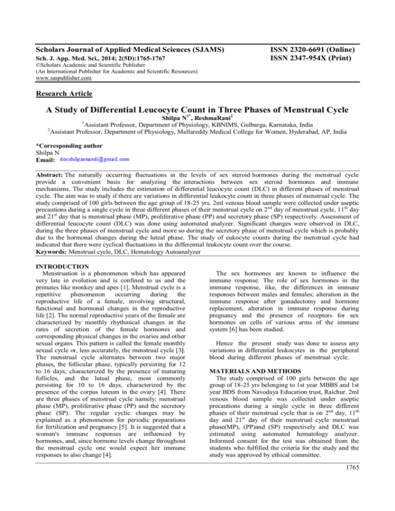
Scholars Journal of Applied Medical Sciences (SJAMS) Sch. J. App. Med. Sci., 2014; 2(5D):1765-1767 ISSN 2320-6691 (Online) ISSN 2347-954X (Print) ©Scholars Academic and Scientific Publisher (An International Publisher for Academic and Scientific Resources) www.saspublisher.com Research Article A Study of Differential Leucocyte Count in Three Phases of Menstrual Cycle Shilpa N1*, ReshmaRani2 Assistant Professor, Department of Physiology, KBNIMS, Gulbarga, Karnataka, India 2 Assistant Professor, Department of Physiology, Mallareddy Medical College for Women, Hyderabad, AP, India 1 *Corresponding author Shilpa N Email: Abstract: The naturally occurring fluctuations in the levels of sex steroid hormones during the menstrual cycle provide a convenient basis for analyzing the interactions between sex steroid hormones and immune mechanisms. The study includes the estimation of differential leucocyte count (DLC) in different phases of menstrual cycle. The aim was to study if there are variations in differential leukocyte count in three phases of menstrual cycle. The study comprised of 100 girls between the age group of 18-25 yrs. 2ml venous blood sample were collected under aseptic precautions during a single cycle in three different phases of their menstrual cycle on 2nd day of menstrual cycle, 11th day and 21st day that is menstrual phase (MP), proliferative phase (PP) and secretory phase (SP) respectively. Assessment of differential leucocyte count (DLC) was done using automated analyzer. Significant changes were observed in DLC, during the three phases of menstrual cycle and more so during the secretory phase of menstrual cycle which is probably due to the hormonal changes during the luteal phase. The study of eukocyte counts during the menstrual cycle had indicated that there were cyclical fluctuations in the differential leukocyte count over the course. Keywords: Menstrual cycle, DLC, Hematology Autoanalyzer INTRODUCTION Menstruation is a phenomenon which has appeared very late in evolution and is confined to us and the primates like monkey and apes [1]. Menstrual cycle is a repetitive phenomenon occurring during the reproductive life of a female, involving structural, functional and hormonal changes in the reproductive life [2]. The normal reproductive years of the female are characterized by monthly rhythmical changes in the rates of secretion of the female hormones and corresponding physical changes in the ovaries and other sexual organs. This pattern is called the female monthly sexual cycle or, less accurately, the menstrual cycle [3]. The menstrual cycle alternates between two major phases, the follicular phase, typically persisting for 12 to 16 days, characterized by the presence of maturing follicles, and the luteal phase, most commonly persisting for 10 to 16 days, characterized by the presence of the corpus luteum in the ovary [4]. There are three phases of menstrual cycle namely; menstrual phase (MP), proliferative phase (PP) and the secretory phase (SP). The regular cyclic changes may be explained as a phenomenon for periodic preparations for fertilization and pregnancy [5]. It is suggested that a woman's immune responses are influenced by hormones, and, since hormone levels change throughout the menstrual cycle one would expect her immune responses to also change [4]. The sex hormones are known to influence the immune response. The role of sex hormones in the immune response, like, the differences in immune responses between males and females; alteration in the immune response after gonadectomy and hormone replacement, alteration in immune response during pregnancy and the presence of receptors for sex hormones on cells of various arms of the immune system [6] has been studied. Hence the present study was done to assess any variations in differential leukocytes in the peripheral blood during different phases of menstrual cycle. MATERIALS AND METHODS The study comprised of 100 girls between the age group of 18-25 yrs belonging to 1st year MBBS and 1st year BDS from Navodaya Education trust, Raichur. 2ml venous blood sample was collected under aseptic precautions during a single cycle in three different phases of their menstrual cycle that is on 2nd day, 11th day and 21st day of their menstrual cycle menstrual phase(MP), (PP)and (SP) respectively and DLC was estimated using automated hematology analyzer. Informed consent for the test was obtained from the students who fulfilled the criteria for the study and the study was approved by ethical committee. 1765 Shilpa N et al., Sch. J. App. Med. Sci., 2014; 2(5D):1765-1767 RESULTS Using Automated analyzer, DLC was estimated and the obtained data was tabulated. Master chart (Appendix IV) showing the baseline parameters ; Age, Height, Weight, BMI, DLC were tabulated. Table 1: Anthropometric measurements of the subjects (n=100) Parameters Minimum Maximum Mean ± SD Age (yrs) 18 19 18.03 ± 0.171 Height (meters) 1 2 1.52 ± 0.018 Weight (Kg) 40 70 51.69 ± 5.462 BMI 18 30 22.39 ± 2.13 Table 2: Repeated ANOVA with post hoc test of results for various leukocyte counts during different phases of menstrual cycle Menstrual Proliferative Secretory Parameters F-value p-value Post hoc test Phase Phase Phase MP vs. PF, q= 9.953, p<0.001 MP vs. SP, q= 2.123, Neutrophil (%) 65.90±6.162 60.79 ± 6.652 66.99±7.201 41.565 p<0.0001 p>0.05 PF vs. SP, q= 12.076, p<0.001 MP vs. PF, q= 8.497, p<0.001 MP vs. SP, q= 0.183, Lymphocyte (%) 27.61±5.952 33.18 ± 6.82 27.49±6.856 24.594 p<0.0001 p>0.05 PF vs. SP, q= 8.68, p<0.001 Post tests were not calculated because the Monocyte (%) 2.34 ± 1.81 2.06 ± 1.34 1.89 ± 1.5 2.10 p=0.124 p value was greater than 0.05. Post tests were not calculated because the Eosinophil (%) 4.15 ± 1.37 3.97 ± 1.73 3.63 ± 1.66 2.736 p=0.06 p value was greater than 0.05. Post tests were not calculated because the Basophil (%) 0.00 ± 0.0 0.00 ± 0.0 0.00 ± 0.0 0.00 p=0.99 p value was greater than 0.05. The decrease in PP was statistically significant when compared to MP and SP ( p< 0.001) There was no significant difference in the rise of neutrophil count in both MP and SP (p>0.05). There was statistically significant increase in lymphocyte counts in PP when compared to MP (p<0.001) and also significant difference between PP and SP. The increase in SP was not significant when compared to MP (p>0.05). There is decrease in monocyte count from MP to SP, but the decrease is non-significant (p>0.05). The eosinophil count decreased from MP to SP which was non- significant (p>0.05) DISCUSSION The study was done to assess the changes in DLC during three phases of regular menstrual cycle. The DLC was estimated during the MP, PP, and SP of regular menstrual cycle in 100 female students. The neutrophil percentage are 65.90 ± 6.162, 60.79 ± 6.652, 66.99 ± 7.201 during MP, PP, SP respectively. The lymphocyte percentage are 27.61 ± 5.952, 33.18 ± 6.82, 27.49 ± 6.856 during MP, PP and SP respectively. Significant changes were observed in, neutrophil %, and lymphocyte% during the three phases of menstrual cycle. Neuroendocrine regulation on immune responses is suggested during an ovarian cycle, which may be critical for embryonic implantation and pregnancy [7]. The percent change increase in total WBC count in the secretory phase found in one study corroborated with earlier studies and is due to increase in all subpopulations (lymphocytes, monocytes and granulocytes). The levels of estrogen or progesterone are important factors in regulating the neutrophil count. Estrogen seems to enhance granulocyte proliferation in vitro [8]. 1766 Shilpa N et al., Sch. J. App. Med. Sci., 2014; 2(5D):1765-1767 Different reproductive processes like ovulation, menstruation, are influenced by the immune system. This results in a sexual dimorphism in the immune response in humans [9]. A significant increase in granulocyte numbers was found during pregnancy and in the luteal phase as compared to the follicular phase of the normal ovarian cycle which suggests a role of progesterone and estrogen in increasing granulocyte numbers from the bone marrow [10] as well as to delayed apoptosis. It has also been shown that progesterone enhanced chemotactic activity of neutrophils, while estrogens decreased the activity [11]. Diseases mediated by excessive antibody production as in case of systemic lupus eryhthematosus (SLE) tends to flare up during pregnancy [12] and in the luteal phase of menstrual cycle [13]. The reason may be that both the luteal phase and pregnancy are associated with a type 2 immune response shifting immunity away from the cell mediated immune (CMI) response. 3. 4. 5. 6. 7. 8. 9. CONCLUSION The study has indicated that there are cyclical fluctuations in the differential leukocyte count over the course of the menstrual cycle and the immune response is influenced by changes in hormone status. It points to the need for determining the role that variation in immune cell levels may play in women’s immune response and may have therapeutic implications in diseases of autoimmunity in women. REFERENCES 1. Tikare SN, Das KK; Blood leukocyte profile in different phases of menstrual cycle. IJPP 2008; 52(2): 201-201. 2. Rajnee, ChawlaVK, Choudhary R, Binawara BK, Choudhary S; Haematological and 10. 11. 12. 13. electrocardiographic variations during menstrual cycle. Pak J Physiology, 2010; 6(1): 18-21. Arthur C Guyton, Textbook of Medical Physiology, 11th edition, Saunders Co., 2005. Begum S, Ashwini S; Study of immune profile during different phases of menstrual cycle. Int J Biol Med Res., 2012; 3(1): 1407-1409. Molina PE; Lange’s Endocrine Physiology, 3rd edition, The Mc Graw Hill Companies, Inc., 2010. King CA; Cytokine expression during different phases of the menstrual cycle, Thesis (M.Sc.), Memorial University of Newfoundland, 1998. Lee S1, Kim J, Jang B, Hur S, Jung U, Kil K et al.; Fluctuation of peripheral blood T, B, and NK cells during a menstrual cycle of normal healthy women. The Journal of Immunology, 2010; 185(1): 756762. Pierre RV; Peripheral blood film review. Clin Lab Med., 2002; 22(1): 279-297. Bouman A, Heineman MJ, Faas MM; Sex hormones and the immune response in humans. Human Reproduction Update, 2005; 11(4): 411– 423. Ahmed SA, Penhale WJ; The influence of testosterone on the development of autoimmune thyroiditis in thymectomized and irradiated rats. Clin Exp Immunol., 1982; 48(2): 367-374. Miyagi M, Aoyama H, Morishita M, Iwamoto Y; Effects of sex hormones on chemotaxis of human peripheral polymorphonuclear leukocytes and monocytes, J Periodontol., 1992; 63(1): 28-32. Varner MW; Autoimmune disorders and pregnancy. Semin Perinatol., 1991; 15(3): 238-250. Pando JA, Gourley MF, Wilder RL, Crofford LJ; Hormonal supplementation as treatment for cyclical rashes in patients with systemic lupus erythematosus, J Rheumatol., 1995; 22(11): 21592162. 1767

