Nutrient Identification Lab: Chemical Tests & Procedures
advertisement
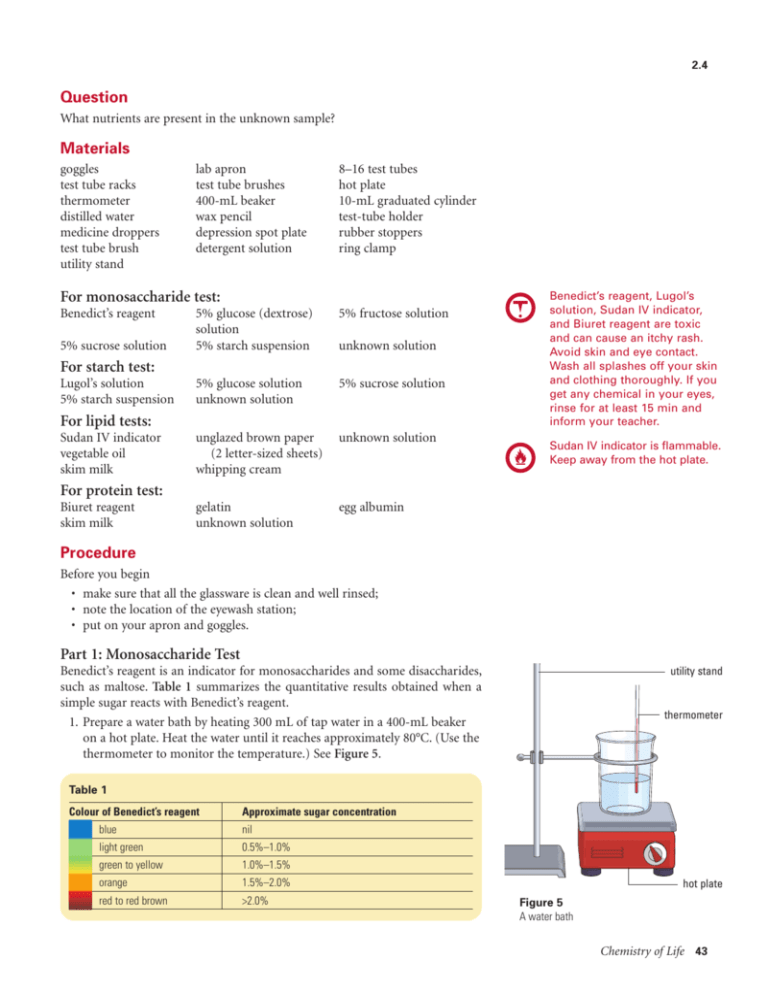
2.4 Question What nutrients are present in the unknown sample? Materials goggles test tube racks thermometer distilled water medicine droppers test tube brush utility stand lab apron test tube brushes 400-mL beaker wax pencil depression spot plate detergent solution 8–16 test tubes hot plate 10-mL graduated cylinder test-tube holder rubber stoppers ring clamp For monosaccharide test: Benedict’s reagent 5% sucrose solution 5% glucose (dextrose) solution 5% starch suspension 5% fructose solution 5% glucose solution unknown solution 5% sucrose solution unglazed brown paper (2 letter-sized sheets) whipping cream unknown solution gelatin unknown solution egg albumin unknown solution For starch test: Lugol’s solution 5% starch suspension For lipid tests: Sudan IV indicator vegetable oil skim milk Benedict’s reagent, Lugol’s solution, Sudan IV indicator, and Biuret reagent are toxic and can cause an itchy rash. Avoid skin and eye contact. Wash all splashes off your skin and clothing thoroughly. If you get any chemical in your eyes, rinse for at least 15 min and inform your teacher. Sudan IV indicator is flammable. Keep away from the hot plate. For protein test: Biuret reagent skim milk Procedure Before you begin • make sure that all the glassware is clean and well rinsed; • note the location of the eyewash station; • put on your apron and goggles. Part 1: Monosaccharide Test Benedict’s reagent is an indicator for monosaccharides and some disaccharides, such as maltose. Table 1 summarizes the quantitative results obtained when a simple sugar reacts with Benedict’s reagent. 1. Prepare a water bath by heating 300 mL of tap water in a 400-mL beaker on a hot plate. Heat the water until it reaches approximately 80°C. (Use the thermometer to monitor the temperature.) See Figure 5. utility stand thermometer Table 1 Colour of Benedict’s reagent Approximate sugar concentration blue nil light green 0.5%–1.0% green to yellow 1.0%–1.5% orange 1.5%–2.0% red to red brown >2.0% hot plate Figure 5 A water bath Chemistry of Life 43 2. Using a 10-mL graduated cylinder, measure 3 mL each of distilled water, fructose, glucose, sucrose, starch, and the unknown solution. Pour each solution into a separate test tube. Clean and rinse the graduated cylinder after pouring each solution. Label each test tube using the wax pencil. Record the code for your unknown sample. Add 1 mL of Benedict’s reagent to each of the test tubes. 3. Using a test tube holder, place each of the test tubes in the hot water bath. Observe for 6 min. Record any colour changes in a chart. Part 2: Starch Test Lugol’s solution contains iodine and is an indicator for starch. Iodine turns blueblack in the presence of starch. 4. Using a medicine dropper, place a drop of water on a depression spot plate and add a drop of Lugol’s solution. Record the colour of the solution. 5. Repeat the procedure, this time using drops of starch, glucose, sucrose, and your unknown solution. Record the colour of the solutions in a chart. Which solutions indicate a positive test? To mix the contents of a stoppered test tube thoroughly, make sure the stopper is on tight. Place your index finger on top of the stopper, gripping the test tube firmly with your thumb and other fingers. Then shake away from others. Part 3: Sudan IV Lipid Test Sudan IV solution is an indicator of lipids, which are soluble in certain solvents. Lipids turn from a pink to a red colour. Polar compounds will not assume the pink colour of the Sudan IV indicator. 6. Using a 10-mL graduated cylinder, measure 3 mL each of distilled water, vegetable oil, skim milk, whipping cream, and the unknown solution. Pour each solution into a separate labelled test tube. Clean and rinse the graduated cylinder after each solution. Add 6 drops of Sudan IV indicator to each test tube. Place stoppers on the test tubes and shake them vigorously for 2 min. Record the colour of the mixtures in a chart. Part 4: Translucence Lipid Test Lipids can be detected using unglazed brown paper. Because lipids allow the transmission of light through the brown paper, the test is often called the translucence test. 7. Draw one circle (10-cm diameter) on a piece of unglazed brown paper. Place 1 drop of water in the circle and label the circle accordingly. Using more sheets, draw a total of 7 more circles (10-cm diameter). Place 1 drop of vegetable oil, skim milk, whipping cream, and unknown solution, each inside its own circle, labelling the circles as you do. When the water has evaporated, hold both papers to the light and observe. In a chart, record whether or not the papers appear translucent. Part 5: Protein Test Proteins can be detected by means of the Biuret reagent test. Biuret reagent reacts with the peptide bonds that join amino acids together, producing colour changes from blue, indicating no protein, to pink (+), violet (++), and purple (+++). The + sign indicates the relative amounts of peptide bonds. 8. Measure 2 mL of water, gelatin, albumin, skim milk, and the unknown solution into separate labelled test tubes. Add 2 mL of Biuret reagent to each of the test tubes, then tap the test tubes with your fingers to mix the contents. Record any colour changes in a chart. Analysis (a) What laboratory evidence suggests that not all sugars are monosaccharides? (b) Explain the advantage of using two separate tests for lipids. 44 Chapter 2
