Sample preparation for fluorescence microscopy
advertisement
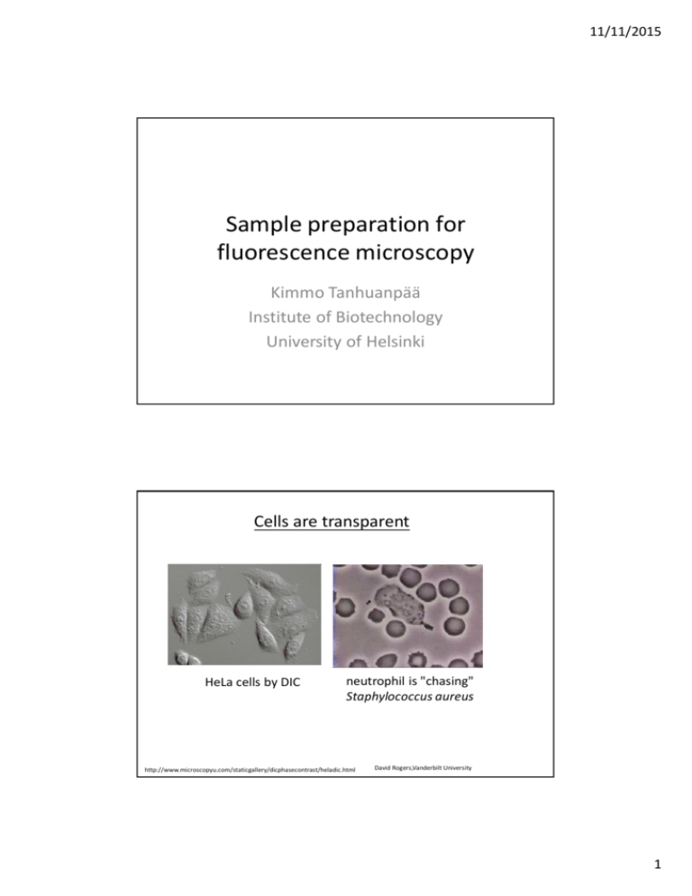
11/11/2015 Sample preparation for fluorescence microscopy Kimmo Tanhuanpää Institute of Biotechnology University of Helsinki Cells are transparent HeLa cells by DIC neutrophil is "chasing" Staphylococcus aureus http://www.microscopyu.com/staticgallery/dicphasecontrast/heladic.html David Rogers,Vanderbilt University 1 11/11/2015 Imaging of proteins and organelles by Fluorescence microscopy methods • To understand the function of proteins, it is essential to know their localization in cells • Similarly, to understand the function of organelles, it essential to visualize them Microtubules Golgi Actin filaments Outline -Ways on labeling proteins and other cellular structures • Immunofluorescence microscopy – specific antibodies • Green Fluorescent Fusion (GFP) –proteins – transfection of DNA constructs • Small fluorescently labeled chemical compounds, which bind with a high specificity to molecules of interest - Choosing the fluorophore – which color? 2 11/11/2015 1. Immunofluorescence microscopy • Requires specific antibodies (primary antibody) that recognize protein of interest • The primary antibody can be directly coupled to a fluorophore, or more commonly a fluorescent secondary antibody that recognizes the primary antibody is used + reveals the localization of the endogenous (cell’s own) protein - finding a good antibody can be difficult - does not permit live-cell imaging Simultaneous visualization of two proteins by immunofluorescence microscopy - two primary antibodies raised in different species (e.g. mouse and rabbit) are used. Each antibody recognizes only one of the proteins of interest - two species-specific secondary antibodies are used (e.g. anti-mouse and anti-rabbit). These secondary antibodies are conjugated with different fluorophores. Rhodamine (red fluorophore) labelled anti-rabbit antibody An antibody against protein X that was raised in rabbits FITC (green fluorophore) labelled anti-mouse antibody An antibody against protein Y that was raised in mouse Protein X Protein Y 3 11/11/2015 Critical steps in immunofluorescense protocol • Grow cells on coverslips • thickness! #1.5 à 0.17 mm • Fixation • cross-linking - aldehydes • (precipitation – organic solvents) • Permeabilization (if using cross-linking) • Incubation with the antibodies (optimized dilutions) • Mounting (remember refractive index!) Mounting media • Preservation of the sample • creating conditions to permit as good imaging as possible • Matching of the immersion medium and mountant • missmatch will cause image degradation due to spherical aberrations and signal loss à Think in advance, which objective you will/can use Cells Water Glycerol Glass Oil Refractive index 1.333 1.466 1.52 1.515 The mountant and the immersion medium should be matched within 0.01-0.05 4 11/11/2015 Mounting media • Base: o aqueous o oil o plastic • Antifade o Most are reactive oxygen scavengers o DABCO o n-Phenylenediamine (PPD) o n-Propyl gallate (NPG) • Hardening and non-hardening mountants • If using a mountant that does not harden, need to seal the coverslip to prevent drying and may also need spacers to prevent squashing of the specimen www.uhnresearch.ca/wcif In general about sample preparation… • After optimizing the protocol, maintain constant sample preparation • If using cells, keep to good cell culture practices • healthy cells • Standardize everything 5 11/11/2015 2. Green Fluorescent Protein (GFP) –fusion proteins • GFP is a fluorescent protein • The protein of interest can be linked to GFP (either to its N- or C-terminus) to make a fusion protein • Also other spectral variants (different colours) are nowadays available, allowing simultaneous visualization of several proteins in the same cell GFP + No need for an antibody, imaging in both fixed and live cells possible à dynamic studies - Overexpression of the protein of interest, sometimes GFP-fusion impairs protein function Nature Biotechnology 22, 1567 - 1572 (2004) Cloning GFP-fusion proteins GFP Protein X Protein X GFP - Clone the cDNA of the protein of your interest in frame with the GFP in either (or both) vectors - Transfect the DNA construct into cells 6 11/11/2015 Studying the sub-cellular localization of a protein by using GFP-fusion proteins • If fixing cells, use coverslips as for immunofluorescense • If doing live-cell imaging, use either Mattek-dishes (inverted microscope), or normal cell culture dishes of suitable size (upright microscope) 1. Cells are transfected with a plasmid that 2. Cell produces a fluorescent fusion protein that localizes similarly to the endogenous (cell’s own) contains the DNA for production of the protein X. fusion protein (GFP-protein X). Endogenous protein X GFP-protein X –fusion protein. Examples of live cell imaging with GFP-fusion proteins Melanoma cell expressing GFP-actin Fibroblasts expressing MAL-GFP and stimulated with serum 7 11/11/2015 Things to consider when using GFP-fusions -Test the functionality of the fusion proteins - compare the localization to endogenous proteins - Try the fusion protein on both ends of your protein - Remember: you always overexpress the protein - try to use as low expression levels as possible Endogenous MAL MALGFP GFPMAL 3. Small Fluorescent Compounds • phalloidin, which is a toxin isolated from mushrooms, binds actin filaments with high specificity Alexa 594 Phalloidin • DAPI (4',6-diamidino-2-phenylindole) is a fluorescent stain that binds strongly to A-T rich regions in DNA Alexa 594 Phalloidin DAPI + high specificity, labels endogenous protein/structure - Compounds available to a limited number of proteins, often not suitable for live-cell imaging (because may impair function of the protein/structure) 8 11/11/2015 Which color??? Choosing the right fluorophore • Depends on the properties of the microscope you intend to use • excitation à what can you excite à light source • emission à what can you detect à filters (if need several colors especially important!) à When planning the experiment, find out the properties of your microscope!!! http://www.edmundoptics.com/images/articles/fluorointrofigure31.jpg 9 11/11/2015 Multicolour imaging • Often several proteins/structures need to be visualized simultaneously - co-localization • Need to be sure that can get a specific signal from each colour! Cross-excitation or Channel bleed-through (emission) http://www.microscopyu.com/articles/livecellimaging/fpimaging.html Controls for Multicolour imaging • Sample that has not been stained at all à to learn to recognize autofluorescence from the cell • If using antibodies, staining with secondary antibody only à to learn to recognize specific antibody staining • Samples with the different colours separately à to set up the imaging conditions such that there is minimal cross-excitation and channel bleed-through à to get an estimate of the crosstalk, which can be used to correct images post-acquisition • If studying co-localization à quantify for example with Pearson´s coefficient 10 11/11/2015 Fluorophore properties • Colour à excitation and emission spectrum • Molar extinction coefficient o ability of the molecule to absorb light • Quantum yield o fluorescence emission efficiency o ratio of the number of photons emitted to the number of photons absorbed o often sensitive to the environment of the fluorophore • Quenching and photobleaching o reduce the levels of emission o molecular structure and environment affect How to connect the antibody/protein to the fluorophore? • Numerous commercial secondary antibodies available • Alexa dyes available as at least anti-mouse/rabbit/sheep/goat/guinea-pig…. • Most of the dyes can also be bought separately to label the antibody/protein yourself • dye can be pre-”activated” • amine-reactive à binds to primary amines • thiol-reactibe à binds to cysteines • etc…. Monoclonal antibody labelling kit http://www.invitrogen.com/site/us/en/home/References/Molecular-Probes-The-Handbook/Fluorophores-and-Their-Amine-Reactive-Derivatives/Kits-for-Labeling-Proteins-and-Nucleic-Acids.html 11 11/11/2015 Choosing the fluorescent protein • The fluorescent protein (FP) should be expressed efficiently and without toxicity in the experimental system o Some FPs may not fold efficiently at + 37 oC • FP should be bright enough to provide sufficient signal above autofluorescence • The FP should have sufficient photostability for the duration of the experiment • If the FP is used as a fusion protein, then the FP should not oligomerize • The FP should be insensitive to environmental effects • In multi-colour imaging, the set of FPs should have minimal crosstalk Choosing the fluorescent protein Properties of Different Green Fluorescent proteins Protein (Acronym) GFP (wt) Excitation Maximum (nm) Emission Maximum (nm) Molar Extinction Coefficient Quantum Yield in vivo Structure Relative Brightness (% of EGFP) 395/475 509 21,000 0.77 Monomer* 48 EGFP 484 507 56,000 0.60 Monomer* 100 Emerald 487 509 57,500 0.68 Monomer* 116 Superfolder GFP 485 510 83,300 0.65 Monomer* 160 Azami Green 492 505 55,000 0.74 Monomer 121 mWasabi 493 509 70,000 0.80 Monomer 167 TagGFP 482 505 58,200 0.59 Monomer* 110 TurboGFP 482 502 70,000 0.53 Dimer 102 AcGFP 480 505 50,000 0.55 Monomer* 82 ZsGreen 493 505 43,000 0.91 Tetramer 117 T-Sapphire 399 511 44,000 0.60 Monomer* 79 Shaner et al. Nature Methods 2005 12 11/11/2015 Choosing the fluorescent protein Cyan: Cerulean Green: Emerald or EGFP Yellow-green: mCitrine, Venus or Ypet Orange: mOrange or mKO Red: mCherry Far red: mPlum Additional info: http://www.microscopyu.com/ http://www.olympusmicro.com/index.html 13

