Description - Allied Biotech, Inc
advertisement
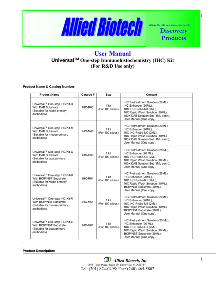
1 Where the life sciences come to life. Discovery Products User Manual Universal TM One-step Immunohistochemistry (IHC) Kit (For R&D Use only) Product Name & Catalog Number: Product Name UniversalTM One-step IHC Kit-R With DAB Substrate (Suitable for rabbit primary antibodies) UniversalTM One-step IHC Kit-M With DAB Substrate (Suitable for mouse primary antibodies) UniversalTM One-step IHC Kit-G With DAB Substrate (Suitable for goat primary antibodies) UniversalTM One-step IHC Kit-R With BCIP/NBT Substrate (Suitable for rabbit primary antibodies) UniversalTM One-step IHC Kit-M With BCIP/NBT Substrate (Suitable for mouse primary antibodies) UniversalTM One-step IHC Kit-G With BCIP/NBT Substrate (Suitable for goat primary antibodies) Catalog # Size Content IHC-R60 1 kit (For 100 slides) IHC-M60 1 kit (For 100 slides) IHC-G60 1 kit (For 100 slides) IHC-R61 1 kit (For 100 slides) IHC-M61 1 kit (For 100 slides) IHC-G61 1 kit (For 100 slides) IHC Pretreatment Solution (20ML), IHC Enhancer (20ML), 10X IHC Probe-R0 (2ML), 10X Rapid Wash Solution (10ML), 100X DAB Solution Set (1ML each), User Manual (One copy). IHC Pretreatment Solution (20ML), IHC Enhancer (20ML), 10X IHC Probe-M0 (2ML), 10X Rapid Wash Solution (10ML), 100X DAB Solution Set (1ML each), User Manual (One copy). IHC Pretreatment Solution (20 ML), IHC Enhancer (20 ML), 10X IHC Probe-G0 (2ML), 10X Rapid Wash Solution (10 ML), 100X DAB Solution Set (1ML each), User Manual (One copy). IHC Pretreatment Solution (20ML), IHC Enhancer (20ML), 10X IHC Probe-R1 (2ML), 10X Rapid Wash Solution (10ML), BCIP/NBT Substrate (20ML), User Manual (One copy). IHC Pretreatment Solution (20ML), IHC Enhancer (20ML), 10X IHC Probe-M1 (2ML), 10X Rapid Wash Solution (10ML), BCIP/NBT Substrate (20ML), User Manual (One copy). IHC Pretreatment Solution (20 ML), IHC Enhancer (20 ML), 10X IHC Probe-G1 (2ML), 10X Rapid Wash Solution (10 ML), BCIP/NBT Substrate (20ML), User Manual (One copy). . Product Description: Allied Biotech, Inc 10075 Tyler Place, Suite 19, Ijamsville, MD 21754 Tel: (301) 874-0495; Fax: (240) 465-5802 1 2 TM Universal One-step IHC is a latest breakthrough technology (patent pending) for performing Immunohistochemistry (IHC) or Immunocytochemistry (ICC). Unlike regular (classical) IHC/ICC, the One-step IHC/ICC technology combines blocking, primary antibody staining, washing and secondary antibody staining into a rapid one-step reaction. Therefore, using UniversalTM One-step IHC kit, your IHC/ICC becomes so simple and is no longer a time-consuming and labor-intensive procedure. The One-step IHC kit is a universal kit, which is suitable for most primary antibodies and no need of secondary antibody. Currently, we provide six types of UniversalTM One-step IHC kits as shown on the above table. Each kit is sufficient to stain 100 slides or 400 wells for 96-well plates. Application: Immunohistochemistry (IHC) or Immunocytochemistry (ICC). Features: Easy, a simple One-step reaction instead of the 4-step procedure: blocking, primary antibody-binding, washing and 2nd antibody-binding. Rapid, just 1~2 hrs instead of 4~5 hrs. Universal, suitable for most primary antibodies and no need of secondary antibody. Sensitive, comparable sensitivity with regular IHC or ICC. Reproducible, highly reproducible results. Storage: Pretreatment Solution and Enhancer: for continuous use, store in 2~8 °C for up to 1 month, or store in -20 °C for up to a year. IHC Probe and Substrate: for continuous use, store in -20 °C for up to 6 months or store in -70 °C manual defrost freezer for up to a year. Repeated freezing and thawing is not recommended. Rapid Wash Solution: store in room temperature for up to one year. Note: Kit will be shipped in ambient temperature. Comparison of One-step IHC with Regular IHC: Protocol: I. Other required materials: Primary antibody: Purified monoclonal or affinity-purified polyclonal antibodies (antigen-specific IgGs) are preferred. Although unpurified antibody or serum can also be used for the One-step IHC, user may need to optimize staining conditions to avoid non-specific binding. Note: We currently provide six types of UniversalTM One-step IHC kits. Kits labeled with -R are suitable for using together with rabbit primary antibodies, with -M suitable for using together with mouse primary antibodies, and with -G suitable for using together with goat primary antibodies. Fixatives: 30% Buffer-neutralized Formalin or Z-fix. Counter staining Reagent: Mayer’s hematoxylin or other proper counter staining reagents. Allied Biotech, Inc 10075 Tyler Place, Suite 19, Ijamsville, MD 21754 Tel: (301) 874-0495; Fax: (240) 465-5802 2 3 II. Reagent preparation: One-step IHC Antibody Mixture: Mix 5-30ul of primary antibody and 100ul IHC Probe (supplied in the kit) with 1ml of IHC Enhancer (supplied in the kit). The amount of primary antibody added may vary depending on the quality of the antibody and it may need optimization by user to obtain best results. 1X Rapid Wash Solution: Dilute 10ml of 10X Rapid Wash Solution (supplied in the kit) with 90ml distilled water. 1X DAB Mixture: Mix 100ul of 100X DAB solution (supplied in the kit), 100ul of 100X DAB buffer (supplied in the kit) with 10 ml of distilled water. III. Procedure: 1. Fix: Incubate sample (tissue or cells) on slide with Formalin (or other proper fixatives) for 20 min and then rinse the slide with 600ul PBS. Note: this step is same to the method for regular IHC or ICC. 2. Pretreatment: Incubate the pre-fixed tissue section or cells on slide with 200ul (per slide) of Pretreatment Solution for 10 minutes at room temperature in order to inactivate the endogenous peroxidase, and then rinse this slide once with 600ul PBS. 3. One-step Reaction: Incubate the above slide (from Step 2) with 200ul of the prepared One-step IHC Antibody Mixture (a mixture of primary antibody, IHC enhancer and IHC Probe) for 30~60 minute at room temperature. 4. Wash: Rinse the slide three times with 600ul of 1X Rapid Wash Solution and once with 2ml of PBS. Development: Apply 200ul of the pre-prepared 1XDAB mixture (for kits IHC-R60, IHC-M60 and IHC-G60) or IHC-R61, IHC-M61 and IHC-G61) to the washed section on slide and incubate for approximate 2 min or an appropriate time until a desired color appears. To stop the development, simply rinse slide with distilled water. 5. BCIP/NBT substrate mixture (for kits 6. Counter-staining: Incubate the slide with the solution of Mayer’s hermatoxyin for 0.5-5 min at room temperature until a desired background color appears. Rinse the slide with distilled water. Note: this step is same to the method for regular IHC or ICC. 7. After Staining: Take photos and/or mount the section with glycerol gelatin and a sealing cover-slip with clear nail polish. Note: This step must be performed according to the standard procedure used for regular IHC or ICC Example: Comparison of One-step ICC with Regular ICC in Detection of the Expression of a Flag-tagged protein in A549 Cells. A549 cells cultured in 96-well plate were directly fixed on the wells of tissue culture plate and then pretreated with the kit-provided pretreatment solution. Expressed Flag-tagged proteins in cells were detected using mouse anti-Flag antibody by a regular ICC procedure (A) or by a one-step IHC procedure (B) or by a one-step IHC procedure but without using primary antibody (C). As showed in the picture, the results by both methods are very similar but the testing time and procedure are greatly different. A: 5 hours (using regular method). B: <1 hour (using One-step IHC kit). C: One-step ICC without primary antibody (as a negative control). Note: the brown color signals indicated the positive stains using anti-Flag antibody and DAB substrate. Allied Biotech, Inc 10075 Tyler Place, Suite 19, Ijamsville, MD 21754 Tel: (301) 874-0495; Fax: (240) 465-5802 3 4 Troubleshooting: Problem Probable Cause Weak Signal or No Staining Inadequate deparaffinization Solution Deparaffinize sections longer or change fresh xylene Inactive primary antibodies Replace with a new batch of antibodies Antibodies do not work due to improper storage Aliquot antibodies into smaller volumes and store in freezer (-20 to 70 C) and avoid repeated freeze and thaw cycles. Or store antibodies according to manufacture's instructions. Increase the concentration of primary and/or One-step IHC probe. Or Concentration of primary antibody or IHC run a serial dilution test to determine the optimal dilution that gives probe was too low the best signal to noise ratio Incorrect primary antibody type Check the label of IHC Kit and select compatible primary antibodies and One-Step IHC Kit. Kits labeled with -R are suitable for rabbit primary antibodies, with -M suitable for mouse primary antibodies, and with -G suitable for goat primary antibodies. Poor specificity of primary antibodies. Use purified monoclonal or affinity-purified polyclonal primary antibodies. Inadequate antibody incubation time Increase antibody incubation time Inadequate or improper tissue fixation Increase duration of postfixation or try different fixatives Tissue overfixation Reduce the duration of postfixation. If the tissue has already been overfixed, perform an appropriate or recommended antigen retrieval antigen retrieval procedure. Defective One-step IHC probe Replace with a new batch of One-step IHC probe. Inadequate substrate incubation time Increase the substrate incubation time. Over-staining The concentration of primary and/or One- Reduce the concentration of antibody or IHC probe, or perform a step IHC probe was too high titration to determine the optimal dilution for primary and IHC probe High Background Incubation time was too long Reduce incubation time Incubation temperature was too high Reduce incubation temperature Substrate incubation time was too long Reduce substrate incubation time Sections dried out Avoid sections being dried out Inadequate washing of sections Wash at least 3 times between steps Increase incubation with pre-treatment solution or 3% hydrogen peroxide to block endogenous enzyme activities prior to incubation of Tissue contains endogenous peroxidase antibody. Non-specific binding of primary Non-specific binding may be reduced by using higher dilution of antibodies to tissue or antibody primary antibodies or by using purified monoclonal or affinity-purified concentration was too high polyclonal primary antibodies. Diffusion of tissue antigen due to inadequate fixation Increase duration of postfixation Sections dried out Avoid sections being dried out Allied Biotech, Inc 10075 Tyler Place, Suite 19, Ijamsville, MD 21754 Tel: (301) 874-0495; Fax: (240) 465-5802 4
