T-cell lymphomas
advertisement
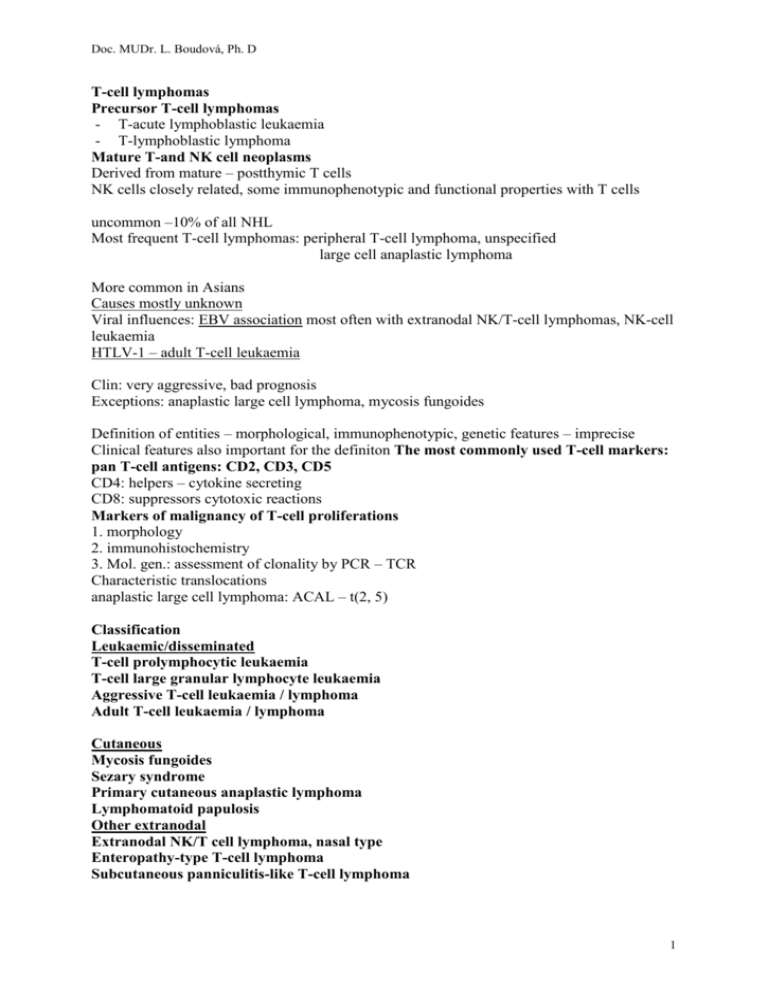
Doc. MUDr. L. Boudová, Ph. D T-cell lymphomas Precursor T-cell lymphomas - T-acute lymphoblastic leukaemia - T-lymphoblastic lymphoma Mature T-and NK cell neoplasms Derived from mature – postthymic T cells NK cells closely related, some immunophenotypic and functional properties with T cells uncommon –10% of all NHL Most frequent T-cell lymphomas: peripheral T-cell lymphoma, unspecified large cell anaplastic lymphoma More common in Asians Causes mostly unknown Viral influences: EBV association most often with extranodal NK/T-cell lymphomas, NK-cell leukaemia HTLV-1 – adult T-cell leukaemia Clin: very aggressive, bad prognosis Exceptions: anaplastic large cell lymphoma, mycosis fungoides Definition of entities – morphological, immunophenotypic, genetic features – imprecise Clinical features also important for the definiton The most commonly used T-cell markers: pan T-cell antigens: CD2, CD3, CD5 CD4: helpers – cytokine secreting CD8: suppressors cytotoxic reactions Markers of malignancy of T-cell proliferations 1. morphology 2. immunohistochemistry 3. Mol. gen.: assessment of clonality by PCR – TCR Characteristic translocations anaplastic large cell lymphoma: ACAL – t(2, 5) Classification Leukaemic/disseminated T-cell prolymphocytic leukaemia T-cell large granular lymphocyte leukaemia Aggressive T-cell leukaemia / lymphoma Adult T-cell leukaemia / lymphoma Cutaneous Mycosis fungoides Sezary syndrome Primary cutaneous anaplastic lymphoma Lymphomatoid papulosis Other extranodal Extranodal NK/T cell lymphoma, nasal type Enteropathy-type T-cell lymphoma Subcutaneous panniculitis-like T-cell lymphoma 1 Doc. MUDr. L. Boudová, Ph. D Nodal Angioimmunoblastic T-cell lymphoma Peripheral T-cell lymphoma, unspecified Anaplastic large cell lymphoma Neoplasms of uncertain lineage and stage of differentiation Blastic NK cell lymphoma Leukaemic - disseminated cases T-cell prolymphocytic leukaemia (not obligatory to learn about) rare, lymphocytosis – over 100 x 10 9 /l, aggressive BM, PB, LN, liver, spleen, skin anaemia, thrombocytopenia In LN – diffuse - more in the paracortex, no pseudofollicles T-cell large granular lymphocyte leukaemia (not obligatory to learn about) Heterogeneous, most cases indolent Tumour cell: large granular lymphocyte - abundant cytoplasm, coarse cytoplasmic granules – azurophilic Main problem: neutropenia often autoimmune disease Aggressive NK – cell leukaemia(not obligatory to learn about) Aggressive, Asian, syoung often coagulopathy, haemophagocytic syndrome, multiorgan failure Adult T-cell leukamia/ lymphoma Tumor cell: highly pleomorphic cells, size medium - large Caused by the human retrovirus, human T-cell leukaemia virus type 1 . Endemic (more important) or sporadic Endemic: in several parts of the world: Japan, in the Caribbean basin, parts of Central Africa only minority who infected develop the leukamia Systemic – widely disseminated – BM. PB, LN, other organs Several clinical variants with different prognosis Survival: weeks - 1y Extranodal Extranodal NK/T cell lymphoma Angiocentric, vascular destruction, prominent necrosis Immunophenotype –/NK or T/ Nasal cavity the most common site Asia, Central and South America Presentation: epistaxis – large mutilating lesions /former name: lethal midline granuloma presents localised, dissemination may occur also outside the nasal cavity Associated with EBV Usually aggressive 2 Doc. MUDr. L. Boudová, Ph. D Enteropathy associated T-cell lymphoma Uncommon, more frequent in areas with high incidence of coeliac dis.(develops in the intestine affected by coeliac dis.) Jejunum or ileum Adults Abdominal pain, often intestinal perforation Multiple ulcers Prognosis poor Hepatosplenic T-cell lymhoma Rare, young male Hepatosplenomegaly Aggressive Cutaneous lymphoma Mycosis fungoides (MF) and Sezary syndrome Most common primary skin lymphoma Elderly Remains limited to the skin for ys At advanced stages: dissemination – LN, liver. PB, spleen, lungs – late event (does not pccur always) Grossly: Patches, plaques, - infiltration enlarges, thickens – tumours trunk Rarely:generalised dis – erythroderma Histologically: Epidermal and dermal infiltration by small to medium.sized T-cells with cerebriform nuclei Pautrier abscess: collections of tumour cells in the epidermis Enlarged LN – dermatopathic LA, later tumor cells Aetiology unknown Prognosis: most important factor: extent of the disease Sezary syndrome Generalised mature T-cell lymphoma Erythroderma, generalised lymphadenopathy, neoplastic T-ly in the PB Pruritus, alopecia, nail diseases Hist. similar to MF Cerebriform nuclei variant of MF, but more aggressive Primary cut. ALCL ALCL – exists: systemic or primary cutaneous cutaneous: At the time of diagnosis limited only to the skin, may disseminate afterwards, diagnosis (primary cut. /systemic) clinically important Grossly: nodules, solitary or multicentric; ulcerated Tumor cell: large, large vesicular nucleus – folded =embryo cell, conspicuous nucleolus Prognosis favourable: 5ys: 90%, often young 3 Doc. MUDr. L. Boudová, Ph. D Lymphomatoid papulomatosis Chronic, recurrent, benign course, may progress to lymphoma NODAL AIBL – angioimmunoblastic lymphadenopathy LN - Polymorphous infiltrate, prominence of venules with high endothelium Rel. common. 50 ys Gen LA, enlarged L+ S, advanced stage, skin rash, Systemic symptoms, arthritis, ascite Immune abnormalities – sec. to the neoplastic process Aggressive – survival less than 3 ys, succumb to infections Peripheral T-cell lymphoma, NOS Common, Adults LN, often generalised - BM, L, S, extranodal, may becomeleukaemic - Often B-symptoms - Causes unknown morphological subtypes T-zone interfollicular pattern of growth Lennert – lymphoepithelioid cell variant Small cells and clusters of epithelioid histiocytes in the infiltrate, EO, other Among the most aggressive of the NHL Overall survival rates less than 30% Anaplastic large cell lymphoma - systemic Tumor cell: large, horseshoe shaped, large vesicular nucleus – folded =embryo cell, conspicuous nucleolus Abundant cytoplasm CD30 (Ki1)+ ALCL kinase protein Young male (also older) Sites of involvement: LN, extranodal sites Present with advanced stage Often B-symptoms /fever); rel. favourable prognosis Translocation t(2, 5) 4
