file - BioMed Central
advertisement
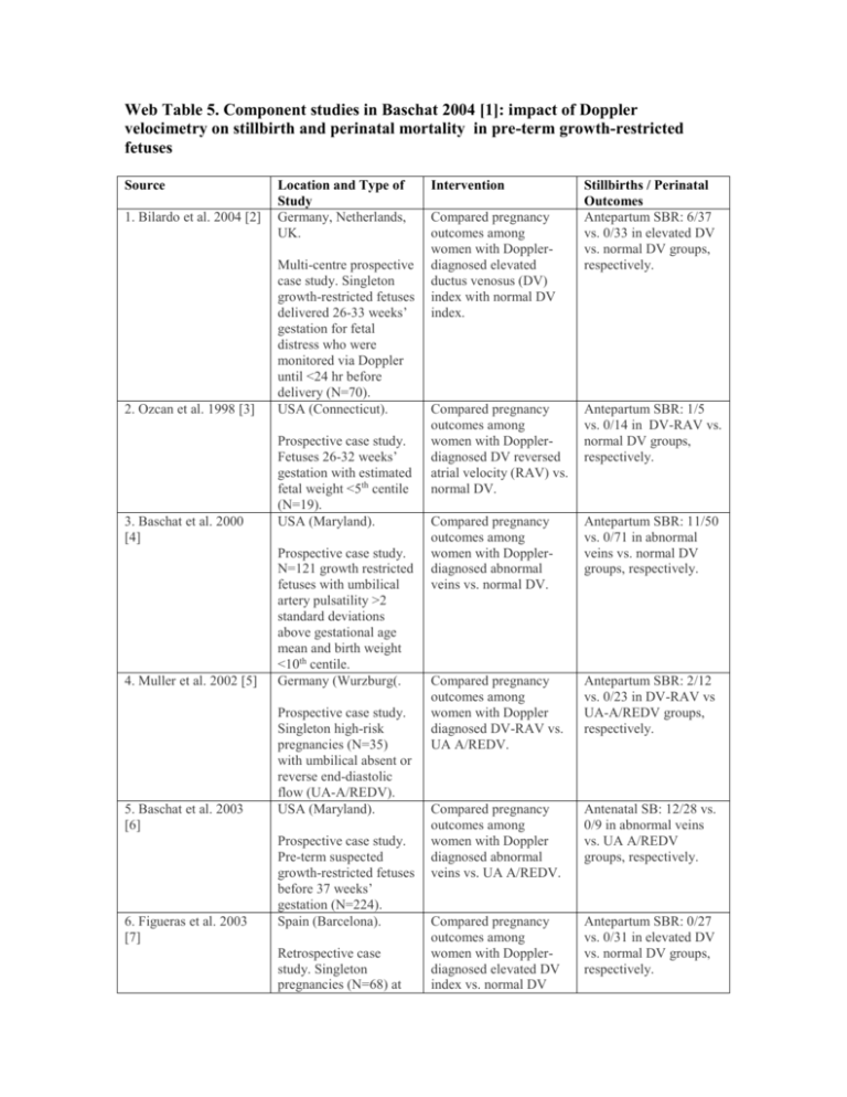
Web Table 5. Component studies in Baschat 2004 [1]: impact of Doppler velocimetry on stillbirth and perinatal mortality in pre-term growth-restricted fetuses Source 1. Bilardo et al. 2004 [2] 2. Ozcan et al. 1998 [3] 3. Baschat et al. 2000 [4] 4. Muller et al. 2002 [5] 5. Baschat et al. 2003 [6] 6. Figueras et al. 2003 [7] Location and Type of Study Germany, Netherlands, UK. Multi-centre prospective case study. Singleton growth-restricted fetuses delivered 26-33 weeks’ gestation for fetal distress who were monitored via Doppler until <24 hr before delivery (N=70). USA (Connecticut). Prospective case study. Fetuses 26-32 weeks’ gestation with estimated fetal weight <5th centile (N=19). USA (Maryland). Prospective case study. N=121 growth restricted fetuses with umbilical artery pulsatility >2 standard deviations above gestational age mean and birth weight <10th centile. Germany (Wurzburg(. Prospective case study. Singleton high-risk pregnancies (N=35) with umbilical absent or reverse end-diastolic flow (UA-A/REDV). USA (Maryland). Prospective case study. Pre-term suspected growth-restricted fetuses before 37 weeks’ gestation (N=224). Spain (Barcelona). Retrospective case study. Singleton pregnancies (N=68) at Intervention Compared pregnancy outcomes among women with Dopplerdiagnosed elevated ductus venosus (DV) index with normal DV index. Stillbirths / Perinatal Outcomes Antepartum SBR: 6/37 vs. 0/33 in elevated DV vs. normal DV groups, respectively. Compared pregnancy outcomes among women with Dopplerdiagnosed DV reversed atrial velocity (RAV) vs. normal DV. Antepartum SBR: 1/5 vs. 0/14 in DV-RAV vs. normal DV groups, respectively. Compared pregnancy outcomes among women with Dopplerdiagnosed abnormal veins vs. normal DV. Antepartum SBR: 11/50 vs. 0/71 in abnormal veins vs. normal DV groups, respectively. Compared pregnancy outcomes among women with Doppler diagnosed DV-RAV vs. UA A/REDV. Antepartum SBR: 2/12 vs. 0/23 in DV-RAV vs UA-A/REDV groups, respectively. Compared pregnancy outcomes among women with Doppler diagnosed abnormal veins vs. UA A/REDV. Antenatal SB: 12/28 vs. 0/9 in abnormal veins vs. UA A/REDV groups, respectively. Compared pregnancy outcomes among women with Dopplerdiagnosed elevated DV index vs. normal DV Antepartum SBR: 0/27 vs. 0/31 in elevated DV vs. normal DV groups, respectively. 7. Hofstaetter C et al 2002 [8] 8. Hofstaetter et al. 1996 [9] or after 26 weeks of pregnancy in which delivery occurred within 3 days of Doppler surveillance. Germany (Bonn). Prospective case study. Growth-restricted fetuses with reversed umbilical artery flow (N=37). Sweden (Malmo). Prospective case study. High-risk complicated pregnancies (N=87) referred for umbilical artery Doppler assessment. index to contraction stress test results. Compared pregnancy outcomes in women with Doppler-diagnosed abnormal veins vs. UA A/REDV. Antepartum SBR: 12/28 vs. 0/9 in abnormal veins vs. UA A/REDV groups, respectively. Compared pregnancy outcomes among women with elevated DV index vs. normal DV index. Antepartum SBR: 0/22 vs. 0/65 in elevated DV vs. normal DV groups, respectively. References 1. 2. 3. 4. 5. 6. 7. Baschat AA: Doppler application in the delivery timing of the preterm growth-restricted fetus: another step in the right direction. Ultrasound Obstet Gynecol 2004, 23(2):111-118. Bilardo CM, Wolf H, Stigter RH, Ville Y, Baez E, Visser GH, Hecher K: Relationship between monitoring parameters and perinatal outcome in severe, early intrauterine growth restriction. Ultrasound Obstet Gynecol 2004, 23(2):119-125. Ozcan T, Sbracia M, d'Ancona RL, Copel JA, Mari G: Arterial and venous Doppler velocimetry in the severely growth-restricted fetus and associations with adverse perinatal outcome. Ultrasound Obstet Gynecol 1998, 12(1):39-44. Baschat AA, Gembruch U, Reiss I, Gortner L, Weiner CP, Harman CR: Relationship between arterial and venous Doppler and perinatal outcome in fetal growth restriction. Ultrasound Obstet Gynecol 2000, 16(5):407-413. Muller T, Nanan R, Rehn M, Kristen P, Dietl J: Arterial and ductus venosus Doppler in fetuses with absent or reverse end-diastolic flow in the umbilical artery: correlation with short-term perinatal outcome. Acta Obstet Gynecol Scand 2002, 81(9):860-866. Baschat AA, Gembruch U, Weiner CP, Harman CR: Qualitative venous Doppler waveform analysis improves prediction of critical perinatal outcomes in premature growth-restricted fetuses. Ultrasound Obstet Gynecol 2003, 22(3):240-245. Figueras F, Martinez JM, Puerto B, Coll O, Cararach V, Vanrell JA: Contraction stress test versus ductus venosus Doppler evaluation for the prediction of adverse perinatal outcome in growth-restricted fetuses with non-reassuring non-stress test. Ultrasound Obstet Gynecol 2003, 21(3):250-255. 8. 9. Hofstaetter C, Gudmundsson S, Hansmann M: Venous Doppler velocimetry in the surveillance of severely compromised fetuses. Ultrasound Obstet Gynecol 2002, 20(3):233-239. Hofstaetter C, Gudmundsson S, Dubiel M, Marsal K: Ductus venosus velocimetry in high-risk pregnancies. Eur J Obstet Gynecol Reprod Biol 1996, 70(2):135-140.
