Gumboro Published on: 03/30/2009 Author/s : Dr Yonatan (Yoni
advertisement
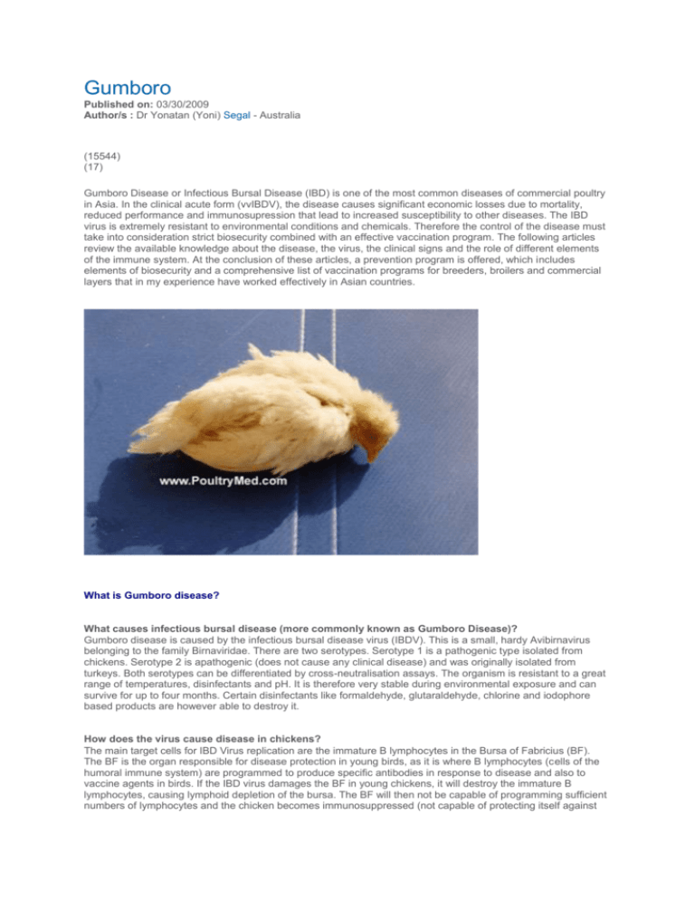
Gumboro Published on: 03/30/2009 Author/s : Dr Yonatan (Yoni) Segal - Australia (15544) (17) Gumboro Disease or Infectious Bursal Disease (IBD) is one of the most common diseases of commercial poultry in Asia. In the clinical acute form (vvIBDV), the disease causes significant economic losses due to mortality, reduced performance and immunosupression that lead to increased susceptibility to other diseases. The IBD virus is extremely resistant to environmental conditions and chemicals. Therefore the control of the disease must take into consideration strict biosecurity combined with an effective vaccination program. The following articles review the available knowledge about the disease, the virus, the clinical signs and the role of different elements of the immune system. At the conclusion of these articles, a prevention program is offered, which includes elements of biosecurity and a comprehensive list of vaccination programs for breeders, broilers and commercial layers that in my experience have worked effectively in Asian countries. What is Gumboro disease? What causes infectious bursal disease (more commonly known as Gumboro Disease)? Gumboro disease is caused by the infectious bursal disease virus (IBDV). This is a small, hardy Avibirnavirus belonging to the family Birnaviridae. There are two serotypes. Serotype 1 is a pathogenic type isolated from chickens. Serotype 2 is apathogenic (does not cause any clinical disease) and was originally isolated from turkeys. Both serotypes can be differentiated by cross-neutralisation assays. The organism is resistant to a great range of temperatures, disinfectants and pH. It is therefore very stable during environmental exposure and can survive for up to four months. Certain disinfectants like formaldehyde, glutaraldehyde, chlorine and iodophore based products are however able to destroy it. How does the virus cause disease in chickens? The main target cells for IBD Virus replication are the immature B lymphocytes in the Bursa of Fabricius (BF). The BF is the organ responsible for disease protection in young birds, as it is where B lymphocytes (cells of the humoral immune system) are programmed to produce specific antibodies in response to disease and also to vaccine agents in birds. If the IBD virus damages the BF in young chickens, it will destroy the immature B lymphocytes, causing lymphoid depletion of the bursa. The BF will then not be capable of programming sufficient numbers of lymphocytes and the chicken becomes immunosuppressed (not capable of protecting itself against any disease agent). The severity of the disease is directly related to the number of susceptible B cells present in the Bursa at the time of infection. The time when chickens are most susceptible is therefore between 3 and 6 weeks, when the Bursa of Fabricius is at its maximum rate of development and the follicles are filled with immature B lymphocytes. Virus replication also occurs in other lymphoid organs like the spleen and cecal tonsils but to a lesser extent. Which birds are at risk of Gumboro disease? The population at risk includes broiler flocks and young pullets destined for breeder and commercial egg laying flocks. Light weight laying breeds are more susceptible then heavy broiler breeds. Males are more susceptible then females. How is the virus transmitted? Chickens infected with the IBD virus shed the virus in their faeces. Virus shedding starts 48 hours after infection and lasts for 14 -16 days. Feed, water, and poultry house litter become contaminated. Other chickens in the house become infected by ingesting the virus. It is also transmitted mechanically among the farms by people, equipment and vehicles, but also by wild birds, flies and other insects. IBDV does not appear to spread through the air. How common is it? Since the first description of the disease in the USA by Cosgrove in the sixties, serological surveys have shown a high prevalence of IBD antibody in breeder flocks, which has been acquired either following vaccination or subclinical infection. This immunity is passively transmitted to the offspring via the egg yolk, protecting them at a young age (for about the first two weeks) against the clinical disease. Before 1984, in most parts of the world IBD was essentially a sub-clinical disease. The highly contagious strains caused less than 5% mortality and there were associated indirect economic losses due to immuno-suppression. This form of IBD was satisfactory controlled by vaccination of the breeders. Since 1984 the virus has gone through an evolutionary process. Two major epidemiological events causing vaccination failures have been described in different parts of the world. In the USA, due to antigenic mutation, new virus “variants” appeared, causing only a slight increase in mortality. From 1987 in Europe and later on in Asia and other parts of the world, the new hyper virulent strains emerged and were characterised by a very high mortality of up to 80% in layer pullets and 25% in broilers. Thus clinical acute IBD became predominant with additional losses due to specific mortality. In Europe and Asia, the new strains still belong to classical serotype 1 but are characterised by a marked increase in pathogenicity. First described in Belgium at the end of the eighties, the hyper-virulent or very virulent forms of the disease (vvIBD) were then described in Japan in the early nineties and have rapidly spread all over the Asiatic and European continents and to major parts of the world. Latin America has been the latest victim of this epidemic. As a probable consequence of strict import controls, Australia and the USA are, so far, still unhurt. What is the incubation period of IBD? The incubation period (time between infection and the appearance of clinical disease) of IBDV in chickens is about 2 to 4 days. How does the disease manifest in chickens? Infectious bursal disease follows one of two courses, depending on the type of virus to infect the chicken, the age of the infected chicken, breed of chicken and the presence or not of maternally derived antibodies (passive immunity). IBDV of classic virulence, which is still prevalent in the USA, Australia and the Scandinavia, causes subclinical disease. When the disease occurs in chickens less than 3 weeks of age, deficient of maternally derived antibodies, the chickens have no clinical signs of disease but experience permanent and severe immunosupression. The affected birds cannot respond properly to any infection, and are not capable of developing effective immunity following vaccination. The reason why young chickens exhibit no clinical signs of disease is related to relatively low levels of circulatory B lymphocytes (which might be destroyed by IBDV) during the first two weeks. However, immunosupression occurs due to damage of the BF. This form is almost absent in commercial production due to the routine vaccination of breeders. When the subclinical form of the disease occurs in chickens older than 3 weeks of age, chickens do not display clinical signs of disease but experience reduction in overall performance, with high feed conversion ratios and slow growth rates. This is the more economically important form of the disease. The clinical acute form of IBD known also as vvIBD usually occurs in chickens from 3 to 6 weeks of age. It is caused by the hyper virulent strains of infectious bursal disease virus. The clinical disease has a sudden onset. The first symptom in infected chickens is an acute onset of depression. Birds are listless and may be reluctant to move with a tendency to sit. Clinical signs of disease include fever, dehydration, trembling and ruffled feathers. Whitish, watery or mucoid diarrhoea may be evident in the flock, with very sticky litter and soiling of vent feathers, often followed by vent pecking. Affected flocks show this depression for 5-7 days. There is also poor feed conversion and deaths. The mortality rate has a typical bell shape curve, rising rapidly for the first two days. Four days after the onset of clinical signs, the mortality peaks and returns to normal within a week. The number of affected birds in a flock (morbidity) is variable and can approach 100%. The mortality pattern may range from 5% to a total of 25% in broilers, and 35% to 80% in layers, but the usual mortality rate is low. Affected chickens experience a transient immunosupression. Secondary disease conditions, such as E. coli infection, Marek’s disease, Gangrenous Dermatitis (Clostridium perfringens, Cl. septicum and Staph. aureus) and Inclusion Body Hepatitis (Adenovirus) may increase in incidence, and condemnation rates at slaughter may be elevated. Sick birds do not die if management is good and stresses kept to a minimum. What effect do the variant strains of IBD have? The clinical nature of IBD has been further complicated by the recognition of new variant strains of the IBD virus in the USA and recently also in Australia. Variant viruses induce damage in the BF in chickens, causing vaccine failure, even when high and uniform antibody titres are present. Variant strains do not cause obvious clinical disease, but induce severe immunosupression. The BF of affected chickens undergoes rapid atrophy (lymphocyte depletion). These variants are not from a different serotype, but are antigenically different enough to cause problems. Recent research from the University of Arkansas has demonstrated a new form of the disease caused by variant strain of IBD virus, characterised by an enlarged and flabby proventriculus, distension of the intestine airsaculitis, dermatitis, reduced body weight gain and immunosupression. What is seen at post mortem? In the clinical acute form of the disease the Bursa of Fabricius is the first internal organ to show lesions. This occurs within 24 hours of infection. During the initial stages of the disease the BF is swollen due to inflammation. By day three it has increased in size and weight, appears oedematous and hyperemic and has a gelatinous yellowish transudate covering the serosal surface. By day four the BF has doubled in weight. The transudate disappears as the bursa returns to its normal size and becomes grey in colour, undergoing atrophy. At day 8, the bursa is usually one-third of its original weight. The infected bursa always shows necrotic foci and petechial haemorrhages on the mucosal surface. Necrosis and depletion of lymphocytes also occurs in the secondary lymphoid organs, including the spleen, Harderian glands, and cecal tonsils although these organs are typically affected less severely than the BF. The spleen therefore appears slightly enlarged with small grey foci dispersed uniformly on the surface. Haemorrhage may be present in the thigh and pectoral muscles, because the IBD virus interferes with the normal blood clotting mechanism. The kidneys may appear swollen and pale with an accumulation of crystalline urate in the tubules in birds that die or that are in the advanced stages of the disease. Such lesions probably result form severe dehydration, not direct viral damage. Isolating and Identifying the IBD virus What techniques are used to cultivate IBDV in the laboratory? A filtered homogenate of the bursa of Fabricius from infected birds, collected 2 to 10 days post infection, is inoculated into an embryonated egg. The homogenate is preferably inoculated into the chorion allantoic membrane (CAM) or into the yolk sac. Embryo death occurs 3 to 7 days post inoculation. Typically an oedematous congested embryo is seen with gelatinous appearance of the skin and haemorrhages in the toes or the encephalon. The liver appears pale, bile stained and with wide necrotic foci. What kind of virus is IBD? IBDV is a small, hardy Avibirnavirus belonging to the family Birnaviridae. a double stranded RNA virus. It’s genome encodes 5 viral proteins: VP1–VP5. VP2 and VP3 are structural proteins which form the viral capsid. The epitopes located on the VP2 protein are capable of eliciting the host’s protective neutralizing antibodies against IBD, they are serotype determinant. But as the hyper-variable region of VP2 is subject to mutations, it often leads to changes in virus antigenicity and pathogenicity. What are the criteria used to describe an isolate of IBDV? IBDV strains vary greatly, as are the control measures they require. It is therefore very important not only to identify an isolate as IBDV, but also to provide detailed information about its relationships with other viruses described previously. Three criteria may be used jointly for this: antigenic relatedness, genetic relatedness and pathogenicity. What is virus antigenicity? Virus antigenicity is the ability of a virus to induce specific antibodies following infection. Two viruses are antigenically related when the antibodies induced by one of them cross react with the other (and possibly protect against it, as far as protective antibodies are concerned). What techniques are used to identify antigenic relatedness? Overall antigenic relatedness between IBDV isolates is traditionally assessed by labour-intensive, crossneutralisation assays. Results can now be obtained more rapidly using new tools based on monoclonal antibodies (Mabs). A Mab is a laboratory reagent that recognises, on the proteins exposed at the surface of a virus particle, one of the tiny structures responsible for the in-vivo induction of antibodies. If one virus versus one Mab shows little reactivity, then this means that the corresponding structure has been modified. Many different antibody-inducing structures exist on the same viral particle, and thus many different Mabs are required to accurately assess the antigenic profile of a studied virus. For IBDV, Mab-based antigen-capture (AC) ELISA was originally developed to characterise North-American "variant" IBDV strains. A similar assay is now applied to vv IBDVs, based on a new range of 7 Mabs. In this assay, vv IBDVs do not react with Mabs 3 and 4, unlike the older "classical" European IBDVs. What techniques are used to identify genetic relatedness? From minute amounts of viral RNA contained in the original sample from the bursa of Fabricius or from homogenate of chicken embryo organs, reverse transcription (RT) followed by polymerase chain reaction (PCR) techniques now make it possible to amplify (produce) sufficient DNA to enable the infecting virus to be characterised. A first approach is provided by restriction analysis. The technique is based on the use of enzymes that specifically cut the amplified nucleic acid wherever it contains a specific stretch of nucleotides (the restriction site) which the enzyme recognises as a target. Nucleotide changes occurring at the restriction site prevent the enzyme from cutting. When the objective is to detect multiple strains of the virus, primers are selected in the highly preserved zones. But when the objective is to identify and characterised the virus strains, the highly variable zone of the VP2 is chosen, and this amplified fragment can be then further characterised by direct sequencing. The simultaneous presence of four amino acids, Alanine in position 222, Isoleucine in positions 256 and 294, and Serine in position 299, is considered to be indicative of a very virulent IBDV. Are antigenic or genomic specific features responsible for enhanced pathogenicity? Although some antigenic and genomic-specific features are observed repeatedly in vv IBDVs, these features have not been shown to be responsible for any enhanced pathogenicity. Until 100 % reliable pathogenicity markers have been identified, pathogenicity testing in susceptible chickens under isolated conditions remains the only definitive method of identifying vv IBDV. Variations in the challenge protocol (breed or age of chicken, virus dose, route of inoculation) are critical, and the results should be compared with relevant references. Other laboratory tests Agar gel immunodiffusion (AGID) test This test can be used to detect viral antigen in the bursa of Fabricius. A portion of the bursa is removed, homogenised, and used as antigen in a test against known positive antiserum. This is particularly useful in the early stages of the infection, before the development of an antibody response. Immunofluorescence test This test can also be used to detect antigen in bursal tissue. Using IBD-virus-specific chicken antiserum that has a fluorescent dye chemically linked to it. When a virus is present the IBD-virus-specific chicken antiserum bind to the bursa tissue or cells and a bright fluorescence can be seen by use of a special microscope. Prevention of Gumboro disease What elements need to be taken into consideration in order to achieve a comprehensive prevention strategy? The IBD virus is extremely resistant to physical and chemical agents. That means, once a farm becomes contaminated, the virus persists in the environment for a long period of time, risking infection and economic losses to successive flocks. A comprehensive prevention program against IBD includes the following components: An effective biosecurity program (in order to reduce environmental contamination) An effective breeder vaccination program (in order to give their progeny passive protection), An effective broiler and pullet vaccination program (in order to confer the progeny with effective active immunity) What are the essential biosecurity measures against IBDV? The infectious bursa disease virus is highly resistant to environmental exposure and is not easily killed. Therefore the development and enforcement of a comprehensive biosecurity program is the most important factor in limiting losses due to IBD. Effective control of IBD in commercial poultry requires that field virus exposure be reduced by proper clean-up and disinfection between flocks and that traffic (people, equipment and vehicles) onto the farm be controlled. The sanitary precautions must include “all in all out” farming methods. In between flocks thorough cleaning and disinfection of the poultry house and equipment must take place. This involves the removal of all organic matter and dust, washing with a high pressure hose with hot water and detergent, followed by the application of disinfectants like formaldehyde, glutaraldehyde, chlorine or iodophore based products. . Efforts at biosecurity (cleaning, disinfecting, traffic control) must be continually practiced, as improvement is gradual and often only seen after 3 or 4 flocks. How does a chicken’s immune system respond to IBDV infection: Humoral immunity or cellular immunity? Humoral immunity plays a very important role in protecting the bird against IBDV. There is a direct correlation between the level of neutralizing antibodies and protection. High levels of neutralising antibodies in the chicken block the virus before infecting the bursa. With lower level of antibodies, sufficient virus is neutralized to prevent clinical disease. The role of the humoral immunity is evident by the passive protection provided to young chicks by the maternally derived antibodies against bursa lesions, mortality and immuno-suppression. In conclusion, the more virulent (pathogenic) strains of IBDV have the capacity to induce strong stimulation of elements of cell mediated immunity. This in turn stimulates the humoral immunity to produce earlier and higher levels of protective antibodies. This has implications for the efficacy of the different vaccine’s types against virulent field strains of IBDV. What kind of vaccines exists against IBDV? Inactivated vaccines, although costly, were used successfully until the emergence of the hypervirulent strains. Indeed, before 1987 it was normal practice in breeders to vaccinate hens with an oil-emulsion adjuvant vaccine just before laying in order to induce a high level of passive immunity in the offspring. This would protect the chicks until an age where infection is less detrimental in terms of immuno-suppression (2-3 weeks of age). This procedure was satisfactory until the emergence of hypervirulent strains of IBDV in 1987 when it was no longer possible to protect the offspring passively during the whole growing period and a live vaccination of the offspring became necessary. Inactivated vaccines are still widely used in countries where clinical acute IBD is prevalent. The vaccine is used in breeders and sometimes in commercial layers, when high, uniform and long lasting antibody titres are required. Inactivated vaccine seems to be less affected by maternally derived antibodies. Classical live attenuated vaccines achieve lifelong and broad protection but possess various degrees of residual pathogenicity. This can cause transitional but reversable damage to the bursa of Fabricius and some risk although very low of reversion to virulence. Four categories of live attenuated vaccines, based on their pathogenicity, have been described: 1) mild, 2) intermediate, 3) intermediate plus and 4) hot. While these vaccines induce similar serological reactions they differ in terms of their invasiveness (ability to replicate in the bursa). The vaccines are classified according to: 1. 2. Their ability to induce immunity in the presence of maternal antibodies. Their residual pathogenicity, which is determined by the B/ B Index. The B/B Index is the ratio between the weight of the Bursa and the body weight of vaccinated birds divided by the same ratio of non-vaccinated birds. The following table shows the B/B index of IBD vaccines. • Mild type: • Low invasiveness. • It is neutralised when a low level of maternal antibodies are present • Mild vaccines are rarely used in broilers, but are used to prime breeders prior to inoculation with an inactivated vaccine. • Intermediate type: • A medium level of invasiveness. • It induces immunity against infectious bursa disease even when average levels of maternal antibodies are present (breaking through the MDA titres ≤ 6 log2 VN or ELISA (IDEXX standard) titre 200). • In the absence of MDA, it can be applied to 1-day old chicks. • Sometimes it is not effective in preventing clinically acute IBD. • Intermediate Plus type: • High invasiveness. • It induces immunity against infectious bursa disease even when high levels of maternal antibodies are present (breaking through the MDA titres ≤ 8 log2 VN or ELISA (IDEXX standard) titre 500). • It should never be applied before 10 days of age in broilers, or before 15 days in layer and breeder pullets, so as to avoid damage to the bursa of Fabricius. • It is most effective in preventing clinically acute IBD. • Hot type: • It has very high invasiveness but also possesses very high residual pathogenicity. • It is likely to induce immunity-related sequelae. • It is rarely used. What contributes to successful vaccinations? Successful vaccinations depend on the choice of a correct vaccine strain and vaccination schedule. Successful vaccinations must take account of: • The existence of certain virus pathotypes. • The local epidemiological situation on the farm in question and on surrounding farms. • The day old chick quality, the presence of different levels and uniformity of interfering maternally derived antibodies. • The type of production, which determines the speed of decay of the maternally derived antibodies and the optimal age for vaccine application. • The best route of application. Which type of vaccine should be selected? The best vaccination program will take account of the type of IBD threatening that particular farm. Due to the use of sophisticated laboratory tools (cross neutralisation studies, monoclonal antibodies, RT-PCR, RFLP), important progress has been made in the understanding of the antigenic and genomic structures of IBDV and their great variability. However, it is still not possible to make a link between the observed structural characteristics of a certain IBDV and its pathogenicity. There is still no recognised virulence factor that helps predict, based on the sole laboratory findings, the pathogenicity (the « pathotype ») of a particular strain of IBDV or the efficacy of a certain type of vaccine (the « protectotype ») compared to another. Only field studies can answer these questions. Poultry veterinarians therefore need to focus on what is happening in the field and try to accurately establish a proper diagnosis. Practically speaking there are three types of IBDV: • Subclinical IBDV (scIBDV), which does not cause mortality but can often indirectly induce economic loss, due to reduced performance. • Clinical acute IBDV or very virulent IBDV (vvIBDV) is responsible for the typical Gumboro mortality outbreaks observed in most Asian countries (see Table One). Although the surviving birds can show good or even excellent growth performance, mortality decreases economic output. • Variant IBDV (var IBDV), does not give rise to significant mortality but is capable of infecting chickens in the presence of Maternally Derived Antibody (MDA) levels that are still protective against scIBDV and vvIBDV. The presence of variant IBDV has mostly been reported in Northern America and Australia. This has necessitated heavy vaccination programmes in breeders and/or the use of inactivated vaccines made from variant IBDV strains. Table 1 shows how to recognise a mortality outbreak attributable to vvIBDV. After conducting the necessary field work the poultry veterinarian should be able to answer the following 3 basic questions: • Is this farm contaminated with IBDV? • Is this IBDV of the very virulent type (vvIBDV; see table 1) or of the subclinical type (scIBDV)? • What is the situation in the surrounding poultry farms? Clinical observations together with, if necessary, simple serological and/or histological laboratory testing are sufficient to answer these questions and put the farm (as well as the surrounding poultry farms) into one of the following 3 categories (see Table Two): • The farm is not contaminated with an IBDV. • The farm is contaminated with a vvIBDV. • The farm is contaminated with a scIBDV. Although this may seem simple, this system of classification is reliable and useful in the field because it allows the selection of the most appropriate vaccine. While the classification of the farm into one of the first two categories is sufficient to decide on a vaccination programme, the third category requires additional information. This is because even though chickens may be infected with scIBDV, it does not necessarily follow that there are associated economic losses. There exist IBDV strains that have few pathogenic properties and against which it is not always necessary to implement a vaccination programme. This is why, in this case, it is necessary to closely look after production performance like feed conversion rate (FCR) and growth rate to evaluate the economic consequences of infection with scIBDV (see table two). Table 2: The selection of a Gumboro vaccine (Intermediate or intermediate plus vaccine) when it is required, according to farm and the area situation. The diagnostic criteria listed in Table 2 helps decide if it is necessary to vaccinate and which type of vaccine is the most appropriate. Then it remains necessary to decide on the other options concerning the prevention programme. The Maternally Derived Antibodies (MDA) and the Vaccination Program What role do maternally derived antibodies play in vaccination programs against IBDV? All live attenuated IBD vaccines are susceptible to virus-neutralising maternally-derived antibodies (MDA), which can neutralise them or delay their action. Serological monitoring is usually necessary to predict the optimal timing for vaccination. Age(s) at vaccination should be decided according to the level and homogeneity (uniformity) of MDA present in the day-old chicks. A quantitative serological test, like the Virus Neutralisation test (VN) or the more commonly used ELISA test, evaluate antibody levels in chicks (expressed in geometric mean titre (GMT)) and calculate the optimal time for vaccine application. This is done using well established calculation formulas like B. Kouwenhoven’s or Deventer’s. It is also possible to utilise existing databases and apply predetermined vaccination ages, but by doing so the risk of vaccine failure is higher. It is important to note that the optimal age for vaccination is rarely identical for all chickens of the same flock. One can therefore use mean values, which is acceptable if MDA titres are homogeneous or increase the number of vaccine applications if MDA titres are heterogeneous. The decay rate of MDA Fast growing birds like broilers have a fast and uniform MDA decay rate with a half life of 3-3.5 days. An application of a single live vaccine is therefore often sufficient to confer good protection to the whole flock. Birds exhibiting a slow growth rate (pullet’s layers or breeders, free range or native type chickens) have a slow and uneven MDA decay rate with a half life of 5-6 days. This results in a greater uncertainty of the optimal time to vaccinate and the number of applications that must be increased to ensure success. This is done by the calculation of the central vaccination time (CVT) and then two vaccine application six days apart, three days before the central vaccination time and three days after (3-CVT+3). Nutrition and environmental conditions also influence the rate of MDA decay The uniformity of MDA The MDA uniformity is the result of the breeder’s vaccination program, exposure to any field challenge and the age of the breeder. Breeders vaccinated with live and inactivated vaccines and/or exposed to field challenge, during their prime production period at around 35 weeks of age, are very likely to present with a high and uniform MDA titre. On the other hand, breeders vaccinated only with live vaccines and at the end of their production cycles (around 60 weeks of age) are very likely to show low and uneven MDA titres. The flock uniformity is expressed as a coefficient of variation (%CV). When the %CV is less than 40% it is considered to be a homogenous flock and usually one vaccine application should provide good and uniform protection to the whole flock. In cases where the %CV is higher than 80%, the flock is heterogeneous and two or more vaccine applications are required for the protection of the entire flock. What is the optimal time for vaccine application? An effective immunisation programme changes the bird from being passively immune by MDA’s into an actively protected bird through the development of its own antibodies. The correct timing of vaccination plays a critical role in this operation. If the vaccine is applied too early, while the maternally derived antibodies titre is too high, the vaccine is neutralised and no protection can be expected. On the other hand, if the vaccine is applied too late, then an exposure to field virus and a consequent disease outbreak may occur. Therefore the accurate identification of the window of opportunity is extremely important. When an ELISA test (Idexx kit), for serological monitoring is used, a calculation of the MDA level at which the vaccine can break through is possible. Based on previous experience, Intermediate plus vaccines are capable of breakthrough at a titre of ELISA ~500. Intermediate vaccines can break through at a titre of ELISA ~200. This means you have to wait longer before being able to apply an Intermediate vaccine. Table 3. An example of how to calculate the vaccination time for broilers with Intermediate plus vaccine.When the MDA level at day one is ELISA 6000 and the MDA decay rate is calculated by half life method, at 3.5 days. Note: during the first three days the MDA level remains unchanged due to the continuous absorption of MDA from the egg yolk into the blood stream How many applications should be made? The number of applications necessary will vary according to the following factors: The spreading capacity of the vaccine. Due to an uneven MDA level in the chicks, several applications of a poorly spreading vaccine are necessary to ensure 100% coverage of the whole chick population. Provided that the optimal age at vaccination is properly selected, Intermediate Plus vaccines require only one application because these vaccines have good spreading capacity. Homogeneity of MDA levels in the baby chicks. The higher the heterogeneity (low uniformity) of MDA levels in the chicks, the higher the number of desired applications; particularly if a poorly spreading mild or intermediate type vaccine is employed. The expected level of protection or the accepted risk of failure. The accepted level of risk has economic consequences. A heavy vaccination programme is more expensive but will help lower the risk of vaccine failure although it cannot eliminate it. The use of serological testing increases the cost of a basic vaccination programmes but considerably reduces the risk of vaccine failure. Designing a vaccination program against vvIBDV What is the preferred vaccination program in an area where vvIBD is prevalent? For breeders Vaccination of breeders is based on a process called "hyper immunisation". This involves induction of very high levels of circulating antibodies in the laying hens. This then results in high levels of passive antibodies (Maternally Derived Antibodies or MDA) being transmitted to the chicks via the egg. The rate of transmission is proportional to the level of maternal antibodies; the higher the antibody titre in the breeder, the stronger the protection transmitted to the chick. The presence of MDA’s protect chicks against immunosuppressive and sub clinical IBD. Early vaccination programmes occurring during the first weeks of age, consist of at least two vaccine applications, six days apart, according to the 3-CVT+3 calculation method. After this, the pullet receives an additional hyper immunisation programme. This consists of a live "primer" at around 8 to 12 weeks of age (with a live vaccine, preferably of the Intermediate Plus type) followed by the injection of an inactivated vaccine before the onset of lay. The strong booster effect which is then observed is responsible for the hyper immunisation process. A second injection of an inactivated vaccine at around 45 weeks of age may be necessary if the level or the homogeneity of the MDA in the chicks is not sufficient. Some breeding and commercial layer companies prefer to use only inactivated vaccines combined with Intermediate vaccines to protect their birds. This avoids the use of Intermediate plus vaccines, which can cause negative post-vaccination effects on birds’ performance. Vaccination program Option One (a) High quality chicks (high and uniform MDA) Intermediate Plus vaccine at 20 and 26 days Intermediate Plus vaccine at 8 to 10 weeks Inactivated vaccine at 15 to 17 weeks (b) Low quality chicks (low and uneven MDA) Intermediate vaccine at 5 to 10 days Intermediate Plus vaccine at 16 and 22 days Intermediate Plus vaccine at 8 to 10 weeks Inactivated vaccine at 15 to 17 weeks Option two (a) High quality chicks (high and uniform MDA) Intermediate vaccine at 5 to 7 days Inactivated vaccine at 5 to 7 days Intermediate vaccine at 22 and 28 days Intermediate vaccine at 8 to 10 weeks • Inactivated vaccine at 15 to 17 weeks (b) Low quality chicks (low and uneven MDA) Intermediate vaccine at 5 to 7 days Inactivated vaccine at 5 to 7 days Intermediate vaccine at 18 and 24 days Intermediate vaccine at 8 to 10 weeks Inactivated vaccine at 15 to 17 weeks For broilers The vaccination programme for broilers in an area where vvIBD is prevalent comprises of the application of Intermediate plus vaccines. When the chicks are of a good quality, in terms of high and uniform MDA titre, a single application at the age of 12 to 16 days, (according to MDA titre), will provide good protection until slaughter. However, when the chicks are of poor quality, mainly due to poor uniformity, particularly when day-old chicks from different breeding flocks, young (high level of Maternally Derived Antibodies) and old ones(low level of Maternally Derived Antibodies), are mixed together at the same poultry house, then two applications are required to protect the whole flock. The first application of Intermediate vaccine is given at 7 to 10 days of age, to correct the flock heterogeneity. The second application of Intermediate plus vaccine is given at 12 to 16 days, to ensure that all birds are protected. Table 4: What happens when you mix day-old chicks from two different breeder flocks For commercial layer pullets The early vaccination programme for commercial layer pullets during the first weeks of age is quite similar to the breeders programme: Option One (a) High quality chicks (high and uniform MDA) Intermediate Plus vaccine at 20 and 26 days Intermediate Plus vaccine at 8 to 10 weeks (b) Low quality chicks (low and uneven MDA) Intermediate vaccine at 5 to 10 days Intermediate Plus vaccine at 16 and 22 days Intermediate Plus vaccine at 8 to 10 weeks Some commercial layer companies prefer to use only inactivated vaccines combined with Intermediate vaccines to protect their birds, eliminating the use of Intermediate plus vaccines from their programme, due to some negative post-vaccination effects on the birds’ performance. Thus the second option below: Option Two (a) High quality chicks (high and uniform MDA) Intermediate vaccine at 5 to 7 days Inactivated vaccine at 5 to 7 days Intermediate vaccine at 22 and 28 days Intermediate vaccine at 8 to 10 weeks (b) Low quality chicks (low and uneven MDA) Intermediate vaccine at 5 to 7 days Inactivated vaccine at 5 to 7 days Intermediate vaccine at 18 and 24 days Intermediate vaccine at 8 to 10 weeks What is the most efficacious route of administration? The primary tropism of IBDV is for lymphoid structures of the gut, this is the reason why it is strongly advised that IBD live vaccines be administered through drinking water. Per-oral (mouth squirting) and ocular routes are also efficacious but not so practical and more expensive (those methods are to be applied to young birds, before 10 days of age). The respiratory route (spray) is responsible for rather high vaccine losses which decrease its efficacy in the field. In certain countries early application of intermediate vaccine by the intramuscular route has been reported. The best vaccination programme is useless if not properly administered. A correct vaccination technique ensures that all the birds receive a full dose of the selected vaccine on the same day. Compounds capable of neutralising chlorine and favouring the survival of the vaccine virus should be utilised. Products using dyes that help to monitor the actual consumption of the vaccine by the chickens are useful. What are the possible reasons for vaccination failure? When faced with apparent vaccine failure the following questions should be asked: Is the problem actually due to IBDV? Was the vaccine handled and administered correctly? Was the vaccination programme properly adapted to the field situation i.e. type of vaccine/s, age(s) at application(s), number of applications? Are immunosuppressive factors (which prevent the chicken’s ability to build up an immune response to vaccination) such as infections with immunosuppressive viruses like Marek, CAV, REO Virus, etc. present? Are there any Mycotoxins in the feed or any other severe stress that may be affecting the flock? What is the physical and immune (level and homogeneity of MDA levels) quality of the baby chicks? Vaccination failure may also suggest an evolution of the disease situation, requiring review of the initial diagnosis and suspicion of the presence of a new type of IBDV. What are the new developments in the field of IBDV vaccines? Virus-Antibody Complex Vaccines or Virus Neutralising Factor (VNF) vaccines Homologous antibodies for the vaccine virus are mixed in an appropriate ratio. This process results in a virus-antibody complex vaccine. The amount of antibody in the complex is so small that it does not provide passive immunity or neutralise the vaccine virus but it is enough to delay by several days the normal course of vaccine virus replication. This allows for the safe administration in-ovo or to day-old chicks, of Intermediate plus vaccine viruses. Those specific antibodies metabolised are in correlation with the decay of the maternally derived antibodies. The vaccine virus starts to replicate and stimulate the immunity system of the chick once the maternally derived antibodies of the individual bird within the flock has reached the level that permits vaccine virus replication without the risk of being neutralised, or causing any damage to the bursa. The advantages of the VNF vaccine over the classic vaccines are due to the fact that: o A single vaccine’s application confers very good protection. o Effective immunity develops irrespective of the MDA level, as the vaccine virus starts to replicate only when it is capable to break through the MDA level of the individual birds o There is no need for serological tests for the prediction of vaccination date o The vaccine application in the hatchery In-Ovo or Sub-Cutaneous at hatch What to do in the case of an outbreak Where an outbreak of IBD occurs several measures can be implemented to reduce morbidity and mortality: 1. 2. 3. 4. Improving the conditions in the chicken house, like better temperature and ventilation control, can reduce any unnecessary stress and assist with reduction of the overall mortality. Sick birds often suffer severe dehydration and damaged kidneys. Administration of electrolytes in drinking water and reduction in protein levels of the ration for four to five days can assist in the birds’ recovery. Antibacterial therapy is sometimes required to minimize secondary infection. Make sure not to use medications which are metabolized or eliminated via the urinary system. Supplementation of multivitamins in drinking water is also recommended. Sources 1. Butcher GD. Infectious Bursa Disease (Gumboro) in Commercial Broilers. Document VM-84, College of veterinary medicine, University of Florida. 2. Gardin Y & Camillo V. The Control of the Gumboro Disease in the Fields. Paper presented at APINCO meeting, Nov 2001,Ceva Veterinaria, Brasil 3. Haddad E.E., Whitfill C.E., Avakian A.P., Ricks C.A., Andrews P.D., Thoma J.A. & Wakenell P.S. (1997). Efficacy of a novel infectious bursal disease virus immune complex vaccine in broiler chickens. Avian Dis., 41, 882-889. 4. Kouwenhoven B. & Van Der Bos J. (1993). Control of very virulent infectious bursal disease (Gumboro disease) in the Netherlands with so called ‘hot’ vaccines. Proceedings of the 42nd Western Poultry Disease Conference, Sacramento, California, USA, 37-39. 5. Lasher H.N. & Shane S.M. (1994). Infectious bursal disease. World’s Poult. Sci., 50, 133-166. 6. Lukert P.D. & Saif Y.M. (1997). Infectious bursal disease. In: Diseases of Poultry, Tenth Edition, Calnek B.W., ed. Iowa State University Press, Ames, Iowa, USA, 721-738. 7. Marquardt W.W., Johnson R.B., Odenwald W.F. & Schlotthoken B.A. (1980). An indirect enzyme-linked immunosorbent assay (ELISA) for measuring antibodies in chickens infected with infectious bursal disease virus. Avian Dis., 24, 375-385. 8. Rosenberger J.K. & Cloud S.S (1986). Isolation and characterization of variant infectious bursal disease viruses. J. Am. Vet. Med. Assoc., 189, 9. Sapats, S. and Ignjatovic, J. (2000) Antigenic and sequence heterogeneity of infectious bursal disease virus strains isolated in Australia. Archives of Virology 145:773-785. 10. Silke Rautenschlein, H.Y. Yeh, J. M. Sharma. (2003) Comparative Immuno pathogenesis of mild, intermediate and virulent strains of classic infectious bursal disease virus. Avian Disease 47:66-78 2003 Acknowledgment: The photos in this article are used with the permission of Dr Nati Elkin (PoultryMed.com)
