Ministry of higher Education and Scientific Research Foundation of
advertisement
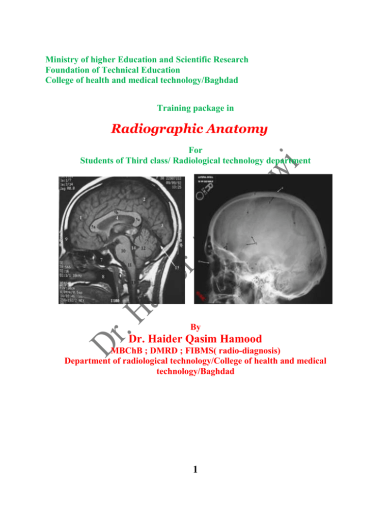
Ministry of higher Education and Scientific Research Foundation of Technical Education College of health and medical technology/Baghdad Training package in Radiographic Anatomy For Students of Third class/ Radiological technology department By Dr. Haider Qasim Hamood MBChB ; DMRD ; FIBMS( radio-diagnosis) Department of radiological technology/College of health and medical technology/Baghdad 1 2 Overview A / Target population :Students of Third class/ Radiological technology department/ College of health and medical technology/Baghdad B/ Rationale :Normal radiological anatomy of the cranial contents is very important subject to be studied by students of diagnostic radiological technologies in order to have a full knowledge, which is important in their work as an operators on diagnostic radiological equipments. C/ Central Idea :Normal radiological cranial anatomy: a. Bones: Skull vault and calvarium, skull sutures, , base of skull, cranial fossae. b. Radiological features of skull bones. c. Facial bones: normal radiographic anatomy. b. Brain: main parts, cavities, radiographic features. D/ Instructions:1.Study over view thoroughly. 2.Identify the goal of this modular unit. 3.Do the pre test and if you :get 9 or more you do not need to proceed . get less than 9 you have to study this unit well . 4.After studying the text of this unit ,do the post test, and if you :get 9 or more , so go on studying next modular unit . get less than 9 , go back and study the first modular unit; or any part of it ; again and then do the post test again . Pre-test Circle the correct answer :1. They are skull bones:a- temporal bones b- mastoid bone c- pubis d- acetabulum 2. Bony host of pituitary is :a- sella turcica b- acetabulum c- pleura d- pericardium 3. It is a part of eye : a - retina b- stabes c- incus d- falanges 4. Bones of middle ear except : a- stabes b- malleus c- incus d- styloid process 5. Structures of chest except : a-uterus b-heart c- lung d- thymus 6. Bones of foot except : a- talus b- cuboid c- calcaneous d- scaphoid 3 7. Carpal bones are, except : a- lunate b- triquetral c- hamate 8. Bones of pelvis, except : a- pubis b- sacrum c- ischium 9. Right lung consists of : a- Three lobes b- Four lobes c- Five lobes 10. Facial bones are : a- 10 b- 13 c- 14 d- incus d- pisiform d- Two lobes d- 12 Performance Objectives After studying the modular unit , the student will be able to:1.Define normal radiological cranial anatomy . 2.Describe the radiological features of the cranial structures in the different radiological modalities (plain radiograph, CT, MRI) which represent human skull parts and cranial organs with their ability to draw them. 3.Determine the ideal radiological modality to reveal human anatomical structure of cranial contents. The text Normal anatomy of cranial contents: The skull consists of the calvarium, facial bones and the mandible. The calvarium: It is the brain case and it consists of skull vault and skull base, the calvarium composed of 8 bones : 1.Two parietal bones 2.Two temporal bones 3.Frontal bone 4.Occipital bone 5.Sphenoi bone 6.Ethmoid bone The bone of calvarium are joined at fibrous immovable joints which are called ( skull sutures ) . Skull: Lateral aspect 4 Facial bones: The skeleton of the face is composed of 14 bones , 6 of them are paired bones : 1.Nasal bones 2.Lacrimal bones 3.Maxillary bones 4.Zygomatic bones 5.Palatine bones 6.Inferior nasal conchae Other 2 bones are single : 1.Vomer bone 2.Mandible bone Many facial cavities are seen including orbits, Paranasal sinuses, nasal cavity and oral cavity. Skull sutures: 1.Sagittal suture at the junction of the parietal bones. 2.Coronal suture at the junction of the frontal bone with parietal bones. 3.Lambdoid suture at the junction of the occipital bone with occipital with parietal bones. The point at which the coronal suture meets the sagittal suture is known as (bregma) . The point at which the lambdoid suture meets the sagittal suture is known as (lambda). The skull vault is composed of three layers outer table, diploic space and inner table. Fetal skull 5 Quiz1: Enumerate Calvarial bones? Skull base : The inner aspect of the skull base is made up of the following from anterior to posterior direction : 1.Orbital plates of the frontal bone 2.Cripriform plate of the ethmoid bone 3.Sphnoid bone with its greater and lesser wings 4.Sella turcica (pituitary fossa) in about the middle of cranial cavity 5.Parts of sequamous and petrous portions of the temporal bone 6.Occipital bone Cranial fossae: Within the skull there are three cranial fossae: 1.Anterior cranial fossa which support the frontal brain lobes 2.Middle cranial fossa which contains the temporal lobes of the brain , pituitary gland and the foramina of the skull base. 3.Posterior cranial fossa which is the largest and deepest fossa and it contains the cerebellum , pons and medulla oblongata. Cranial fossae Radiological features of the skull vault and base: Plain radiograph: several projections are required for full assessment , standard projections are lateral , occipito-frontal (OF20) and Towns projections . Pituitary fossa is visible on OF 20 caudal angulation, OF30 and sub-mentovertical view (SMV) , but the lateral view is the most frequently used . The thickness of skull vault is not uniform. The inner and outer tables of skull are thickened at internal and external occipital protuberances respectively. Intracranial calcification on plain radiographs may be normal finding as pineal gland and basal ganglia calcifications . 6 Quiz2 : Enumerate the facial bones. Brain: It is a semisolid structure which conform the shape of the skull. Brain consists of cerebrum, cerebellum and brain stem . The main parts of the brain are: 1.Forebrain : which include the cerebrum and diencephalons (thalamus and hypothalamus). 2.Midbrain: is narrow and connecting the forebrain to the hind brain. 3.Hindbrain: which include cerebellum, pons and medulla oblongata which connecting the brain to spinal cord.The cerebrum is the largest brain part. It consists of two hemispheres which are connected by mass of white matter called (corpus callusum). The surface layer of each hemisphere called ( cortex ) and it is composed of gray matter . The brain cortex is thrown into folds called ( gyri ) and these separated by fissures called ( sulci ). Each cerebral hemisphere divided into 4 lobes: frontal , parietal, occipital and temporal lobes. Brain cavities (ventricles): The brain ventricles are: 1.Lateral ventricle which are right and left lateral ventricles 2.Third ventricle 3.Fourth ventricle The lateral ventricle has anterior, body , inferior and posterior horns. The trigone is the location of junction of body with posterior and inferior horns of the lateral ventricle , in trigone the glomus is situated which is the largest tuft of choroid plexus which produce the CSF. The lateral ventricle communicates with the third ventricle via interventricular foramen (foramen of Monro). The third ventricle communicates with fourth ventricle via cerebral aqueduct which communicates with the central canal of the spinal cord. The ventricles filled with cerebro- spinal fluid (CSF) which produced by choroid plexus within the ventricles. Post-test Circle the correct answer from the following:1. It is facial bone:a- sacral bone b- Lacrimal bone c- pubis d- acetabulum 2. Bones of skull base are, except :a- sella turcica b- ethmoid bone c- Sphnoid bone d- acetabulum 3. standard skull x-Ray projections are, except : a- lateral b- AP view c- OF20 d- Towns 4. Pituitary fossa is visible on: a- AP view b- lateral view c- OF20 d- Towns 5. It is part of brain stem : a-Pons b-putamen c- corpus d- fornix 6. Bones of face except : 7 a- maxillary b- vomer c- inferior choncae d- occipital bone 7. It is fore-brain part : a- cerebellum b- diencephalon c- Pons d- Medulla 8. Hind-brain parts, except : a- Pons b- cerebrum c- cerebellum d- Medulla 9. The brain consists of : a- 3 lobes b- 2 lobes c- 5 lobes d -4 lobes 10. Main brain ventricles are : a- 6 b- 3 c-5 d- 4 key answer Pre-test Post-test 1. a 1. b 2. a 2. d 3. a 3. b 4. d 4. b 5. a 5. a 6. d 6. d 7. c 7. c 8. d 8. b 9. a 9. d 10. c 10. d Quiz No. 1 / 1.Two parietal bones 2.Two temporal bones 3.Frontal bone 4.Occipital bone 5.Sphenoi bone 6.Ethmoid bone Quiz No. 2 / 1.Nasal bones 2.Lacrimal bones 4.Zygomatic bones 5.Palatine bones Other 2 bones are single : 1.Vomer bone 3.Maxillary bones 6.Inferior nasal conchae 2.Mandible bone Sources Stephanie Ryan: Textbook anatomy for diagnostic imaging. 8 9 Overview A / Target population :Students of Third class/ Radiological technology department/ College of health and medical technology/Baghdad B/ Rationale :Cross-sectional radiological anatomy of the cranial contents is very important subject to be studied by students of diagnostic radiological technologies in order to have a full knowledge, which is important in their work as an operators on diagnostic radiological equipments. C/ Central Idea :Normal cross-sectional radiological cranial anatomy: a. Radiological features of skull bones in cross-sectional imaging modalities. b. Facial bones: normal radiographic anatomy in cross-sectional imaging modalities. c. Brain: main parts, cavities, cross-sectional radiographic features. D/ Instructions:1.Study over view thoroughly. 2.Identify the goal of this modular unit. 3.Do the pre test and if you :get 9 or more you do not need to proceed . get less than 9 you have to study this unit well . 4.After studying the text of this unit ,do the post test, and if you :get 9 or more , so go on studying next modular unit . get less than 9 , go back and study the first modular unit; or any part of it ; again and then do the post test again . Pre-test Circle the correct answer from the following:1. It is facial bone:a- maxillary bone b- Ethmoid bone c- pubis d- acetabulum 2. Bones of skull base are, except :a- Ethmoid bone b- sella turcica c- Sphnoid bone d- acetabulum 3. standard skull x-Ray projections are, except : a- AP view b- lateral c- OF20 d- Towns 4. Pituitary fossa is visible mostly on: a- AP view b- Towns c- OF20 d- lateral view 5. It is part of brain stem : a- putamen b- Pons c- corpus d- fornix 6. Bones of face except : 10 a- maxillary b- vomer c- inferior choncae d- occipital bone 7. It is fore-brain part : a- cerebellum b- medulla c- Pons d- cerebrum 8. Hind-brain parts, except : a- Pons b- Medulla c- cerebellum d- cerebrum 9. The brain consists of : a- 4 lobes b- 2 lobes c- 5 lobes d -3 lobes 10. Main brain ventricles are : a- 6 b- 3 c-4 d- 5 Performance Objectives After studying the modular unit , the student will be able to:1.Define normal cross-sectional radiological cranial anatomy . 2.Describe the radiological features of the cranial structures in the different radiological modalities (CT, MRI) which represent human skull parts and cranial organs with their ability to draw them. 3.Determine the ideal radiological modality to reveal cross-sectional structure of cranial contents. The text Cross sectional cranial anatomy: Computed tomography (CT) provides excellent visualization of the skull base and foramina when narrow high resolution images are obtained. MRI with narrow section thickness slices is an excellent imaging modality for demonstration of the soft tissue contents of the cranial foramina, in particular the cranial nerves. The plane of imaging can be chosen to demonstrate the structure of interest , for example imaging in several planes are necessary to demonstrate the course of the facial nerve through the skull base from its entry into the internal auditory canal to its exit through the stylomastoid foramen. Axial section through the posterior cranial fossa , pons and temporal lobes: At this level , the anterior cranial fossa is bounded anteriorly by the frontal bone which contains the frontal sinuses. The middle cranial fossa is separated from anterior cranial fossa by greater wing of sphenoid bone. In the middle cranial fossa , the temporal lobes are seen and temporal horns of the lateral ventricles. The occipital bone surrounds the posterior cranial fossa . Basilar artery separate the pons from the posterior clinoid process and clivus and internal carotid artery is seen in cavernous sinus. The pons and cerebellum occupy the reminder of posterior cranial fossa. 11 Quiz1:enumerate the structures in CT of brain at posterior cranial fossa level Axial section at level of brain stem (pons): At this level , the frontal lobes are seen to be separated by interhemispheric fissure and falx cerebri. The temporal lobe lies in the middle third of the section and separated from the frontal lobe by sylvian fissure. The pons is visible in midline , separated by interpeduncular cistern. Anterior to the mid-brain , the suprasellar cistern is seen which contains the internal carotid artery and its branches which will form the circle of Willis . In the posterior part of the section , the cerebellum and fourth ventricle are seen . Axial section through third ventricle, thalami and internal capsule: At this section , the frontal horns of lateral ventricles are seen , separated by septum pellucidum. The third ventricle is seen as a narrow slit-like structure in the midline . Pineal gland lies within the quadrigeminal cistern. Basal ganglia , thalami and internal capsule are seen at middle third of this section. The head of caudate nucleus lies between the frontal horns of the lateral ventricles and the anterior limb of internal capsule. Lentiform nucleus lies lateral to the internal capsule with globus pallidus medially and putamen laterally . Claustrum separated from the putamen by external capsule. Insula is the floor the sylvian fissure. 12 Axial section through third ventricle Quiz2: enumerate the structures in CT of brain at 3rd ventricle level Sagittal section at pituitary level: In this section the pituitary gland is seen above the sphenoid sinuses . the corpus callusum and its parts are visualized . the septum pellucidum which separate the lateal ventricles from each other is seen.the ponsand medulla oblongata and cerebellum are seen. Brain CT sagittal section: 1.frontal lobe 2.parietal lobe 3.occipital lobe 4.septum pellucidum 5.corpus callusum 6.pituitary gland 7.sphenoid sinus 8.oral cavity 9.frontal sinus 10.pons 11.medulla oblongata 12.cerebellum 13.spinal cord 14.fourth ventricle 13 Post-test Circle the correct answer from the following:1. facial nerve entry is: a-foramen magnum b-inner auditory canal c-orbit d-cochlea 2. facial nerve exit through : a- stylomastoid foramen b- inner auditory canal c- cochlea d- orbit 3. middle cranial fossa is separated from anterior cranial fossa by: a- sella b-sphenoidal greater wing c- crista galli d- iris 4. Bone of middle ear : a- scaphiod bc- cocyx d- styloid process 5. Structures of abdomen except : ab-liver c- gall bladder d- spleen 6. Bones of face except : a- maxillary b- vomer c- inferior choncae d7. They are upper limb bones , except : ab- triquetral c- radius d- ulna 8. Bones of hand, except : a- lunate bc- hamate d- pisiform 9. Right lung consists of : a- 2 lobes b- 4 lobes c- 5 lobes d10.Tarsal bones are : a- 10 b- 13 cd- 8 key answer Pre-test Post-test 1.a 1.b 2.a 2.d 3.a 3.b 4.d 4.b 5.a 5.a 6.d 6.d 7.c 7.c 8.d 8.b 9.a 9.d 10.c 10.d 14 Quiz No. 1 / Basilar artery separate the pons from the posterior clinoid process and clivus and internal carotid artery is seen in cavernous sinus. The pons and cerebellum occupy the reminder of posterior cranial fossa. Quiz No. 2 / The head of caudate nucleus lies between the frontal horns of the lateral ventricles and the anterior limb of internal capsule. Lentiform nucleus lies lateral to the internal capsule with globus pallidus medially and putamen laterally . sources Stephanie Ryan: Textbook anatomy for diagnostic imaging. 15 16 Overview A / Target population :Students of Third class/ Radiological technology department/ technology/Baghdad B/ Rationale :Radiological anatomy of the orbit is very important subject to be radiological technologies in order to have a full knowledge, which operators on diagnostic radiological equipments. C/ Central Idea :Normal radiological anatomy of orbit: a. Bones: orbital bones (walls). b. Radiological features of orbital bones. c. Eye: normal radiographic anatomy. D/ Instructions:1.Study over view thoroughly. 2.Identify the goal of this modular unit. 3.Do the pre test and if you :get 9 or more you do not need to proceed . get less than 9 you have to study this unit well . 4.After studying the text of this unit ,do the post test, and if you :get 9 or more , so go on studying next modular unit . get less than 9 , go back and study the first modular unit; or any post test again . College of health and medical studied by students of diagnostic is important in their work as an part of it ; again and then do the Pre-test Circle the correct answer :1. It is orbital roof :a- frontal bone b- mastoid bone c- pubis d- acetabulum 2. Bony host of pituitary is :a- sella turcica b- acetabulum c- pleura d- pericardium 3. It is a part of eye : a - retina b- stabes c- incus d- falanges 4. Bones of middle ear except : a- stabes b- malleus c- incus d- styloid process 5. Structures of chest excet : a-uterus b-heart c- lung d- thymus 6. Bones of foot except : 17 a- talus b- cuboid c- calcaneous 7. Carpal bones are, except : a- lunate b- triquetral c- hamate 8. Bones of pelvis, except : a- pubis b- sacrum c- ischium 9. Right lung consists of : a- Three lobes b- Four lobes c- Five lobes 10. Facial bones are : a- 10 b- 13 c- 14 d- scaphoid d- incus d- pisiform d- Two lobes d- 12 Performance Objectives After studying the modular unit , the student will be able to:1.Define normal radiological orbital anatomy . 2.Describe the radiological features of the orbital structures in the different radiological modalities (plain radiograph, CT, MRI). 3.Determine the ideal radiological modality to reveal human anatomical structure of orbital contents. The text The orbit: The orbit is four sided pyramidal bony cavity whose skeleton is contributed to several bones of the skull. The base of pyramid is open and points anteriorly to form the orbital rim. The lateral, medial, superior and inferior walls converge posteromedially to an apex . At the apex the optic foramen is opened and transmitting the optic nerve and ophthalmic artery. The orbit has a supero-medial depression for the Lacrimal gland. Walls of the orbit: 1.lateral wall: is formed by zygomatic bone and greater wing of sphenoid bone. 18 2.superior wall(roof):is formed by orbital plate of the frontal bone and lesser wing of sphenoid. 3.medial wall: is thin and formed by lamina papyracea of ethmoid which separates the orbit from the nasal cavity , ethmoid air cells and sphenoid. 4.inferior wall : is formed by Zygomatic and maxillary bones . Quiz1:Discuss the walls of the orbit. Radiological features of the orbit: 1.Plain radiograph: the orbit may be assessed on OF20 and OM projections. The difference between the right and left optic foramen dimensions , should not exceed 1 mm. . the infraorbital foramen is seen on OF 20 and OM views and the floor of the orbit is best visualized on OM and OM30 projections. 2.CT and MRI : The bony orbit and its soft tissue contents are demonstrated by CT scan in coronal and axial planes. Coronal imaging with reconstruction in midsagittal plane of the orbit shows the floor of the orbit and it is useful for assessment of the orbit in trauma ,when the fracture is suspected. CT scan is an excellent imaging modality for demonstration of the extra-ocular contents of the orbit. The orbital contents usually visualized in 4 mm. slice sections intervals. MRI is more valuable than CT in demonstration of the orbital soft tissue contents. It may be performed in any plane. It particularly demonstrate the optic nerve and internal anatomy of the eye. Contents of the orbit: the orbit contains eye globe, Lacrimal gland, extra-ocular muscles, optic nerve and ophthalmic vessels. The whole embedded in fat. Eye globe: the eye globe composed of three layers: 1.outermost layer: it consists of sclera posteriorly and cornea anteriorly. The junction point of sclera and cornea is known as limbus. 2.middle layer: it is vascular layer and it consists of choroid posteriorly and ciliary body with iris anteriorly. 3.innermost layer: it consists of retina which contains rods and cons. Quiz2: enumerate the eye globe components. Post-test Circle the correct answer from the following:1. Skull bone :a- sacral bone b- mastoid bone c- pubis 2. Bony host of eye is :a- sella turcica b- acetabulum c- pleura 3. It is not a part of eye : 19 d- acetabulum d- orbit a- retina b- stabes c- sclera d- iris 4. Bone of middle ear : a- scaphiod b- malleus c- cocyx d- styloid process 5. Structures of abdomen except : a-uterus b-liver c- gall bladder d- spleen 6. Bones of face except : a- maxillary b- vomer c- inferior choncae d- occipital bone 7. They are not carpal bones , except : a- talus b- triquetral c- radius d- ulna 8. Bones of hand, except : a- carpal b- cuieniform c- lunate d- pisiform 9. Left lung consists of : a- 3 lobes b- 4 lobes c- 5 lobes d- 2 lobes 10. Calvarial bones are : a- 10 b- 13 c- 14 d- 8 key answer Pre-test Post-test 1.a 1.b 2.a 2.d 3.a 3.b 4.d 4.b 5.a 5.a 6.d 6.d 7.c 7.c 8.d 8.b 9.a 9.d 10.c 10.d Quiz No. 1 / Walls of the orbit: 1.lateral wall: is formed by zygomatic bone and greater wing of sphenoid bone. 2.superior wall(roof):is formed by orbital plate of the frontal bone and lesser wing of sphenoid. 3.medial wall: is thin and formed by lamina papyracea of ethmoid which separates the orbit from the nasal cavity , ethmoid air cells and sphenoid. 4.inferior wall : is formed by Zygomatic and maxillary bones . 20 Quiz No. 2 / the eye globe composed of three layers: 1.outermost layer: it consists of sclera posteriorly and cornea anteriorly. The junction point of sclera and cornea is known as limbus. 2.middle layer: it is vascular layer and it consists of choroid posteriorly and ciliary body with iris anteriorly. 3.innermost layer: it consists of retina which contains rods and cons. sources Stephanie Ryan: Textbook anatomy for diagnostic imaging. 21 22 Overview A / Target population :Students of Third class/ Radiological technology department/ College of health and medical technology/Baghdad B/ Rationale :Radiological anatomy of temporal bone is very important subject to be studied by students of diagnostic radiological technologies in order to have a full knowledge, which is important in their work as an operators on diagnostic radiological equipments. C/ Central Idea :Normal radiological temporal bone anatomy: a. Temporal Bone: anatomical features. b. Radiological features of temporal bones. D/ Instructions:1.Study over view thoroughly. 2.Identify the goal of this modular unit. 3.Do the pre test and if you :get 9 or more you do not need to proceed . get less than 9 you have to study this unit well . 4.After studying the text of this unit ,do the post test, and if you :get 9 or more , so go on studying next modular unit . get less than 9 , go back and study the first modular unit; or any part of it ; again and then do the post test again . Pre-test Circle the correct answer :1. They are skull bones :a- temporal bones b- mastoid bone c- pubis d- acetabulum 2. Bony host of eye is:a- orbit b- acetabulum c- pleura d- pericardium 3. It is a part of eye : a - choroid b- stabes c- incus d- falanges 4. Bones of middle ear except : a- stabes b- malleus c- incus d- clavicle 5. Structures of chest excet : a-testis b-heart c- lung d- thymus 6. Bones of foot except : a- talus b- cuboid c- calcaneous d- lunate 7. Carpal bones are, except : 23 a- lunate b- triquetral c- hamate 8. Bones of pelvis, except : a- pubis b- sacrum c- ischium 9. Right lung consists of : a- Three lobes b- Four lobes c- Five lobes 10. Facial bones are : a- 10 b- 13 c- 14 d- incus d- pisiform d- Two lobes d- 12 Performance Objectives After studying the modular unit , the student will be able to:1.Define normal temporal bone anatomy . 2.Describe the radiological features of the temporal bone in the different radiological modalities (plain radiograph, CT, MRI) . 3.Determine the ideal radiological modality to reveal human anatomical features of the temporal bone. The text Normal anatomy of the Temporal bone: The temporal bone consists of four parts: 1.Sequamous portion :which forms a part of the skull vault and base and it articulates with parietal and sphenoid bones. 2.Petrous portion : which houses the middle and inner ears and it forms a part of skull base. 3.Mastoid portion: it is aerated part (contains mastoid air cells) . 4.Styloid process Zygomatic process projects from the outer part of the of the sequamous portion of the temporal bone and it is continuous with the Zygomatic arch. The greater wing of sphenoid bone and sequamous portion of the temporal bone form the side of the skull vault below the frontal and parietal bones. 24 Temporal bone :external aspect Temporal bone :internal aspect The middle ear : is a slit-like cavity housing the petrous portion of the temporal bone and it lies between the tympanic membrane laterally and inner ear medially. The lower opart of the middle ear contains the ossicles (malleus, incus and stapes) . The middle ear continuous inferiorly with Eustachian tube which opens into the nasopharynx , this tube is 3.5 cm. long and it consists of proximal bony and distal cartilaginous portions. Inner ear is a membranous labyrinth of fluid filled sacs concerned with hearing and balance. The membranous labyrinth housing in dense bony labyrinth lies in the petrous portion of the temporal bone. Quiz1: discuss the anatomical features of temporal bone. Radiological features of temporal bone: 1.Plain radiograph: the internal acoustic meatus and bony labyrinth of inner ear may be identified on straight OF skull projection. 2.CT scan: images are obtained in axial and coronal planes .The internal auditory meati are readily assessed . High rsolusion scanning of the temporal bone can image the cochlea of the inner ear, ossicles and bony canal of facial nerve in axial and coronal planes. 3.MRI: it used to study the contents of the temporal bone and it has an advantage of ability to image the temporal bone at and desired plane . coronal plane demonstrate the contents of internal auditory canal on T1WI. Quiz2: discuss the radiological features of temporal bone. Post-test 25 Circle the correct answer from the following:1. Skull bone :a- sacral bone b- mastoid bone c- pubis d- acetabulum 2. Bony host of eye is :a- sella turcica b- acetabulum c- pleura d- orbit 3. It is not a part of eye : a- retina b- stabes c- sclera d- iris 4. Bone of middle ear : a- scaphiod b- malleus c- cocyx d- styloid process 5. Structures of abdomen except : a-uterus b-liver c- gall bladder d- spleen 6. Bones of face except : a- maxillary b- vomer c- inferior choncae d- occipital bone 7. They are not carpal bones , except : a- talus b- triquetral c- radius d- ulna 8. Bones of hand, except : a- carpal b- cuieniform c- lunate d- pisiform 9. Left lung consists of : a- 3 lobes b- 4 lobes c- 5 lobes d- 2 lobes 10. Calvarial bones are : a- 10 b- 13 c- 14 d- 8 key answer Pre-test Post-test 1.a 1.b 2.a 2.d 3.a 3.b 4.d 4.b 5.a 5.a 6.d 6.d 7.c 7.c 8.d 8.b 9.a 9.d 10.c 10.d 26 Quiz No. 1 / The temporal bone consists of four parts: 1.Sequamous portion :which forms a part of the skull vault and base and it articulates with parietal and sphenoid bones. 2.Petrous portion : which houses the middle and inner ears and it forms a part of skull base. 3.Mastoid portion: it is aerated part (contains mastoid air cells) . 4.Styloid process Quiz No. 2 / 1.Plain radiograph: the internal acoustic meatus and bony labyrinth of inner ear may be identified on straight OF skull projection. 2.CT scan: images are obtained in axial and coronal planes .The internal auditory meati are readily assessed . High rsolusion scanning of the temporal bone can image the cochlea of the inner ear, ossicles and bony canal of facial nerve in axial and coronal planes. 3.MRI: it used to study the contents of the temporal bone and it has an advantage of ability to image the temporal bone at and desired plane . coronal plane demonstrate the contents of internal auditory canal on T1WI. Sources Stephanie Ryan: Textbook anatomy for diagnostic imaging. 27 28 Overview A / Target population :Students of Third class/ Radiological technology department/ College of health and medical technology/Baghdad B/ Rationale :Radiological anatomy of paranasal sinuses is very important subject to be studied by students of diagnostic radiological technologies in order to have a full knowledge, which is important in their work as an operators on diagnostic radiological equipments. C/ Central Idea :Radiological anatomy of paranasal sinuses: a. Anatomical features of paranasal sinuses b. Radiological features of paranasal sinuses. D/ Instructions:1.Study over view thoroughly. 2.Identify the goal of this modular unit. 3.Do the pre test and if you :get 9 or more you do not need to proceed . get less than 9 you have to study this unit well . 4.After studying the text of this unit ,do the post test, and if you :get 9 or more , so go on studying next modular unit . get less than 9 , go back and study the first modular unit; or any part of it ; again and then do the post test again . Pre-test Discus the anatomical features of the middle ear. Performance Objectives After studying the modular unit , the student will be able to:1.Define normal Anatomical features of paranasal sinuses. 29 2.Describe the radiological features of the paranasal sinuses in the different radiological modalities (plain radiograph, CT, MRI) . The text The nasal cavity and paranasal sinuses : A nasal cavity is a passage from the external nose anteriorly to the nasopharynx posteriorly. The nasal cavity divides by the sagittal nasal septum into two portions. The floor of the nasal cavity s formed by the roof of the oral cavity. Lateral walls are formed by maxillary, palatine, Lacrimal and ethmoid bones. These lateral walls bearing three , superior, middle and inferior curved extensions known as (turbinates or conchae), under each turbinate there is a meatus so, we have superior, middle and inferior meati. Paranasal sinuses: There are four paired paranasal sinuses arranged around and drain into the nasal cavity named frontal, ethmoid, sphenoid and maxillary sinuses . 1.Frontal sinuses: these lying between the inner and outer tables of the frontal bone above the nose . They are vary in size and often asymmetrical . These sinuses drain into middle meatus in the nasal cavity. 2.Ethmoid sinuses: these are pair of bony cavities or aerated cells located between the medial walls of the orbits and the lateral walls of upper nasal cavity. These sinuses divided into anterior and posterior groups of air cells. 30 3.Sphenoid sinuses: These paired cavities located in the body of the sphenoid bone and they are partially separated from each other . The sella turcica of pituitary gland located superior to the sphenoid sinuses and the roof of nasopharynx forms the floor of the sphenoid sinuses. 4.maxillary sinuses: these are the largest paranasal sinuses . Maxillary ostium opens superiorly into the channel called infundibulum . The region of ostium , infundibulum and middle meatus known as (osteomeatal complex). Quiz1: Discuss the frontal sinuses. Radiological features of paranasal sinuses: 1.Plain radiograph: The maxillary sinuses are the first to appear and visible on plain film in the firs weeks of age. The frontal sinuses are not visible on PA skull radiograph until the age of 2 years and usually they are asymmetrical. Pneumatization of the sphenoid sinuses commence at 3rd year of age. Paranasal Sinuses: waters view 2. CT scan: Axial and coronal planes provide an excellent visualization of the paranasal sinuses. Particular attention is paid to the osteomeatal complex region into which the maxillary, frontal and anterior ethmoid sinuses are drain . 3.MRI: MRI demonstrate the lining mucosa of the sinuses of high signal intensity and the air of low signal intensity values. MRI is useful in identification of the soft tissue masses particularly. 31 CT coronal section:1.frontal lobe 2.temporalis muscle 3.nasal septum 4.middle concha 5.inferior conha 6.ethmoid sinus 7.mandible 8.dental filling 9.maxillary sinus 10.eye globe Quiz2: Discuss the sphenoid sinuses anatomy. Post-test Discuss the radiological features of the paranasal sinuses. key answer Pretest: The middle ear : is a slit-like cavity housing the petrous portion of the temporal bone and it lies between the tympanic membrane laterally and inner ear medially. The lower opart of the middle ear contains the ossicles (malleus, incus and stapes) . The middle ear continuous inferiorly with Eustachian tube which opens into the nasopharynx , this tube is 3.5 cm. long and it consists of proximal bony and distal cartilaginous portions. 32 Quiz No. 1 / Frontal sinuses: these lying between the inner and outer tables of the frontal bone above the nose . They are vary in size and often asymmetrical . These sinuses drain into middle meatus in the nasal cavity. Quiz No. 2 / Sphenoid sinuses: These paired cavities located in the body of the sphenoid bone and they are partially separated from each other . The sella turcica of pituitary gland located superior to the sphenoid sinuses and the roof of nasopharynx forms the floor of the sphenoid sinuses. sources Stephanie Ryan: Textbook anatomy for diagnostic imaging. 33 34 Overview A / Target population :Students of Third class/ Radiological technology department/ College of health and medical technology/Baghdad B/ Rationale :Radiological anatomy of the cervical spine is very important subject to be studied by students of diagnostic radiological technologies in order to have a full knowledge, which is important in their work as an operators on diagnostic radiological equipments. C/ Central Idea :Normal radiological anatomy of the cervical spine: a. Normal anatomical features of the cervical spine. b. Radiological features of cervical spine. D/ Instructions:1.Study over view thoroughly. 2.Identify the goal of this modular unit. 3.Do the pre test and if you :get 9 or more you do not need to proceed . get less than 9 you have to study this unit well . 4.After studying the text of this unit ,do the post test, and if you :get 9 or more , so go on studying next modular unit . get less than 9 , go back and study the first modular unit; or any part of it ; again and then do the post test again . Pre-test Discuss the anatomy of nasal cavity. Performance Objectives After studying the modular unit , the student will be able to:1.Define normal anatomical features of the cervical spine. 2.Describe the radiological features of the cervical spine in the different radiological modalities (plain radiograph, CT, MRI) . 3.Determine the ideal radiological modality to reveal human anatomical structure of cervical spine. 35 The text The vertebral column: The vertebral column consisting of 33 vertebrae, 7 cervical vertebrae, 12 thoracic vertebrae , 5 lumbar vertebrae, 5 sacral fused vertebrae and 4 coccygeal fused vertebrae. The vertebra has body anteriorly and neural arch posteriorly. The neural arch consists of pedicles laterally and laminae posteriorly. Transverse process arising from the junction of pedicles with laminae and extend laterally on the vertebral sides. Both laminae fuse posteriorly to form spinous process. The superior and inferior articular facets on the laminae, the part of the lamina between the superior and inferior articular facets called pars interarticularis. Cervical vertebra: The most distinctive feature is the presence of the foramen tranversarium in the transverse process. This transmits the vertebral artery (except C7) and its accompanying veins and sympathetic nerves. Small lips are seen on the posterolateral side of the superior surface of the C3-C7 vertebral bodies, with corresponding bevels on the inferior surface. Small joints, called neurocentral joints (of Luschka) or uncovertebral joints, are formed between adjacent cervical vertebral bodies at these sites. These are not true synovial joints although often called so, but are due to degenerative changes in the disc. The cervical vertebral canal is triangular in cross-section. The spinous processes are small and bifid, whereas the articular facets are relatively horizontal. The atlas-C1 : The atlas has no body as it is fused with that of the axis to become the odontoid process. A lateral mass on each side has a superior articular facet for articulation, with the occipital condyles in the atlanto-occipital joint, also an inferior articular facet for articulation with the axis in the atlantoaxial joint. The anterior arch of the atlas has a tubercle on its anterior surface and a facet posteriorly for articulation with the odontoid process. The posterior arch is grooved behind the lateral mass by the vertebral artery as it ascends into the foramen magnum. The axis - C2: The odontoid process, which represents the body of the atlas, bears no weight. Like the atlas, the axis has a large lateral mass on each side that transmits the weight of the skull to the vertebral bodies of the remainder of the spinal cord. Sloping articular facets on each side of the dens are for articulation in the atlantoaxial joint. Quiz1: Discuss the anatomical features of the atlas vertebra. 36 Vertebra prominens - C7: This name is derived from its long, easily felt, non-bifid spine. Its foramen tranversarium is small or absent and usually transmits only vertebral veins Cervical vertebra Atlas vertebra: superior view Axis vertebra: Lateral view Radiological features of vertebral column: the component parts of the vertebra (body, pedicles, laminae, transverse process, articular processes and spinous processes) are seen on plain radiographic films. Oblique view (plain film) is used for better visualization of the neural foramina and pars-interarticularis. MRI is widely used to visualize the spinal column and its contents . T1WI reveals the vertebrae and intervertebral discs . Quiz2: discuss the anatomical features of the axis vertebra. 37 MRI of the cervical spine, mid-sagittal section 1. Pons 2. Medulla oblongata 3. Spinal cord 4. Cisterna magna 5. Prepontine cistern 6. Interpeduncular cistern 7. Cerebellum 8. Cerebellar tonsil 9. Occipital lobe 10. Arch of atlas 11. Odontoid process 12. Basivertebral vein of C3 13. C3/C4 disc space 14. Soft palate 15. Tongue 16. Epiglottis 17. Clivus 18. Sphenoid sinus Intervertebral disc: The vertebral bodies are joined by fibrocartilagenous discs which adherent to the end-plates of the vertebral bodies. Each disc has a central nucleus pulposus which is gelatinous in young subjects and become more fibrous with age and it is surrounded by tough fibrous tissue called annulus fibrosus. Radiological features of intervertebral disc: 1.Plain radiograph: the intervertebral disc space rather than the disc it self is visible on plain film. 2.CT scan: on CT , the intervertebral discs are seen as structures of low attenuation values between the vertebral bodies. 3.MRI: the annulus fibrosis can be seen and distinguish from the central nucleus pulposus. Sagittal plane used to visualize multiple discs and their situation. Post-test Discuss the radiological features of the cervical spine. 38 key answer A nasal cavity is a passage from the external nose anteriorly to the nasopharynx posteriorly. The nasal cavity divides by the sagittal nasal septum into two portions. The floor of the nasal cavity s formed by the roof of the oral cavity. Lateral walls are formed by maxillary, palatine, Lacrimal and ethmoid bones. These lateral walls bearing three, superior, middle and inferior curved extensions known as (turbinates or conchae), under each turbinate there is a meatus so, we have superior, middle and inferior meati. Pretest: Quiz No. 1 / The atlas has no body as it is fused with that of the axis to become the odontoid process. A lateral mass on each side has a superior articular facet for articulation, with the occipital condyles in the atlanto-occipital joint, also an inferior articular facet for articulation with the axis in the atlantoaxial joint. The anterior arch of the atlas has a tubercle on its anterior surface and a facet posteriorly for articulation with the odontoid process. The posterior arch is grooved behind the lateral mass by the vertebral artery as it ascends into the foramen magnum. Quiz No. 2 / The odontoid process, which represents the body of the atlas, bears no weight. Like the atlas, the axis has a large lateral mass on each side that transmits the weight of the skull to the vertebral bodies of the remainder of the spinal cord. Sloping articular facets on each side of the dens are for articulation in the atlantoaxial joint. sources Stephanie Ryan: Textbook anatomy for diagnostic imaging. 39 40 Overview A / Target population:Students of Third class/ Radiological technology department/ College of health and medical technology/Baghdad B/ Rationale :Radiological thoracic anatomy is very important subject to be studied by students of diagnostic radiological technologies in order to have a full knowledge, which is important in their work as an operators on diagnostic radiological equipments. C/ Central Idea :Normal radiological thoracic anatomy: a. Normal thoracic anatomy. b. Radiological features of the thorax. D/ Instructions:1.Study over view thoroughly. 2.Identify the goal of this modular unit. 3.Do the pre test and if you :get 9 or more you do not need to proceed . get less than 9 you have to study this unit well . 4.After studying the text of this unit ,do the post test, and if you :get 9 or more , so go on studying next modular unit . get less than 9 , go back and study the first modular unit; or any part of it ; again and then do the post test again . Pre-test Discuss the anatomical features of the vertebra. Performance Objectives After studying the modular unit , the student will be able to:1.Define normal thoracic anatomy . 2.Describe the radiological features of the thoracic structures in the different radiological modalities (plain radiograph, CT, MRI) which represent human chest parts and thoracic organs with their ability to draw them. 3.Determine the ideal radiological modality to reveal human anatomical structure of thorax. 41 The text Thorax: Thoracic cage : the roof of thoracic cage is formed by supra-pleural membrane and its floor is formed by the diaphragm , thoracic spine posteriorly , the walls are made by the skeleton and attached muscles. The thoracic cage skeleton (bones) consists of are 12 thoracic vertebrae , 12 ribs and costal cartilage and sternum. Thoracic cage anterior aspect Thoracic cage lateral aspect Ribs: There are 12 pairs of ribs: 7 pairs are true , 3 pairs are false and 2 pairs are floating ribs. Each rib has head , neck, tubercle and shaft. The intercostals space is bridged by external, internal and innermost inter-costal muscles. Costal cartilage are un-ossified anterior ends of ribs and they form synovial sterno-chondral joints with the sternum except 1st costal cartilage which forms primary cartilaginous joint with sternum. Sternum: The sternum consists of 3 parts : 1.Manubrium 2. Body of sternum , sternal angle located between manubrium and body of sternum 3.Xiphoid process 42 Sternum : anterior aspect sternum : lateral aspect Radiological features of sternum: Plain radiograph, oblique view is necessary to project away from the heart. Depression of the lower end of sternum known as (pectus exacavatum) and prominent mid-sternal part known as (pectus carinatum). Xiphoid process fuses with the body of sternum at 40 years old. Diaphragm: The diaphragm forms the convex floor of the thoracic cage. It arises from the vertebral , costal and sternal origins. The motor nerve supply of the diaphragm from the right and left phrenic nerves which arising from C3-C5 nerve roots in the spinal cord . Openings of the diaphragm: Orifice Location or Spinal level 1 Aortic T12 2 Esophageal T10 3 Caval (IVC) T8 4 Sympathetic trunk Behind medial arcuate ligament 5 subcostal nerves and vessels Behind lateral arcuate ligament 6 superior epigastric vessels Between sternal and costal origins Radiological features of the diaphragm: 1.Plain chest radiograph: PA chest film shows that the highest point of the right dome of diaphragm at 6th intercostals space anteriorly. The right dome of diaphragm is higher than the left dome by 2 cm.. Lateral chest radiograph shows the heart shadow obliterates part of left diaphragmatic dome . Air within the gastric fundus lies under left diaphragmatic dome. 2.Fluoroscopy: provide direct and real time appreciation of diaphragmatic movement. 43 3.CT scan: the diaphragm usually is not visible as a structure discrete from the liver or other abdominal organs , unless there is a lot of fat on its abdominal aspect. 4.MRI: diaphragm appears as thin muscular septum of intermediate signal intensity on sagittal and coronal planes. The Pleura: It is a serous membrane that covers the lung (visceral pleura) and lining the thoracic cavity and mediastinum (parietal pleura) with a potential space between two layers (pleural space). Quiz1: Discuss the anatomical features of the sternum. Trachea: Begins at lower border of the cricoid cartilage at C6 vertebral level. It extends to the (carina ) where it divides into right and left main bronchi at sternal angle level and at T5 vertebral level. The trachea is 15 cm. long and 2 cm wide and it is made –up of incomplete rings of cartilages. Bronchi: Right main bronchus is 2.5 cm. long and 1.5 cm. wide , it is wider , shorter and more vertical than the left main bronchus which is 5 cm. long and 1.2 cm wide. The right main bronchus divides into upper, middle and lower lobe bronchi while the left main brnchus divides into upper and lower lobe bronchi. The Lungs: The lungs are described as having costal, mediastinal , apical and diaphragmatic surfaces. The right lung has three lobes and the left lung has two lobes with lingual of upper lobe corresponding to the right middle lobe. Inter-lobar fissures are oblique (major) fissure and transverse (minor ) fissure . The oblique fissure is similar in right and left lungs and it extends from T4/T5 posteriorly to the diaphragm anteriorly. The transverse (minor ) fissure seen in right lung only and it separates the upper and middle lobes of right lung , it runs from the hilum of right lung to the anterior and lateral surfaces of right lung at the 4 th costal cartilage level. Radiological features of the lung: 1. Plain radiograph: In PA film, oblique fissures are only seen if tangential to the beam . Transverse fissures seen in about 50% of chest radiograph. Trachea is seen as mid-line translucency enters the thorax mid-way between sternum and vertebral column, it is 1.5-2 cm in diameter. PA chest radiograph: 1.Rt.lung 2.heart 3.Rt.cardiac border 4.Rt. dome of diaphragm 5.Trachea 6.Lt. lung 7.Lt. cardiac border 8.Cardiac apex 44 In lateral chest radiograph , the left oblique fissure is more vertical than right and reaches the diaphragm more posteriorly than the right fissure. 2. CT scan: The fissures are seen less visible than plain radiograph. Narrow slices improve visualization of the bronchi. The right pulmonary artery passes anterior to the right bronchus and the left pulmonary artery passes anterior to the left main bronchus. Axial CT scan: 1.trachea 2.Rt. middle lobe 3. Rt. main bronchus 4.Rt.oblique fissure 5. Rt.lower lobe 6.lingula 7.Lt.upper lobe 8. Lt.upper lobe broncus 9. Lt.main bronchus 10.Lt.oblique fissure 3.MRI: the lungs have very low proton density and thus they are poorly visualized by MRI . Mediastinum: It is a space between the lungs and their pleura. It is divided into four sub-divisions: 1.superior mediastinum above the line drown from lower border of T4 to the sternal angle . 2.middle mediastinum occupied by the heart and great vessels. 3.anterior mediastinum between anterior surface of the heart and sternum. 4.posterior mediastinum between posterior surface of the heart and vertebral column(thoracic spine). Contents of the mediastinum: I. Superior mediastinum contents are: 1. Aortic arch and its branches 2. Superior vena cava (SVC) and brachiocephalic veins 3. Trachea 4. Oesophagus 5. Nerves and Lymph nodes II. Middle mediastinum contents are: 1. Heart and pericardium 2. Great vessels 3. Nerves and lymph nodes III. Anterior mediastinum contents are: 1.Thymus 2. Mammary vessels 3. Lymph nodes IV. Posterior mediastinum contents are : 1.Descending aorta 45 2.Oesophagus 3.Azygus veins 4.Thoracic duct 5.Lymph nodes The Heart: It is pyramidal in shape and lies obliquely in the chest . Its square base points posteriorly and its apex pointed inferiorly and to the left. The left atrium with pulmonary veins form the posterior border of the heart. The right atrium with SVC and IVC form the right border of the heart . The right ventricle forms the anterior border of the heart. The left ventricle and apex form the left border of the heart. The tricuspid valve located between the right atrium and ventricle and the mitral valve located between left atrium and ventricle. Radiological features of the heart: 1. Plain chest radiograph: the cardiac contour is assessed on PA chest film . PA radiograph is preferred to AP radiograph as the heart in PA view being closer to the film so it will not magnified as in AP view. If the coronary arteries calcified , they may be visualized in normal people. 2. Fluoroscopy: it is mainly for assessment of the heat and its valves. 3. Echocardiography: It is 2 dimensional ultrasound to image the heart and its internal anatomy including walls, chambers and valves . 4. Angiocardiography: injection of the contrast medium via catheter introduced through femoral artery into the heart chambers. 5.CT and MRI : their application in cardiac cross-imaging is steadily increasing. Axial chest CT scan: 1.R.atrium 2.Lt.atrium 3.Rt.ventricle 4.Lt.ventricle 5.descending aorta 6.transverse process 7.Rt.bronchus 8.Lt.bronchus Great vessels of the thorax: Aorta: I. The ascending aorta begins at aortic valve at level of lower border of third costal cartilage. It ascends to the right, arching over the pulmonary trunk to lie behind the second right costal cartilage. Sternum and right lung are anterior to the ascending aorta. The coronary arteries which supplying the heart arising from aortic sinuses. 46 Great vessels II. Arch of aorta passes posteriorly anterior to the trachea and then arches over the left main bronchus and pulmonary artery to lie on the left side of body of fourth thoracic vertebra (T4). Branches of the arch of aorta are : 1.Brachiocephalic (innominate) artery which bifurcates into a. Right common carotid artery b. Right subclavian artery 2.Left common carotid artery . 3.Left subclavian artery . III. Descending aorta passes inferiorly through the posterior mediastinum on the left side of the vertebral column , then it passes through the diaphragm at T12 vertebral level. Branches of descending aorta are: 1.Nine pairs of intercostals arteries. 2.Oesophegeal arteries. 3.Bronchial arteries . 4.Mediastinal branches . 5.Phrenic branches. 6.pericardial branches. Subclavian artery: The right subclavian artery arises from the brachiocephalic trunk behind the right sternoclavicular joint. The left subclavian artery arises from the arch of aorta infront the trachea at T3-T4 vertebral level. It divides into three parts ,1st part arches over the apex of lung. 2nd part passes behind the scalenus anterior muscle. 3rd part passes from lateral border of scalenus anterior to the lateral border of the first rib then it becomes axillary artery. Pulmonary artery: The pulmonary trunk begins at pulmonary valve and it is about 5 cm. long. At first it lies anterior to the aorta and it passes posteriorly to lies in the arch of aorta where it bifurcates into right and left main pulmonary arteries. Great veins of thorax: The right and left brachiocephalic veins are formed by union of internal jugular veins with subclavian veins on both sides behind the medial end of the clavicle. The left brachiocephalic vein is longer than 47 the right one. Then the right and left brachiocephalic veins unite behind the junction of 1st costal cartilage with manubrium to form superior vena cava (SVC). Radiological features of thoracic vessels: Thoracic great vessels may be imaged by angiography. Echocardiography , CT and MRI. MRI can image in any plane with no need for the contrast media , sagittal oblique plane is particularly useful for imaging of thoracic aorta. Reconstructed chest CT( sagittal plane):1.Arch of aorta 2.Descending aorta 3.Lt.subclavian artery 4.Brachiocephalic artery 5.Lt.common carotid artery 6.Ascending artery 7.Pulmonary artery 8.Heart Oesophagus: It is 25 cm. long . it begins at C5-C6 vertebral level as a continuation of the oropharynx. It descends behind the trachea and thyroid gland. In the chest , it lies from above downward behind the trachea , left main bronchus, left atrium and upper part of left ventricle .it passes through the diaphragm at T10 vertebral level. The oesophagus divided into three parts cervical, thoracic and abdominal parts . Radiological features of oesophagus: 1.plain radiograph: the right esophageal wall and azygus vein outlined by the lung and this is seen on frontal view as azygo-oesophageal line. 2.Barium studies(barium swallow): it is main radiological method used for assessment of the oesophagus. Use of antispasmodic paralyzing agents as (buscopan) will diminish the intrinsic esophageal contractions and this allowing better appreciation of the esophageal anatomy and lining mucosa. Thoracic esophagus is best demonstrated in right posterior oblique position. Four impressions are seen in the course of esophagus during barium swallow : 1.Cricopharyngeus muscle at C5-C6 level. 2.Arch of aorta at T4 level. 3.Left main bronchus . 4.Diaphragm at T10 level. 3.Cross-sectional imaging : the esophagus imaged by CT and MRI for pathological assessment and to identify its relationship to the surrounding structures. Quiz2: discuss the radiological features of great thoracic vessels. 48 Post-test Discuss the radiological features of the esophagus. key answer The vertebra has body anteriorly and neural arch posteriorly. The neural arch consists of pedicles laterally and laminae posteriorly. Transverse process arising from the junction of pedicles with laminae and extend laterally on the vertebral sides. Both laminae fuse posteriorly to form spinous process. The superior and inferior articular facets on the laminae, the part of the lamina between the superior and inferior articular facets called pars interarticularis. Pretest: Quiz No. 1 / Sternum: The sternum consists of 3 parts : 1.Manubrium 2. Body of sternum , sternal angle located between manubrium and body of sternum 3.Xiphoid process Quiz No. 2 / Thoracic great vessels may be imaged by angiography. Echocardiography , CT and MRI. MRI can image in any plane with no need for the contrast media , sagittal oblique plane is particularly useful for imaging of thoracic aorta. sources Stephanie Ryan: Textbook anatomy for diagnostic imaging. 49 50 Overview A / Target population :Students of Third class/ Radiological technology department/ College of health and medical technology/Baghdad B/ Rationale :Radiological anatomy of abdominal contents is very important subject to be studied by students of diagnostic radiological technologies in order to have a full knowledge, which is important in their work as an operators on diagnostic radiological equipments. C/ Central Idea :Normal radiological abdominal anatomy: a. Abdominal organs b. Radiological features of abdominal organs. c. Cross sectional imaging of abdomen. D/ Instructions:1.Study over view thoroughly. 2.Identify the goal of this modular unit. 3.Do the pre test and if you :get 9 or more you do not need to proceed . get less than 9 you have to study this unit well . 4.After studying the text of this unit ,do the post test, and if you :get 9 or more , so go on studying next modular unit . get less than 9 , go back and study the first modular unit; or any part of it ; again and then do the post test again . Pre-test Enumerate the contents of the mediastinum Performance Objectives After studying the modular unit , the student will be able to:1.Define normal radiological abdominal anatomy . 2.Describe the radiological features of the abdominal structures in the different radiological modalities (plain radiograph, CT, MRI) which represent human abdominal parts and organs with their ability to draw them. 3.Determine the ideal radiological modality to reveal human anatomical structure of abdominal contents. 51 The text Abdomen: Anterior abdominal surface is divided by four planes: 1. Transpyloric plane which passes between two ninth costal cartilages at L1 vertebral level. 2. Intertubercular plane which passes between two iliac tubercles (5 cm. above anterior superior iliac spine) and it passes at L5 vertebral level . 3.Two vertical mid-clavicular planes . These planes divide the anterior abdominal surface into nine anatomical regions : 1.Right and left hypochondrial regions 2.Epigastric region 3.Umbilial region 4.Right and left flanks 5.Right and left iliac regions 6.Hypogastric (suprapubic) region The stomach: It is ( J ) shaped organ , its size and shape varies with the volume of its contents , with body posture (supine and erect) and even with respiration. Parts of the stomach: 1.The stomach has two orifices cardiac orifice at gastro-esophageal junction and pyloric orifice at gastro-duodenal junction. 2.Fundus of stomach located above the cardiac orifice and below the left diaphragmatic dome. 3.Greater and lesser curvatures . incisura angularis at lesser curvature. 4.Body of stomach located between the cardia and incisura angularis. 5.Antrum of stomach located between incisura angularis and pylorus of stomach. Radiological features of stomach: 1.Plain radiograph: plain film of the abdomen in erect position shows fluid levels in the fundus and it may shows soft tissue masses. 2.Double contrast barium meal: demonstrates the anatomical parts of the stomach and gastric mucosa and gastric folds (area gastricae and gastric rugae) and filling defects in the gastric lumen . 3.CT scan: axial Ct scan at T10-T12 levels demonstrate the gastric relationship to the surrounding structures (pancreas , aorta, spleen, left kidney and left adrenal gland) and it demonstrates the different gastric pathological conditions . Quiz1: enumerate the anatomical abdominal regions. Duodenum: It extends from pylorus of stomach to duodeno-jejunal flexure. It is 25 cm. long and it curves in ( C ) shape around the head of pancreas . The duodenum is described as having four parts : 1.First (superior) part at L1 vertebral level 52 2.Second (descending) part at L2 vertebral level 3.Third (horizontal) part at L3 vertebral level 4.Fourth (ascending) part at L2 vertebral level. Radiological features of duodenum: 1.Barium studies: Double contrast barium meal demonstrates the anatomical features of the duodenum. 2.CT scan: The gastro-duodenal junction is marked by increased pyloric muscular thickness posterior to the left hepatic lobe. Abdominal CT with oral contrast at L3 vertebral level :1. transverse colon 2.second part of duodenum 3. third part of duodenum 4.IVC Small intestine: It begins at duodeno-jejunal flexure and it ends at ileoceacal junction. It varies in length from 3-10 meters, average length is 6 meters. The small intestine is very mobile . The proximal two-fifths of small intestine is known as ( jejunum ) and the distal three-fifths of small intestine is known as ( ileum ). Radiological features of small intestine: 1.Plain radiograph: plain abdominal film may demonstrate gas and fluid levels . up to 5 fluid levels within intestinal loops less than 2.5 cm. is considered normal . 2.Barium studies: barium follow-through is used for imaging of small intestine . In this procedure , the barium is taken orally and the images are taken when a single barium column is seen in small intestine until it reaches the ceacum. Small bowel enema (Enteroclysis) examination , a tube is introduced into the duodeno-jejunal flexure and then the barium is injested into the small intestine and images are taken. Normal upper limit of small intestine on small intestine barium enema is up to 4 cm. for jejunum and 3 cm for ileum. Valvulae conniventes may be absent in the ileum especially when it is distended. To improve the visualization of small intestine by introduction of air into the colon through the rectum ( Pneumocolon technique) . 3.CT scan: oral contrast medium is used to discriminate and distinguish the normal intestinal loops from abdominal masses. 53 Enteroclysis ( small bowel enema) Ileocaecal valve: it demarcates the junction of caecum and the ascending colon where the distal ileum opens into the posteromedial aspect of the large intestine. In large bowel barium enema , the ileocaecal valve may be seen as filling defect in posteromedial aspect of caecal wall. Quiz2: discuss the radiological features of the duodenum. Post-test Discuss the radiological features of the small intestine. key answer Pretest: I. Superior mediastinum contents are: 1. Aortic arch and its branches 2. Superior vena cava (SVC) and brachiocephalic veins 3. Trachea 4. Oesophagus 5. Nerves and Lymph nodes II. Middle mediastinum contents are: 1. Heart and pericardium 2. Great vessels 3. Nerves and lymph nodes III. Anterior mediastinum contents are: 1.Thymus 54 2. Mammary vessels 3. Lymph nodes IV. Posterior mediastinum contents are : 1.Descending aorta 2.Oesophagus 3.Azygus veins 4.Thoracic duct 5.Lymph nodes Quiz No. 1 / 1.Right and left hypochondrial regions 2.Epigastric region 3.Umbilial region 4.Right and left flanks 5.Right and left iliac regions 6.Hypogastric (suprapubic) region Quiz No. 2 / 1.Barium studies: Double contrast barium meal demonstrates the anatomical features of the duodenum. 2.CT scan: The gastro-duodenal junction is marked by increased pyloric muscular thickness posterior to the left hepatic lobe. sources Stephanie Ryan: Textbook anatomy for diagnostic imaging. 55 56 Overview A / Target population :Students of Third class/ Radiological technology department/ College of health and medical technology/Baghdad B/ Rationale :Radiological anatomy of liver is very important subject to be studied by students of diagnostic radiological technologies in order to have a full knowledge, which is important in their work as an operators on diagnostic radiological equipments. C/ Central Idea :Normal radiological anatomy of liver: a. Liver: normal anatomical features. b. Radiological features of liver. c. Biliary system: normal radiographic anatomy. D/ Instructions:1.Study over view thoroughly. 2.Identify the goal of this modular unit. 3.Do the pre test and if you :get 9 or more you do not need to proceed . get less than 9 you have to study this unit well . 4.After studying the text of this unit ,do the post test, and if you :get 9 or more , so go on studying next modular unit . get less than 9 , go back and study the first modular unit; or any part of it ; again and then do the post test again . Pre-test Discuss the radiological features of the stomach Performance Objectives After studying the modular unit , the student will be able to:1.Define normal hepatic and biliary system anatomy . 57 2.Describe the radiological features of the liver structures and biliary system in the different radiological modalities (plain radiograph, CT, MRI) which represent human liver with their ability to draw them. 3.Determine the ideal radiological modality to reveal human anatomical structure of liver. The text Liver: The Liver is the largest organ in the body. It is found in the right upper quadrant of the abdomen. It has two surfaces , diaphragmatic and visceral surfaces. Anatomically , the liver is described as having two lobes, larger right hepatic lobe and small left hepatic lobe , in addition there are two small lobes , caudate lobe which located between inferior vena cava(IVC) and the fissure of ligamentum venosum and quadrate lobe which is located between the gall bladder and the fissure of ligamentum teres. Morphologically , the liver is divided into eight segments . Radiological features of the liver: 1.Plain radiograph: hepatic margins are visible especially when they are outlined with fat. 2.CT scan: the hepatic relationship to the surrounding structures as diaphragm. Pleura, lung, Rt. kidney can be demonstrated on CT scan. The caudate lobe located to the left of IVC and posterior to the hilum of liver (porta hepatis)while the quadrate lobe is located to the left of the gall bladder . 3.Ultrasound: the is used as acoustic window for visualization of other surrounding abdominal organs. 4.MRI: the liver is of signal intensity equal to that of pancreas . hepatic signal intensity is higher on T1WI and lower on T2WI than that of spleen. 5.Hepatic arteriography: introduction of the contrast media through the aorta or celiac trunk. 6.Portal venography Biliary system: Gall bladder: It is a pear shaped sac attached to the extra-hepatic bile ducts by cystic duct. It measures about 10 cm. long and 3 cm. wide and it has about 50 ml. capacity. It is described as having fundus, body and neck and it hangs on its bed on the visceral surface of the liver with its neck lying superiorly and the fundus lying inferiorly and it covers with peritoneum on the fundus. Bile ducts: Intrahepatic bile ducts follow the same pattern of hepatic arteries distribution. Bile ducts that emerge from hepatic segments are unite to form right and left hepatic ducts ( 3 mm. in diameter) and these in turn unite at porta-hepatis to form common hepatic duct (CHD) (it is up to 7 mm in diameter). The CHD joins the cystic duct of the gall bladder to form common bile duct (CBD). 58 Quiz1: what are the radiological features of the liver. Radiological features of biliary system: 1. Plain radiograph: Normal gall bladder or bile ducts are not visible on plain film . 2. Ultrasound: intrahepatic bile ducts are seen accompany the portal vein branches. The gall bladder is seen located between the liver and right kidney. 3. Contrast studies: a. Intravenous cholangiography b. Oral cholecystography c. T-tube cholangiography d. Percutaneous transhepatic choleangiography (P.T.C) . e. Endoscopic Retrograde Cholangiopancreaticography (E.R.C.P). 4. CT scan: Cross sectional imaging at T11 and T12 levels demonstrate the gall bladder between the right hepatic lobe and quadrate lobe. The CBD is seen at same level anterior to the portal vein. 5. Scintigraphy of biliary system: Using isotopic materials as Tc99m which accumulates in the liver parenchyma and it is excreted by biliary system to outline the intrahepatic ducts, extrahepatic ducts, GB. and cystic duct. Post-test Discuss the radiological features of the biliary system key answer Pretest: Radiological features of stomach: 1.Plain radiograph: plain film of the abdomen in erect position shows fluid levels in the fundus and it may shows soft tissue masses. 2.Double contrast barium meal: demonstrates the anatomical parts of the stomach and gastric mucosa and gastric folds (area gastricae and gastric rugae) and filling defects in the gastric lumen . 3.CT scan: axial Ct scan at T10-T12 levels demonstrate the gastric relationship to the surrounding structures (pancreas , aorta, spleen, left kidney and left adrenal gland) and it demonstrates the different gastric pathological conditions . Quiz No. 1 / 1.Plain radiograph: hepatic margins are visible especially when they are outlined with fat. 2.CT scan: the hepatic relationship to the surrounding structures as diaphragm. Pleura, lung, Rt. kidney can be demonstrated on CT scan. The caudate lobe located to the left of IVC and posterior to the hilum of liver (porta hepatis)while the quadrate lobe is located to the left of the gall bladder . 59 3.Ultrasound: the is used as acoustic window for visualization of other surrounding abdominal organs. 4.MRI: the liver is of signal intensity equal to that of pancreas . hepatic signal intensity is higher on T1WI and lower on T2WI than that of spleen. 5.Hepatic arteriography: introduction of the contrast media through the aorta or celiac trunk. 6.Portal venography Quiz No. 2 / 1.Plain radiograph: hepatic margins are visible especially when they are outlined with fat. 2.CT scan: the hepatic relationship to the surrounding structures as diaphragm. Pleura, lung, Rt. kidney can be demonstrated on CT scan. The caudate lobe located to the left of IVC and posterior to the hilum of liver (porta hepatis)while the quadrate lobe is located to the left of the gall bladder . 3.Ultrasound: the is used as acoustic window for visualization of other surrounding abdominal organs. 4.MRI: the liver is of signal intensity equal to that of pancreas . hepatic signal intensity is higher on T1WI and lower on T2WI than that of spleen. 5.Hepatic arteriography: introduction of the contrast media through the aorta or celiac trunk. 6.Portal venography sources Stephanie Ryan: Textbook anatomy for diagnostic imaging. 60 61 Overview A / Target population :Students of Third class/ Radiological technology department/ College of health and medical technology/Baghdad B/ Rationale :Radiological anatomy of male pelvis is very important subject to be studied by students of diagnostic radiological technologies in order to have a full knowledge, which is important in their work as an operators on diagnostic radiological equipments. C/ Central Idea :Normal radiological male pelvic anatomy: a. Bones: bones of pelvis. b. Radiological features of pelvic bones. c. Male reproductive system: normal radiographic anatomy. D/ Instructions:1.Study over view thoroughly. 2.Identify the goal of this modular unit. 3.Do the pre test and if you :get 9 or more you do not need to proceed . get less than 9 you have to study this unit well . 4.After studying the text of this unit ,do the post test, and if you :get 9 or more , so go on studying next modular unit . get less than 9 , go back and study the first modular unit; or any part of it ; again and then do the post test again . Pre-test Discuss the anatomical features of the liver Performance Objectives After studying the modular unit , the student will be able to:1.Define normal pelvic anatomy . 2.Describe the radiological features of the male pelvic structures in the different radiological modalities (plain radiograph, CT, MRI) which represent human pelvic organs with their ability to draw them. 3.Determine the ideal radiological modality to reveal human anatomical structure of pelvic contents. 62 The text The Pelvis: The bony pelvis is a bony ring comprised of paired innominate bones, sacrum and coccyx. Each innominate bone composed of 3 bones , fuse at acetabulum and these bones are: 1.Ilium: is flat curved bone which bears the iliac crest superiorly . the anterior and posterior superior iliac spines are on either sides of the iliac crest superiorly and anterior and posterior inferior iliac spines inferiorly. 2.Pubis: consists of body , inferior and superior rami. The body articulates with the other at pubic symphysis. 3.Ischium: composed of body and inferior ramus which joins the inferior ramus of pubic bone. Sacrum: It is a triangular bone composed of five fused vertebrae. The anterior prominent upper part called sacral promontory. Anteriorly, the sacrum has four pairs of sacral foramina which transmit the nerves. Coccyx: it composed of 3-5 fused vertebrae. Sacroiliac joint (SIJ): It is a synovial joint. The joint surface is flat and uneven and this irregularity increases the joint stability. The ligaments support the front and the back of SIJ. Radiological features of the pelvis: 1.Plain radiograph: the bony landmarks may be identified on plain radiograph. The sacral promontory and superior part of pubic bone define the pelvic inlet and separate the true pelvis below from false pelvis above. The male pelvis different from female pelvis radiographically : 1.Obturator foramen is oval shaped in female pelvis and rounded in male pelvis. 2.Pelvic inlet is oval in females and heart shaped in male pelvis. 3.Infrapubic angle is wider in females and narrower in male. 4.Sacrum is more accentuated in male pelvis. male pelvis Female pelvis 2.Cross sectional imaging: the muscles of the pelvis may be seen on CT and MRI . the MRI of the pelvis gives clear appreciation of the pelvic anatomy. CT is an excellent imaging modality of the SIJ. Blood supply of the pelvis: 63 The aorta bifurcates at L4-L5 level into right and left common iliac arteries. At level of iliac crest, the common iliac artery is situated anterior to the common iliac vein. Each common iliac vessel divides into internal and external iliac vessel, at level of pelvic inlet, the internal iliac vessel runs medially and posteriorly while the external iliac vessel passes downward under the inguinal ligament to enter the thigh as femoral vessel. The right and left common iliac veins unite at L5 level to form inferior vena cava (IVC). Pelvic great vessels Radiological features of pelvic vessels: 1. Angiography: the iliac arteries demonstrated by injection of the contrast medium under high pressure into distal aorta via a pigtail catheter which inserted into common femoral artery .images are obtained either by plain radiograph or digitally by digital subtraction angiography (DSA). 2. Venography: the external iliac veins are demonstrated by injection of the contrast medium into the leg veins. 3. CT and MRI :CT and MRI with administration of the intravenous contrast gives excellent crosssectional information about pelvic vessels. Pelvic arteriogram: 1.aorta 2.internal iliac artery 3.external iliac artery Quiz1: discuss the pelvic blood vessels anatomy. Pelvic organs : Sigmoid colon : Is variable in length (12-75 cm). It is covered by a double layer of peritoneum .It may appear featureless in older subjects. It has the same sacculated pattern as the rest of colon in young people. 64 Rectum : It is about 12 cm. long and it ends in the anal canal 2-3 cm. in front the tip of the coccyx. The rectum has no sacculated pattern as that in colon. The lower part of rectum is dilated into the rectal ampulla. Anal canal: It directed posteriorly at right angle to the rectum. It is narrow, muscular canal. It has internal involuntary and external voluntary sphincters. Radiological features: 1. plain radiograph: The sigmoid colon and the rectum may be identified on plain radiograph outlined by air, faeces or both. The sigmoid colon has characteristic (S) shaped curve as it joins the descending colon to the rectum. 2. Barium enema: It the main radiological examination used for assessment of these structures. The best detail is attained by double contrast technique by using a small amount of barium to coat the mucosa and sufficient air to distend the bowel. The post-rectal space is measured between the posterior wall of rectum and the anterior aspect of the sacrum at S4 level and it should not exceed 1 cm.. Barium enema: 1.Barium in sigmoid colon 2.sigmoid colon 3. ischial tuberosity 4.Sacrum 5.femur 6.rectum 4. CT : the rectum and perirectal tissues are assessed by CT scan and the visualization may be improved by administration of dilated rectal contrast medium. CT usually performed to assess the neoplastic or inflammatory diseases. 5. MRI: it is used to image the sigmoid colon and the rectum. The ability to obtain coronal and sagittal sections in addition to axial section , is of great advantages , particularly in assessment of tumor spread. Urinary bladder: It is pyramidal muscular organ when it is empty. It has a base posteriorly and the apex behind pubic symphysis . The ureters enter the posterolateral angles and the urethra leaves inferiorly. The urinary bladder is extraperitoneal structure . In female , the body of uterus rests on its postero-superior surface and the rectum lies behind the uterus. The peritoneum is reflected between the urinary bladder anteriorly and the uterus posteriorly ,the form anterior cul de sac space (pouch of Douglas) . The peritoneum is reflected between the uterus and the rectum to form posterior cul de sac. In male , the seminal vesicles are located between the urinary bladder and the rectum. 65 Radiological features of the urinary bladder : 1. Plain radiograph: the urinary bladder may be identified when it is full and seen as rounded soft tissue density. 2. Contrast studies (cystography): the urinary bladder filled with the contrast medium retrogradely via a catheter or by intravenous rout during excretory urography (EU). 3. Cross-sectional imaging: The urinary bladder is readily identified by ultrasound, CT and MRI especially when it is distended . Urethra : In Male, the urethra is divided into anterior and posterior urethra . The posterior urethra comprises prostatic and membranous urethra. The anterior urethra comprises bulbous and penile urethra . Male urethrogram: 1.Urinary bladder 2.prostatic urethra 3.membranou urethra 4.obturator foramen In female , the urethra is 4cm. long and the external urethral sphincter is located at urogenital diaphragm. The involuntary internal urethral sphincter is located at urinary bladder neck. Male reproductive system : Prostate gland : It is gland of truncated cone shape and it surrounds the base of the urinary bladder and proximal (prostatic) urethra. It has a base superiorly and apex inferiorly. Anteriorly there is the pubic bone , laterally there are the pelvic wall muscles and posteriorly there are the seminal vesicles. The structure of the prostate gland is arranged into three lobes: Right and left lobes posteriorly and median lobe anteriorly. Seminal vesicles: They are a paired sacculated diverticula lying transversely behind the prostate gland and urinary bladder and they store the seminal fluid. 66 Quiz2: Discuss the anatomical features of the urinary bladder. Radiological features: 1.Ultrasound: the best visualization of the prostate is achieved by transrectal ultrasound(TRUS) and the seminal vesicle are seen as sacculated structure posterolateral to the prostate . 2. MRI: is an excellent method of imaging the prostate . the prostatic urethra is relatively of high signal intensity in T2WI. Zonal anatomy is seen on T2WI . Zonal anatomy is not seen on CT scan and also it cant be identified on T1WI MRI. Testis and epididymis: The testis is an oval sperm producing gland and it is described as having upper and lower poles . the testis is separated from its fellow by incomplete scrotal septum. The epididymis is a convoluted duct which measures about 6 m. when unraveled and it is described as having a head, body and tail which continues as spermatic cord that extends through the scrotum , inguinal canal and then joins with the seminal vesicle just above the prostate gland. Radiological features: 1.Ultrasound: the testes by ultrasound seen as oval structure of uniform low echogenic level and having upper and lower poles. The epididymis is seen posterior to the testis. 2. MRL: in MRI imaging , use surface coil to improve the resolution . the normal testicular tissue is of high signal intensity and the epididymis is of variable signal intensity values. Post-test Discuss the anatomical and radiological features of the testis. key answer Pretest: 67 The Liver is the largest organ in the body. It is found in the right upper quadrant of the abdomen. It has two surfaces , diaphragmatic and visceral surfaces. Anatomically , the liver is described as having two lobes, larger right hepatic lobe and small left hepatic lobe , in addition there are two small lobes , caudate lobe which located between inferior vena cava(IVC) and the fissure of ligamentum venosum and quadrate lobe which is located between the gall bladder and the fissure of ligamentum teres. Morphologically , the liver is divided into eight segments . Quiz No. 1 / Blood supply of the pelvis: The aorta bifurcates at L4-L5 level into right and left common iliac arteries. At level of iliac crest, the common iliac artery is situated anterior to the common iliac vein. Each common iliac vessel divides into internal and external iliac vessel, at level of pelvic inlet, the internal iliac vessel runs medially and posteriorly while the external iliac vessel passes downward under the inguinal ligament to enter the thigh as femoral vessel. The right and left common iliac veins unite at L5 level to form inferior vena cava (IVC). Quiz No. 2 / Urinary bladder: It is pyramidal muscular organ when it is empty. It has a base posteriorly and the apex behind pubic symphysis . The ureters enter the posterolateral angles and the urethra leaves inferiorly. The urinary bladder is extraperitoneal structure . In female , the body of uterus rests on its postero-superior surface and the rectum lies behind the uterus. The peritoneum is reflected between the urinary bladder anteriorly and the uterus posteriorly ,the form anterior cul de sac space (pouch of Douglas) . The peritoneum is reflected between the uterus and the rectum to form posterior cul de sac. In male , the seminal vesicles are located between the urinary bladder and the rectum. sources Stephanie Ryan: Textbook anatomy for diagnostic imaging. 68 69 Overview A / Target population :Students of Third class/ Radiological technology department/ College of health and medical technology/Baghdad B/ Rationale :Radiological anatomy of female reproductive system is very important subject to be studied by students of diagnostic radiological technologies in order to have a full knowledge, which is important in their work as an operators on diagnostic radiological equipments. C/ Central Idea :Normal radiological female reproductive system anatomy: a. anatomical features of female reproductive system . b. Radiological features of female pelvic organs. c. Female reproductive system: normal radiographic anatomy. D/ Instructions:1.Study over view thoroughly. 2.Identify the goal of this modular unit. 3.Do the pre test and if you :get 9 or more you do not need to proceed . get less than 9 you have to study this unit well . 4.After studying the text of this unit ,do the post test, and if you :get 9 or more , so go on studying next modular unit . get less than 9 , go back and study the first modular unit; or any part of it ; again and then do the post test again . Pre-test Discuss the differences between male and female pelvises. Performance Objectives After studying the modular unit , the student will be able to:1.Define normal radiological female pelvic anatomy . 2.Describe the radiological features of the female pelvic structures in the different radiological modalities (plain radiograph, CT, MRI) which represent human pelvic organs with their ability to draw them. 3.Determine the ideal radiological modality to reveal human anatomical structure of pelvic contents. 70 The text Female reproductive system: Vagina: It is muscular canal extends from the uterus to the vestibule . opening behind the urethra externally. Superiorly the cervix of uterus projects into its anterior wall. Uterus: It is a pear-shaped muscular organ lying between the urinary bladder anteriorly and the rectum posteriorly. It described as having fundus, body and cervix. It lies on the postero-superior surface of the urinary bladder . The uterus leads to vagina via the cervical canal and the internal os located at upper end of the cervical canal while the external os located at its lower end. The uterus lining with endometrium which undergoes a cyclical changes of proliferation and desquamation in during the reproductive life. Fallopian tubes: The fallopian (uterine) tubes lie in the free edge of the broad ligament of the uterus and they convey the ovulated mature ova from from the ovary to the uterus. They are open into the uterine cornua and it is 5-6 cm. long and it has four parts, uterine part within the uterine wall, isthmus , ampulla and infundibulum. Quiz1:Discuss the anatomical features of fallopian tubes. Ovaries: They are paired oval organs measuring about 3x2x2 cm. . they lie on the posterior surface of the broad ligament of uterus in close contact with infudibulum of fallopian tubes. The ovary is very mobile organ and it is frequently found behind the uterus in cul de sac space. Radiological features: 1.Ultrasound: is the commonest method of imaging of female reproductive system. The filled urinary bladder will provide an excellent acoustic window for visualization of pelvic organs in addition it will lifts the bowel loops away from the imaging field. 2.CT and MRI : the MRI is better than CT in imaging of uterus because it can image in various planes as well as having superior soft tissue contrast. On T2WI , the endometrium , cervical canal and vaginal canal are of high signal intensity. Oral contrast medium on CT scanning helps to isolate the bowel loops. 3.Hystrosalpingeography (HSG): It is an imaging technique in which the contrast medium is used to outlines the uterine cavity and fallopian tubes . the contrast medium is introduced through the cervical canal. The uterine isthmus is seen as 71 narrow area above the cervix, and the internal os may sometimes identified as a constriction of lumen of the isthmus. The uterine cavity is seen as triangular in shape on frontal view . the contrast medium finally will spills freely into the peritoneal cavity. Hystrosalpingeography: Note the uterine cavity ( U ), fallopian tubes ( short arrows) and spillage of the contrast (long arrows) into the peritoneal cavity Quiz2: Discuss the anatomical features of the ovary. Post-test Discuss the radiological features of the female reproductive system. Key answer Pretest: The male pelvis different from female pelvis radiographically : 1.Obturator foramen is oval shaped in female pelvis and rounded in male pelvis. 2.Pelvic inlet is oval in females and heart shaped in male pelvis. 3.Infrapubic angle is wider in females and narrower in male. 4.Sacrum is more accentuated in male pelvis. Quiz No. 1 / Fallopian tubes: The fallopian (uterine) tubes lie in the free edge of the broad ligament of the uterus and they convey the ovulated mature ova from from the ovary to the uterus. They are open into the 72 uterine cornua and it is 5-6 cm. long and it has four parts, uterine part within the uterine wall, isthmus , ampulla and infundibulum. Quiz No. 2 / Ovaries: They are paired oval organs measuring about 3x2x2 cm. . they lie on the posterior surface of the broad ligament of uterus in close contact with infudibulum of fallopian tubes. The ovary is very mobile organ and it is frequently found behind the uterus in cul de sac space. sources Stephanie Ryan: Textbook anatomy for diagnostic imaging. 73 74 Overview A / Target population :Students of Third class/ Radiological technology department/ College of health and medical technology/Baghdad B/ Rationale :Radiological anatomy of Knee joint is very important subject to be studied by students of diagnostic radiological technologies in order to have a full knowledge, which is important in their work as an operators on diagnostic radiological equipments. C/ Central Idea :Normal radiological anatomy of Knee joint: a. Bones: bones of Knee joint. b. Radiological features of Knee joint. c. normal radiographic anatomy of Knee joint contents. D/ Instructions:1.Study over view thoroughly. 2.Identify the goal of this modular unit. 3.Do the pre test and if you :get 9 or more you do not need to proceed . get less than 9 you have to study this unit well . 4.After studying the text of this unit ,do the post test, and if you :get 9 or more , so go on studying next modular unit . get less than 9 , go back and study the first modular unit; or any part of it ; again and then do the post test again . Pre-test Discuss the anatomical features of the uterus. Performance Objectives After studying the modular unit , the student will be able to:1.Define normal anatomy of the Knee. 2.Describe the radiological features of the Knee joint structures in the different radiological modalities (plain radiograph, CT, MRI) which represent human Knee parts with their ability to draw them. 3.Determine the ideal radiological modality to reveal human anatomical structure of Knee joint. 75 The text Knee joint: It is a synovial hinge joint. Articular surfaces: these are the condyles and patellar surfaces of the femur. Tibial articular surfaces on the tibial platue and the deep surface of thepatella. Capsule: the joint capsule attached at margins of the articular surfaces except superiorly where the joint cavity communicates with suprapatellar bursa (between quadriceps femoris muscle and the femur) and posteriorly where it communicates with the bursa under the medial head of gastrocnemius muscle. Synovium lines the capsule and its associated bursa , fat pad in the joint deep to the ligamentum patellae is called infrapatellar fat pad. Ligaments: 1.medial (tibial) collateral ligament 2.lateral collateral ligament 3.ligamentum patellae 4.oblique popliteal ligament Knee :lateral aspect knee: posterior aspect Internal structures of the knee joint: 1.Anterior and posterior cruciate ligaments: These arising from anterior and posterior parts of the intercondylar area of tibia and they inserted into the inner aspects of lateral and medial femoral condyles respectively. 76 2.Medial and lateral menisci(semilunar) cartilages: They are two crescentic structures , appearin triangular in shape in cross section, they deepen the articular csurface of the tibia . Each is described as having anterior and posterior horn attached the intercondylar area of the tibia. Lateral meniscus is less well attached to the capsule than the medial and it is smaller than the medial meniscus. Quiz1: Describe the cruciate ligaments of the knee joint. Radiological features of the knee joint: 1.Plain radiograph: the ligamentum patellae is visible . the suprapatellar bursa is seen on radiographs when distended with fluid as soft tissue density above the patella. 2.Arthrography:the contrast medium and air are introduced into the joint deep to the patella, the arthrography will allow visualization of the synovial cavity. In arthrography , the menisci are seen as filling defects. 3.MRI: is used in evaluation of the internal derangements of the knee . sagittal section is used to view the menisci and cruciate ligaments. Axial section is used to image the patellofemoral joint and coronal section for collateral ligaments. QuiZ2: Describe the mensci of the knee joine. Post-test Discuss the radiological features of the knee joint. 77 key answer Pretest: It is a pear-shaped muscular organ lying between the urinary bladder anteriorly and the rectum posteriorly. It described as having fundus, body and cervix. It lies on the postero-superior surface of the urinary bladder . The uterus leads to vagina via the cervical canal and the internal os located at upper end of the cervical canal while the external os located at its lower end. The uterus lining with endometrium which undergoes a cyclical changes of proliferation and desquamation in during the reproductive life. Quiz No. 1 / Anterior and posterior cruciate ligaments: These arising from anterior and posterior parts of the intercondylar area of tibia and they inserted into the inner aspects of lateral and medial femoral condyles respectively Quiz No. 2 / Medial and lateral menisci(semilunar) cartilages: They are two crescentic structures , appear in triangular in shape in cross section, they deepen the articular csurface of the tibia . Each is described as having anterior and posterior horn attached the intercondylar area of the tibia. Lateral meniscus is less well attached to the capsule than the medial and it is smaller than the medial meniscus. sources Stephanie Ryan: Textbook anatomy for diagnostic imaging. 78 79 Overview A / Target population :Students of Third class/ Radiological technology department/ College of health and medical technology/Baghdad B/ Rationale :Radiological anatomy of the leg is very important subject to be studied by students of diagnostic radiological technologies in order to have a full knowledge, which is important in their work as an operators on diagnostic radiological equipments. C/ Central Idea :Normal radiological leg anatomy: a. Bones: tibia and fibula, anatomical features. b. Radiological features of leg bones. c. Normal anatomy of the leg muscles. D/ Instructions:1.Study over view thoroughly. 2.Identify the goal of this modular unit. 3.Do the pre test and if you :get 9 or more you do not need to proceed . get less than 9 you have to study this unit well . 4.After studying the text of this unit ,do the post test, and if you :get 9 or more , so go on studying next modular unit . get less than 9 , go back and study the first modular unit; or any part of it ; again and then do the post test again . Pre-test Discuss the ligaments of the knee joint. Performance Objectives After studying the modular unit , the student will be able to:1.Define normal leg bones anatomy . 2.Describe the radiological features of the leg bones and muscles in the different radiological modalities (plain radiograph, CT, MRI) with their ability to draw them. 3.Determine the ideal radiological modality to reveal human anatomical structure of the leg. 80 The text The patella: Small bone in quadriceps tendon which continuous at its apex as ligamentum patellae. Radiological features of patella: Plain radiograph: the outer surface of patella is seen on tangential (skyline)view. Lateral dislocation of patella is less common than medial dislocation because the lateral femoral condyle is more prominent than medial condyle. Ossification of patella begins at 3rd years of age and is complete at puberty. Fractures of patella : transverse fracture associated with separation of the fragments. Comminuted fracture result from direct trauma. Tibia: The upper end of tibia is expanded as (tibial platue) which articulates with larges medial femoral condyle and smaller lateral condyle . The intercondylar eminence or tibial spine located between the medial and lateral tibial condyles . upper anterior surface of tibia has a prominent part called tibial tubercle (tuberosity) into which the ligamentum patellae is inserted . lower tibial end has medial malleolus . inferior tibial surface is flattened and articulates with the talus in ankle joint. Fibula: The fibula is mainly the site of origin of muscles and it has no weight bearing function. It has a head , neck, shaft and its lower end expanded as lateral malleolus which is more distal than the medial malleolus . Radiological features of tibia and fibula: Plain radiograph: the tibial tuberosity is very variable in appearance especially during growth period . Asymmetry and irregularity of tibial tuberosity may be normal. Ossification of tibia and fibula: Primary ossification center appears in the tibial shaft at 7th fetal week and in fibula T 8th fetal week. Secondary ossification center of tibia appears in upper end at birth and in lower end at 2nd year of life , the upper center fuses at 20th year , lower center fuses at 18th year of age. In fibula , secondary ossification centers appear at 1-3 years and fuse at 16-18 years of age. Quiz1: Discuss the anatomical features of the Tibia. 81 Muscles of the leg: In the lower leg , the tibia lies anteromedially subcutaneously. The fibula lies laterally and is more centrally placed. The interosseus membrane between these separates anterior and posterior compartments. Anterior to the interosseus membrane lie the extensors (dorsiflexors) of the ankle and toes: tibialis anterior and extensor digitorum longus. Peroneus tertius is also part of this group (it extends the ankle in addition to everting the ankle) and is found below the lower third of the fibula. Posterior to the interosseus membrane lie the flexors (plantar flexors) of the ankle and toes. A deep group includes the tibialis posterior and, in the lower two-thirds, the flexor digitorum and flexor hallucis longus muscles. More superficially lie the soleus and the gastrocnemius muscle with its medial and lateral heads. These latter two muscles unite inferiorly to form the calcaneal (Achilles) tendon. A lateral compartment contains two peroneal muscles: peroneus longus and, in the distal two-thirds, peroneus brevis. These are everters of the ankle. The anterior tibial vessels lie anterior to the interosseus membrane deep to the muscles of this compartment. The posterior tibial and peroneal vessels lie between the deep and the superficial muscles of the posterior compartment. At first close together, these vessels separate as they descend and the peroneal vessels come to lie closer to the fibula in the same muscle plane, while the posterior tibial vessels become closer to the tibia. Quiz2: Discuss the radiological features of tibia and fibula. Post-test Discuss the muscles of the leg. key answer Pretest: knee Ligaments: 1.medial (tibial) collateral ligament 2.lateral collateral ligament 3.ligamentum patellae 4.oblique popliteal ligament Quiz No. 1 / The upper end of tibia is expanded as (tibial platue) which articulates with larges medial femoral condyle and smaller lateral condyle . The intercondylar eminence or tibial spine located between the medial and lateral tibial condyles . upper anterior surface of tibia has a 82 prominent part called tibial tubercle (tuberosity) into which the ligamentum patellae is inserted . lower tibial end has medial malleolus . inferior tibial surface is flattened and articulates with the talus in ankle joint. Quiz No. 2 / Plain radiograph: the tibial tuberosity is very variable in appearance especially during growth period . Asymmetry and irregularity of tibial tuberosity may be normal. sources Stephanie Ryan: Textbook anatomy for diagnostic imaging. 83 84 Overview A / Target population :Students of Third class/ Radiological technology department/ College of health and medical technology/Baghdad B/ Rationale :Radiological anatomy of foot is very important subject to be studied by students of diagnostic radiological technologies in order to have a full knowledge, which is important in their work as an operators on diagnostic radiological equipments. C/ Central Idea :Normal radiological foot anatomy: a. Bones: bones of the foot normal anatomical features. b. Radiological features of the foot. D/ Instructions:1.Study over view thoroughly. 2.Identify the goal of this modular unit. 3.Do the pre test and if you :get 9 or more you do not need to proceed . get less than 9 you have to study this unit well . 4.After studying the text of this unit ,do the post test, and if you :get 9 or more , so go on studying next modular unit . get less than 9 , go back and study the first modular unit; or any part of it ; again and then do the post test again . Pre-test Discuss the radiological features of the patella. Performance Objectives After studying the modular unit , the student will be able to:1.Define the bones of the foot and its normal anatomical features. 2.Describe the radiological features of the foot in the different radiological modalities (plain radiograph, CT, MRI) . 85 The text Foot: The bones of the foot are 7 tarsal bones, 5 metatarsals and 14 phalanges. The tarsal bones are talus, calcaneus, navicular, cuboid and 3 cuneiform bones, the talus and calcaneus are most important bones radiologicaly. The talus has body, neck and head. Inferior surface of the talus articulates with calcaneus. The calcaneus is the biggest tarsal bone and it lies under the talus. Foot :dorsal view Ossification of the bones of the foot: Except for the calcaneus, the tarsal bones ossify from one centre each - the calcaneus and talus ossify in the sixth fetal month and the cuboid is ossified at birth; the cuneiforms and navicular ossify between 1 and 3 years of age. The secondary centre of the calcaneus ossifies in the posterior aspect of the bone at 5 years and its density may be very irregular in the normal foot. It fuses at puberty. The navicular may ossify from many ossification centres. This should not be confused with fragmentation of osteochondrosis or with fracture. Similarly, the epiphysis at the base of the proximal phalanx of the hallux may be bipartite in the normal foot. 86 Quiz1: Describe the anatomical features of the foot. Radiological features of bones of the foot: Plain radiograph: Bohler’s angle is located between a line drawn from the anterior end to the posterior end of superior surface of the calcaneus and the 2nd line drown from the latter point to the postero-superior border of calcaneus. This angle is normally 30ْ-35ْ , if it is measured less than 28ْ , this indicates a significant damage to the bones. Heel pad thickness is measured on the lateral radiograph of the foot, measure the distance between the skin surface and the calcaneal tuberosity, normally is 21mm. in females and 23 mm. in males. 1. Fibula 2. Tibia 3. Talus 4. Neck of talus 5. Head of talus 6. Sinus tarsi 7. Posterior process of talus 8. Calcaneum 9. Sustentaculum tali 10. Navicular 11. Cuboid 12. Medial cuneiform (superimposed) 13. Intermediate cuneiform (superimposed) 14. Lateral cuneiform (superimposed) 15. Styloid process of fifth metatarsal 16. Shaft of first metatarsal 17. Head of first metatarsal 18. Proximal phalanx, first toe 19. Distal phalanx, fifth toe 20. Base of fourth metatarsal Foot :AP view 87 Quiz2:Discuss the ossification of the foot bones. Post-test Discuss the radiological features of the foot. key answer Pretest: Plain radiograph: the outer surface of patella is seen on tangential (skyline)view. Lateral dislocation of patella is less common than medial dislocation because the lateral femoral condyle is more prominent than medial condyle. Ossification of patella begins at 3rd years of age and is complete at puberty. Fractures of patella : transverse fracture associated with separation of the fragments. Comminuted fracture result from direct trauma. Quiz No. 1 / Foot: The bones of the foot are 7 tarsal bones, 5 metatarsals and 14 phalanges. The tarsal bones are talus, calcaneus, navicular, cuboid and 3 cuneiform bones, the talus and calcaneus are most important bones radiologicaly. The talus has body, neck and head. Inferior surface of the talus articulates with calcaneus. The calcaneus is the biggest tarsal bone and it lies under the talus. Quiz No. 2 / Except for the calcaneus, the tarsal bones ossify from one centre each - the calcaneus and talus ossify in the sixth fetal month and the cuboid is ossified at birth; the cuneiforms and navicular ossify between 1 and 3 years of age. The secondary centre of the calcaneus ossifies in the posterior aspect of the bone at 5 years and its density may be very irregular in the normal foot. It fuses at puberty. The navicular may ossify from many ossification centres. This should not be confused with fragmentation of osteochondrosis or with fracture. Similarly, the epiphysis at the base of the proximal phalanx of the hallux may be bipartite in the normal foot. sources Stephanie Ryan: Textbook anatomy for diagnostic imaging. 88 89
