Lung
advertisement
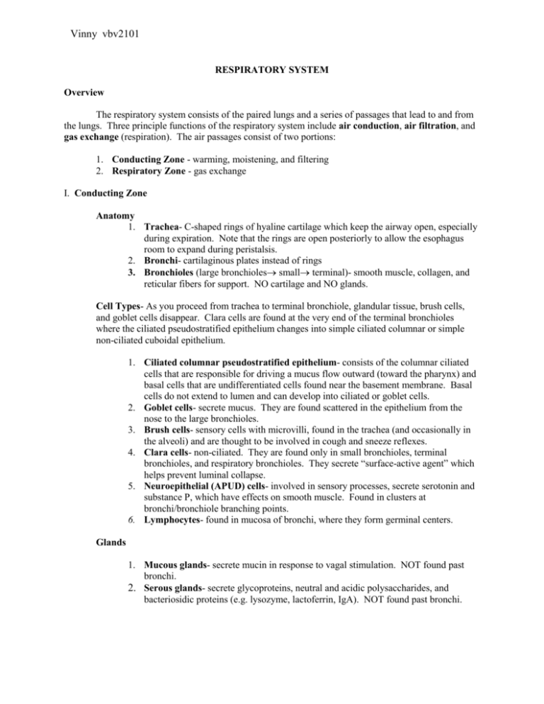
Vinny vbv2101 RESPIRATORY SYSTEM Overview The respiratory system consists of the paired lungs and a series of passages that lead to and from the lungs. Three principle functions of the respiratory system include air conduction, air filtration, and gas exchange (respiration). The air passages consist of two portions: 1. Conducting Zone - warming, moistening, and filtering 2. Respiratory Zone - gas exchange I. Conducting Zone Anatomy 1. Trachea- C-shaped rings of hyaline cartilage which keep the airway open, especially during expiration. Note that the rings are open posteriorly to allow the esophagus room to expand during peristalsis. 2. Bronchi- cartilaginous plates instead of rings 3. Bronchioles (large bronchioles small terminal)- smooth muscle, collagen, and reticular fibers for support. NO cartilage and NO glands. Cell Types- As you proceed from trachea to terminal bronchiole, glandular tissue, brush cells, and goblet cells disappear. Clara cells are found at the very end of the terminal bronchioles where the ciliated pseudostratified epithelium changes into simple ciliated columnar or simple non-ciliated cuboidal epithelium. 1. Ciliated columnar pseudostratified epithelium- consists of the columnar ciliated cells that are responsible for driving a mucus flow outward (toward the pharynx) and basal cells that are undifferentiated cells found near the basement membrane. Basal cells do not extend to lumen and can develop into ciliated or goblet cells. 2. Goblet cells- secrete mucus. They are found scattered in the epithelium from the nose to the large bronchioles. 3. Brush cells- sensory cells with microvilli, found in the trachea (and occasionally in the alveoli) and are thought to be involved in cough and sneeze reflexes. 4. Clara cells- non-ciliated. They are found only in small bronchioles, terminal bronchioles, and respiratory bronchioles. They secrete “surface-active agent” which helps prevent luminal collapse. 5. Neuroepithelial (APUD) cells- involved in sensory processes, secrete serotonin and substance P, which have effects on smooth muscle. Found in clusters at bronchi/bronchiole branching points. 6. Lymphocytes- found in mucosa of bronchi, where they form germinal centers. Glands 1. Mucous glands- secrete mucin in response to vagal stimulation. NOT found past bronchi. 2. Serous glands- secrete glycoproteins, neutral and acidic polysaccharides, and bacteriosidic proteins (e.g. lysozyme, lactoferrin, IgA). NOT found past bronchi. II. Respiratory Zone Function- gas exchange. No need for mucus glands or big cartilage support. Anatomy 1. Respiratory bronchioles- occasional alveoli open directly into the bronchiolar lumen. More typically, the respiratory bronchiole branch into the alveolar duct. 2. Alveolar duct- elongate airways with almost no walls, except for scattered rings of smooth muscle and the openings of alveoli. 3. Alveolar sac- spaces into which clusters of alveoli open 4. Alveoli- site of gas exchange 5. Alveolar septa- walls of tissue found between adjacent alveoli. They consist of 2 thin epithelial layers (of 2 alveoli) which surround capillaries, fibroblasts, elastic/collagen fibers, and the macrophages of the interstitium. -Blood-air barrier: the thinnest possible alveolar septa for gas exchange, has 4 layers: 1) surfactant 2) type I pneumocyte 3) basal lamina- single fused lamina of pneumocyte and capillary endothelium 4) capillary endothelial cell. Cell Types 1. Clara cells- these are the predominant cells in the respiratory bronchioles, forming the epithelial lining of these airways. They are nonciliated cells. 2. Smooth muscle- cords of smooth muscle are found in the non-respiratory portions of the respiratory bronchioles (that is those portions that serve a structural function). Clara cells still line the lumen here. 3. Type I pneumocyte- extremely thin squamous cells that line most of the alveolar surface. There are tight junctions between these and neighboring cells. 4. Type II pneumocyte- secretory cells, produce surfactant that prevents alveolar collapse (this is a different agent than the one the Clara cells secrete). These cells bulge into the alveolar lumen. They have apical granules resolved on TEM or oil that are referred to as lamellar bodies. 5. Endothelial cells- non-fenestrated, squamous cells that line the alveolar capillaries. Easily mistaken for type I pneumocytes. 6. Dust cells (aka macrophages)- you’ve seen these before. They go out into the lumen to degrade surfactant, dust particles, and bacteria that happen to enter the alveoli. 7. Fibroblasts- found in the connective tissue septa. Produce collagen and elastic fibers. Hyaline Cartilage Smooth Muscle Rings Plates Yes No No Yes Yes No Yes Respiratory Respiratory Bronchioles -initial No Yes -distal No Yes Alveolar ducts No Yes Squamous cells (Type I) No Alveoli No Yes Squamous cells (Type I) No Conducting Trachea Bronchi Bronchiole -large -small -terminal Epithelium Glands Special Cells Elastic Tissue Pseudostratified ciliated Pseudostratified ciliated Yes Yes Goblet cells Goblet cells Yes Yes Pseudostratified ciliated Simple columnar or cuboidal, ciliated Simple columnar or cuboidal, ciliated No No Goblet cells Yes Yes No Clara cells Yes Simple columnar or cuboidal, ciliated No Clara cells Yes No Mostly Clara cells Type II Pneumocytes, Macrophages Type II Pneumocytes (Surfactant cells); Macrophages (Dust cells) Yes Cilia = nine doublets surrounding a pair of microtubules Goblet cells secrete: loads of rER and Golgi Clara Cells: produce surfactant-associated protein and contain extensive sER Type I Pneumocytes: epithelial cells Type II Pneumocytes (Surfactant Cells): secrete surfactant from corners of alveoli Macrophages (Dust Cells): within the alveolar space Yes Yes RESPIRATORY SYSTEM QUESTIONS: 1. Which of the following is a correct pairing of the structure at the pointer and its secretory product? a. mucous cells: mucins b. serous gland: non-glycosylated proteins, bacteriostatic proteins c. goblet cell: mucus d. neuroepithelial cells: serotonin and substance P 2. What is the function of the cell type at the pointer? a. secretes surface-active agent b. structural support c. gas exchange d. secretes surfactant 3. The lumen at the pointer a. requires cartilage to maintain its patency b. is lined by stratified squamous epithelium c. contains areas where gas exchange occurs d. is lined by mucus glands but not serous glands 4. The segment of the respiratory system shown at the pointer a. contains c-shaped cartilage rings b. contains Clara cells c. is lined by pseudostratified epithelium d. contains serous glands RESPIRATORY SYSTEM ANSWERS: 1. Lab 13, slide 7 The pointer is on a serous gland in the submucosa of the trachea. 2. Lab 13, slide 17 The pointer is on a type 2 pneumocyte that secretes surface-active agent. This agent forms a monomolecular layer over the alveolar epithelium to reduce surface tension at the air-epithelium interface. Without adequate secretion of surfactant, the alveoli would collapse on exhalation. 3. Lab 13, slide 12 The pointer is on a respiratory bronchiole. A is wrong because there is no cartilage in the bronchioles. B is wrong because respiratory bronchioles are lined by simple cuboidal epithelium. C is correct because alveolar ducts, and thus alveoli, come off of respiratory bronchioles. D is wrong because neither mucus nor serous glands are found beyond the bronchi. 4. Lab 13, slide 11 The pointer is on the bronchiole. A is wrong because there is no cartilage in the bronchioles (rings are found in the trachea). B is correct because the bronchiole contains Clara cells. C is wrong because there is transition from ciliated simple columnar to cuboidal epithelium in the bronchioles. Choice D is wrong because there are no glands found in the bronchioles.
