cheng
advertisement
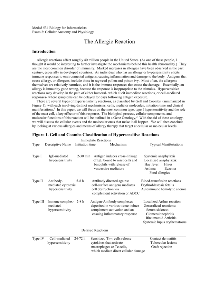
Meded 534 Biology for Informaticists Exam 2: Cellular Anatomy and Physiology The Allergic Reaction Introduction Allergic reactions affect roughly 40 million people in the United States. (As one of these people, I thought it would be interesting to further investigate the mechanisms behind this health abnormality.) They are the most common disorder of immunity. Marked increases in allergies have been observed in the past century, especially in developed countries. An individual who has an allergy or hypersensitivity elicits immune responses to environmental antigens, causing inflammation and damage to the body. Antigens that cause allergy, or allergens, include those in ragweed pollen and poison ivy. Most often, the allergens themselves are relatively harmless, and it is the immune responses that cause the damage. Essentially, an allergy is immunity gone wrong, because the response is inappropriate to the stimulus. Hypersensitive reactions may develop in the path of either humoral- which elicit immediate reactions, or cell-mediated responses- where symptoms can be delayed for days following antigen exposure. There are several types of hypersensitivity reactions, as classified by Gell and Coombs (summarized in Figure 1), with each involving distinct mechanisms, cells, mediator molecules, initiation time and clinical manifestations.1 In this paper, we will focus on the most common type, type I hypersensitivity and the role of the mast cell, a key effector of this response. The biological process, cellular components, and molecular functions of this reaction will be outlined in a Gene Ontology, 2 With the aid of these ontology, we will discuss the cellular events and the molecular ones that make it all happen. We will then conclude by looking at various allergies and means of allergy therapy that target at cellular or molecular levels. Figure 1. Gell and Coombs Classification of Hypersensitive Reactions Type Descriptive Name Immediate Reactions Initiation time Mechanism Type I IgE-mediated hypersensitivity 2-30 min Type II Antibody5-8 h mediated cytotoxic hypersensitivity Type III Immune complex- 2-8 h mediated hypersensitivity Antigen induces cross-linkage of IgE bound to mast cells and basophils with release of vasoactive mediators Typical Manifestations Systemic anaphylaxis Localized anaphylaxis: Hay fever Hives Asthma Eczema Food allergies Antibody directed against Blood-transfusion reactions cell-surface antigens mediates Erythroblastosis fetalis cell destruction via Autoimmune hemolytic anemia complement activation or ADCC Antigen-Antibody complexes deposited in various tissue induce complement activation and an ensuing inflammatory response Localized Arthus reaction Generalized reactions: Serum sickness Glomerulonephritis Rheumatoid Arthritis Systemic lupus erythematosus ______________________________________________________________________________________ Delayed Reactions ______________________________________________________________________________________ Type IV Cell-mediated 24-72 h Sensitized TDTH cells release Contact dermatitis hypersensitivity cytokines that activate Tubercular lesions macrophages or Tc cells, Graft rejection which mediate direct cellular damage Overview of Events at the Cellular Level Biological Process Hypersensitivity Reaction I-III. Types II-IV. IV. Type I: IgE-mediated immediate hypersensitivity A. Sensitization B. Receptor cross-linking with IgE C. Signal transduction 1. Activation of Syk a. Control of calcium flux b. Activation of GTPase/kinase cascades i. Synthesis of mediators -transcription -enzymatic breakdown. ii. Release of mediators in granules D. Late-phase reaction: Infiltration of leukocytes To develop a particular allergy, a genetically predisposed person must first be exposed to the allergen. This initial exposure causes sensitization and it is the subsequent exposures that elicit the damaging immune responses we recognize as the disease. The allergen induces a humoral response, resulting in the generation of antibody-secreting plasma cells and memory cells that later provoke a more powerful response if contact is made again with the allergen. For the signal transduction pathways that lead to B-cell differentiation into plasma cells, take a look at http://www.whfreeman.com/kuby/con_index.htm?11. (This animation has cells secreting immunoglobin A, but it is the same idea for other immunoglobins.) In contrast to a normal individual, differentiated B-cell plasma cells in an individual prone to have allergies, secrete large amounts of IgE as shown in http://www.cat.cc.md.us/courses/bio141/lecguide/unit3/allerg.html. There is a switch from IgM to IgE isotype in the B-cell plasma cell.3 Type I hypersensitivity is also termed IgE-mediated hypersensitivity because of this reason. The Fc region of IgE molecules binds with unusually high affinity (K a ~1010 liters/mole or Kd ~ 1 x 10¹⁰ M) to a class of Fc receptors: FcRI. These receptors are located on the surface of mast cells in tissues and on basophils in the blood, and the IgE molecules bound to them in turn serve as receptors for antigen. Here is a site that shows a molecular model of the Fc receptor of IgE: http://www.cc.ukans.edu/~micro/mhc.html#IgEFcReceptor. IgE antibodies are heterotetramers of two heavy and light chains (H 2L2), with the FcRI-binding domains located in the carboxy-terminal Fc region of the heavy chain. The dimeric Fc fragment of the IgE antibody has full binding activity for FcRI. IgE antibodies bind to the receptor in the absence of antigen, and thus the receptor adopts the antigenic specificity of the prevalent IgE repertoire. Crosslinking of the receptor through antigen/antibody interactions leads to the initiation of a signal transduction cascade mediated by Lyn and Syk kinases, analagous to that induced by T-cell and B-cell receptors. To learn more about immunoglobin E structure and properties (as well as the other immunoglobins, check out http://www.whfreeman.com/kuby/con_index.htm?04 or to look at it directly as a molecular model, look into http://www.whfreeman.com/kuby/content/ANM/ige.html. It is the binding of IgE cross-linked by allergen that triggers the release of inflammatory mediators such as histamine: http://www.ncbi.nlm.nih.gov:80/books/mboc/mboc.cgi?action=figure&fig=23-23 or as shown in an animation, http://www.cat.cc.md.us/courses/bio141/lecguide/unit3/mast.html. Histamines cause dilation and increased permeability of blood vessels and are largely responsible for the clinical manifestations of such allergic reactions as hay fever, asthma, and hives. In normal circumstances, the blood vessel changes are thought to help white blood cells, antibodies, and complement components enter sites of inflammation. The initiation time of this overall reaction is immediate, sometimes requiring as little as two minutes for a response to occur after exposure to the allergen (Figure 1). In addition to releasing these preformed inflammatory mediators stored in mast-cell vesicles, the binding of antigen to IgE triggers the mast cell to synthesize various eicosanoids and potent inflammatory mediators. All these mediators then initiate a local inflammatory response. This also occurs with basophils, the mast cell counterpart in the circulation. Following the immediate hypersensitive response, this late-phase reaction occurs, lasting many hours or days, during which large numbers of leukocytes, especially eosinophils, migrate into the inflamed area due to chemokines released by mast cells as well as other cells. Eosinophils are white blood cells that kill a variety of parasites, especially the parasites coated with IgE or IgA antibodies. The eosinophils, once in the area, secrete mediators that prolong the inflammation and sensitize the tissues, so that less allergen is needed the next time to evoke a response. Biological Techniques Used for Analysis of the Process at Different Levels Physiologically, the mouse is the indispensable model of choice for most immunologists. Scientists can choose from a variety of strains of mice that fit their study. Because mice and humans have similar immunology, many human antibodies are cross-reactive with mice antibodies and vice versa. In addition, chimeric mouse/ human receptors and antibodies have been constructed to study various immunological responses. Extremely valuable models are transgenic mice. This technique introduces foreign genes into mouse embryos. Viable offspring are then bred for the particular gene. Also important, are knockout mice which are transgenic mice whose targeted gene is disrupted and therefore does not express the targeted gene product. By observing these phenotypes, major advances have been made in understanding how that particular gene product affects the immune system. Studies of ‘mast cell knockin’ mice, where in vitro derived mast cell are inserted in to mast cell knockout mice, have showed that mast cells are critical for essentially all of the leukocyte infiltration in the skin.4 These and other reasons render mice a valuable model for studying human immunity. Various techniques can be used to analyze the allergic response on a cellular level. In terms of immunological techniques, various assays can be used. To measure the level of inflammation, cytokine analysis or white blood cell count can be performed via ELISA or flow cytometry. ELISA, or enzymelinked immunosorbent assay, quantitate either antibody or antigen by use of an enzyme-linked antibody and a substrate. Through subsequent washes and antibody bindings, a colored reaction forms, with an optical density that correlates with the amount of antibody or antigen. Using this method, one can measure the amount of cytokine secretion by the cells. Another method that can also quantitate secretion as well as identify the type of cell, is flow cytometry by use of FACS, or fluorescence-activated cell sorter. Cells are labeled with fluorochrome-conjugated antibody, then sorted based on the intensity of staining. The antibody chosen can be a marker for the specific cell type. For example, fluorochrome conjugated antiCD3 can be used to identify T-cells or an anti-CD88 can be used to identify mast cells, eosinophils, macrophages, and neutrophils. Cytokine expression can also be measured and another method used with flow cytometry, intracellular staining, is possible to identify the source of secretion. At the molecular level, several techniques can be used. To identify proteins in terms of size and type, one can administer Western blotting. In this method, protein can be separated according to size via electrophoresis, then transferred onto a membrane can then be identified with a monoclonal antibody incubation, subsequent washes, then a secondary antibody that binds the monoclonal antibody that is enzyme-linked. The protein can be subsequently visualized via substrate addition. This method can be used to characterize novel proteins. In a specific case, for characterization of kinases, kinase assays can be performed. In this case, phosphorylation activity of a kinase can be measured via radiolabelled 32P. Immunohistochemistry or immunofluorescence staining are also techniques that can be used to identify cellular structures. These techniques use antibodies to identify the anatomic distribution of an antigen within a tissue or within compartments of a cell. Using antibodies labeled with a fluorescent moiety or coupled to enzymes that convert colorless substrates to colored insoluble substances, antigens can be localized within the cell when observed through special microscopes. To obtain specific antibody and receptor structure, X-ray crystallographic methods can be performed. Examples of antibody structure can be found: http://www.whfreeman.com/kuby/content/mv/ig/molmast.htm or the antigen-antibody complex: http://www.whfreeman.com/kuby/content/mv/epitope/molmast.htm. Major advances have been made in the x-ray crystallographic structure of the Fc fragment of IgE bound to its high-affinity receptor FcRI.5 Mast Cell: Cellular Components that Participate in Response Cellular Component I. Mast cell A. Cell Membrane 1. FceRI complex a. ITAM (with protein tyrosine kinases) 2. CD45 3. SOCC –calcium channel 4. GTPase complex B. Cytoplasm 1. Endoplasmic Reticulum a. IP3 receptors on calcium channels b. ribosome -translation complex 2. Granules (containing mediators) 3. GTPase complex C. Cytoskeleton 1. Microfilaments 2. Microtubules D. Nucleus 1. Nuclear membrane 2. Nucleoplasm a. transcription complex Mast cells are derived from hematopoetic progenitor cells in the bone marrow. Human mast cells are usually round, oval or sometimes spindle-shaped, with a typically round nuclei. The cytoplasm contains membrane-bound granules and lipid bodies.7 As noted previously, upon appropriate stimulation via FcRI, mast cells can secrete mediators. The human FcRI receptor, can form either a trimeric 2 or tetrameric 2 structure on cell surfaces, with the extracellular domains of the -chain conferring the ability to bind IgE with affinity (Kd~10-9-10-10 M). Theses receptors belong to a family of antibody-binding receptors that also mediate interactions of soluble IgG antibodies with cells of the immune system. 5,8 It has been clearly established that IgE upregulates the expression of FcRI, most probably by protecting it from internalization and degradation.8 This has important clinical implication because mast cells that express high numbers of FcRI release mediators at lower concentrations of antigen and secrete greatly enhanced amount so histamines leukotrienes and Th2 cytokines.6 The mediators are either preformed and granule-associated (e.g. histamine, proteoglycans and neutral proteases) or are synthesized de novo (e.g. leukotriene C4 (LTC4), platelet activating factor [PAF] and prostaglandin D2 (PGD2). In addition, mast cells are potential sources of many cytokines associated with inflammation, immunity, hematopoiesis, tissue remodeling and diverse other biological processes (e.g. IL1, IL-3, IL-4, IL-5, IL-6, IL-8, IL-10, IL-13, IL-16, TNF-, bFGF, VPF/VEGF, TGF- and several C-C chemokines, including MIP-1 and MCP-1).7 The cytoplasmic granules of mast cells stain metachromatically with certain basic dyes, probably primarily reflecting their content of proteoglycans such as chondroitin sulfates and heparin, produced exclusively by mast cells. The biological functions of proteoglycans are not fully understood; however, in mice, heparin is required for the normal packaging of certain neutral proteases in mast cell cytoplasmic granules.7 Mast cell mediators will be discussed further in this paper. Molecular Mechanism behind IgE-mediated Immediate Hypersensitivity: Link of its Enzyme Commission classification are indicated for some enzymes. Molecular Function I. Cell Cycle Regulator II. Microtubule binding III. Actin binding IV. Transcription factor binding V. Nucleic acid binding A. Elk-1 B. NFAT C. AP-1 D. NFB VI. Chaperone VII. Enzyme A. Lyn http://expasy.cbr.nrc.ca/cgi-bin/niceprot.pl?LYN_HUMAN B. Syk http://expasy.cbr.nrc.ca/cgi-bin/niceprot.pl?KSYK_HUMAN C. Csk http://expasy.cbr.nrc.ca/cgi-bin/niceprot.pl?CSK_HUMAN D. PI(3)K http://expasy.cbr.nrc.ca/cgi-bin/nicezyme.pl?2.7.1.137 E. Btk http://expasy.cbr.nrc.ca/cgi-bin/niceprot.pl?BTK_HUMAN F. PKC http://expasy.cbr.nrc.ca/cgi-bin/nicezyme.pl?2.7.1.37 G. PKA http://expasy.cbr.nrc.ca/cgi-bin/niceprot.pl?P54646 H. PLC http://expasy.cbr.nrc.ca/cgi-bin/nicezyme.pl?3.1.4.3 I. MAPK http://expasy.cbr.nrc.ca/cgi-bin/nicezyme.pl?2.7.1.37 J. MEK http://expasy.cbr.nrc.ca/cgi-bin/nicezyme.pl?2.7.1.37 K. ERK http://expasy.cbr.nrc.ca/cgi-bin/nicezyme.pl?2.7.1.37 L. cPLA2 http://expasy.cbr.nrc.ca/cgi-bin/nicezyme.pl?3.1.1.4 M. Jnk http://expasy.cbr.nrc.ca/cgi-bin/nicezyme.pl?2.7.1.37 N. adenylate cyclase http://expasy.cbr.nrc.ca/cgi-bin/nicezyme.pl?4.6.1.1 O. SHP-1 http://expasy.cbr.nrc.ca/cgi-bin/nicezyme.pl?3.1.3.48 P. SHP-2 http://expasy.cbr.nrc.ca/cgi-bin/nicezyme.pl?3.1.3.48 Q. SHIP http://expasy.cbr.nrc.ca/cgi-bin/nicezyme.pl?3.1.3.56 R. Ras/Rac/ Rho http://expasy.cbr.nrc.ca/cgi-bin/get-enzyme-entry-unprecise?2.7.4.S. PAF hydrolase http://expasy.cbr.nrc.ca/cgi-bin/nicezyme.pl?3.1.1.47 VIII. Defense/Immunity Protein A. Mediators 1. IL-1 2. IL-3 3. IL-4 4. IL-5 5. IL-6 6. IL-8 7. IL-10 8. IL-13 9. IL-16 10. TNF- 11. bFGF 12. VPF 13. VEGF 14. TGF- 15. MIP-1 16. MCP-1 17. Histamine 18. Prostaglandins 19. Arachidonic acid 20. leukotrienes 21. PAF IX. Adapter Protein A. LAT B. Slp-76 C. Grb-2 X. Motor Proteins XI. Receptors A. FcRI The Signal Cascade Initial activation The cytoplasmic domains of the and chains of FceRI are associated with protein tyrosine kinases, as mentioned before. After the cross-linking of FcRI, information is passed by unknown mechanisms to the and chains signaling subunits, which results in activation of Syk and Lyn protein tyrosine kinases in a process involving the ITAMs (immunoreceptor tyrosine-base activation motif) of the FcR chain. ITAMs are 18-residue motifs that associate with the Src family of tyrosine kinases and Syk tyrosine kinase. Phospho-ITAMs provide a docking site for cytoplasmic proteins that contain the Src-homology-2 (SH2) domain and hence link receptor and signal transduction cascades, as the SH2 domain has high affinity for phosphorylated tyrosine residues.8 Lyn activation which occurs around five seconds after cross-linkage, is the result of the opposing actions of CD45 phosphatase and the Csk kinase. Activated Lyn phosphorylates the and chain ITAMs, the latter is then able to recruit Syk, which is in turn activated by Lyn. Lyn is found associated with FcRIwhereas Syk is found associated with FcRand FceRI with a higher affinity for FcRI Targets of Lyn and Syk, include phosphorylation of the BTK (Bruton’s tyrosine kinase), the phosphorylation of the membrane localized adapter protein LAT (linker for activation of T-cells), the phosphorylation and activation of isoforms of a phosphatidylinositol-specific phospholipase C (PI-PLC) that catalyze phosphatydylinositol biphosphate breakdown to inositol 1,4,5-triphosphate (IP3) and diacylglycerol (DAG), and the activation of PI(3)K (phosphoinositol-3-OH kinase) to produce phosphotidylinositol (3,4,5) triphosphate (PIP3).9 FcRI control of calcium BTK, which is localized at the membrane by binding to PIP 3, contributes to activation of PLC1, which in turn has been brought to the membrane by the phosphorylated LAT adapter. PLC1 acts on membrane inositol phospholipids to generate Ins(1,4,5)P 3 (IP3) and DAG. The intracellular targets of the latter include PKC isoforms, whereas IP3 binds to the IP3 receptor (IP3R) on the surface of the endoplasmic reticulum calcium stores, leading to store depletion and elevation of cytoplasmic calcium levels. Depletion of calcium stores are coupled to openings of plasma membrane channels for calcium influx; the calcium release activated current (ICRAC) is mediated by these store-operated calcium channels (SOCC), and is responsible for sustained elevations in cytosolic calcium and refilling of endoplasmic reticulum stores. Within 15 seconds of cross-linkage of FcRI, methylation of various member phospholipids is observed, resulting in an increase in the membrane fluidity and formation of calcium channels. The influx of calcium reaches a peak within two minutes of cross-linkage. The calcium increase is due to both a release of calcium from intracellular stores in the endoplasmic reticulum and from the uptake of extracellular calcium. The calcium influx eventually leads to the formation of arachidonic acid, which is converted into two classes of potent mediators: prostaglandins and leukotrienes. 8,9 Release of granules The influx of calcium also promotes the assembly of microtubules and the contraction of microfilaments, both of which are necessary for movement of granules to the plasma membrane. The activated PKC phosphorylates myosin light chains, leading to the disassembly of actin-myosin complexes beneath the plasma-membrane, thereby allowing granules to come in contact with the plasma membrane. Crosslinking of FcRI also activates the enzyme adenylyl cyclase through a heterotrimeric GTP-binding protein. This, in turn, elevates cyclic adenosime monophosphate (cAMP) levels reaching a rapid peak about one minute after cross-linkage of FcRI. The effects of cAMP are exerted through the activation of cAMP-dependent protein kinases, which phosphorylate the granule-membrane proteins, thereby changing the granules’ permeability to water and calcium. Fusion of the granule membrane with the plasma membrane is mediated by interactions between proteins associated with the granule membrane and proteins associated with the plasma membrane. The consequent swelling of the granules facilitate their fusion with the plasma membrane, releasing their contents. The increase in cAMP is transient and is followed by a drop in cAMP to levels below baseline. The drop in cAMP appears to be necessary for degranulation to proceed. cAMP also activates protein kinase A, which inhibits degranulation, suggesting that this pathway is a negative feedback loop. To look at protein kinase A in action, take a look at: http://www.auhs.edu/netbiochem/movies/a_kinase.mov or http://www.chem.umass.edu/~cmartin/Courses/BioStruct/Proj/Glenn/CHIME.htm http://www.nih.go.jp/mirror/Kinases/pkr/3D/text/2cpk/2cpk.html These events are regulated by GTP-binding (activated) form of the Ras-related proteins Rab 3b or d, which are present in mast cells. 8,9 Activation of transcription factors IP3 causes this elevation of cytoplasmic calcium, which, in combination with DAG, activates protein kinase C (PKC). For a movie about PKC, look into http://www.auhs.edu/netbiochem/movies/c_kinase.mov. FcRI-regulated adapter molecules contribute to the activation of GTPase/kinase cascades. Syk targets include the LAT adapter molecule, which anchors the Grb-2 and Slp-76 adapters. These in turn contribute to the recruitment/localization of guanine nucleotide exchange factors for Ras family GTPases. GTP loading of GTPases initiates a wide range of effector responses. These include the induction of serial kinases cascades such as Ras/Raf-1/MEK/ERK cascade, where the terminal targets are transcription factors, and the rearrangement of the actin cytoskeleton to produce changes in cell morphology.8,9 The synthesis of lipid mediators is controlled by activation of the enzyme cytosolic phospholipase A 2 (cPLA2). This enzyme is activated by two signals: elevated cytoplasmic calcium, released by IP 3, and phosphorylation catalyzed by a mitogen-activated protein (MAP) kinase. MAP kinases are activated as a consequence of a kinase cascade initiated through the receptor ITAMs, probably utilizing the same pathways as T-cells. Cytokine transcription in mast cells is also presumed to be similar to the events that occur in T-cells. Syk appears to activate various adapter molecules, leading to nuclear translocation of NFAT (nuclear factor of activated T-cells) and NFB (nuclear factor-B), as well as activation of AP-1 (activator protein) by stress-activated protein kinases, such as c-Jun N-terminal kinase (Jnk). Overall, this response leads to transcription of several cytokines (IL-3, IL-4, IL-5, IL-6, and tumor necrosis factor (TNF), among others) but in contrast to T cells, not IL-2. 8,9 Mediators Found in the Mast Cell Preformed Mediators The primary mediator, histamine, acts by binding to target cell receptors, initiating intracellular events, such as phosphatidylinositol biphosphate breakdown to IP 3 and DAG, which cause changes in different cell types as mentioned earlier. In vascular endothelial cells, binding of histamine leads to endothelial cell contraction and leakage of plasma into the tissues. Histamine also causes endothelial cells to synthesize vascular smooth muscle cell relaxants, such as prostacyclin (PGL2) and nitric oxide, which cause vasodilation. Another result is contraction of intestinal and bronchial smooth muscle, which may contribute to increased peristalsis or bronchospasm associated with ingested allergens or asthma, respectively. However, bronchoconstriction in asthma is more prolonged than the effects of histamine, which is rapidly removed from the extracellular milieu by amine-specific transport systems. 1,7 In addition to vasoactive amines, mast cell granules contain several enzymes, such as serine proteases and aryl sulfatase, as well as proteoglycans such as heparin or chondroitin sulfate. The enzymes may cause tissue injury when released upon mast cell degranulation. One function of the negatively charged proteoglycans may be to bind and store the positively charged biogenic amines. However, it is not clear how important these substances are as mediators of immediate hypersensitivity reactions. 1,7 Newly Synthesized by Activated Mast Cells: Lipid Mediators Prostaglandin D2 (PGD2). Released PGD2 binds to receptors on smooth muscle cells and acts as a vasodilator and as a bronchoconstrictor. Moreover, PGD 2 is released from lung mast cells during asthmatic bronchoconstriction. As mentioned earlier, PGD2 is synthesized from arachidonic acid derived from phospholipid by the sequential actions of enzymes. The synthesis of PGD 2, like that of other prostaglandins, depends upon the enzyme cyclooxygenase, and PGD 2 synthesis can be prevented by inhibitors of cyclooxygenase, such as aspirin and nonsteroidal anti-inflammatory agents.1,7 Leukotrienes. Mast cells convert arachidonic acid by action of 5-lipoxygenase and other enzymes, into leukotriene C4 (LTC4) which can be subsequently degraded into LTD4 and LTE4. Mast cell-derived leukotrienes bind to specific receptors on smooth muscle cells, different from the receptors for PGD 2, and cause prolonged bronchoconstriction. When injected into skin, these leukotrienes produce a characteristic long-lived wheal and flare reaction. Collectively, LTC4, LTD4, and LTE4 constitute what was once called “slow-reacting substance of anaphylaxis” (SRS-A) and are now thought to be major mediators of asthmatic bronchoconstriction.1,7 Platelet activating factor (PAF) PAF is synthesized by acylation of lysoglyceryl ether phosphorylcholine, which is derived from a membrane phospholipid by cPLA2-mediated release of fatty acid from the sn2 position. PAF has direct bronchoconstricting actions. It also causes retraction of endothelial cells and can relax vascular smooth muscle. However PAF is very hydrophobic and is rapidly destroyed by a plasma enzyme called PAF hydrolase, limiting its biologic actions. 1,7 Cytokines TNF, IL-4, IL-1, IL-5, IL-6 and various CSF (colony-stimulating factor) such as IL-3 and granulocytemonocyte CSF are secreted. Cytokines released upon IgE mediated mast cell activation are predominantly responsible for the late phase reaction. IL-4 through its selective induction of endothelial VCAM-1 and stimulation of eosinophil-selective transendothelial migration, is responsible for selective recruitment of eosinophils, and IL-5 is important for eosinophil activation. Disease and Treatment of Allergy The symptoms of allergy are a result of the various effects of these mediators and the body site in which the antigen-IgE-mast cell combination occurs. As inferred from above, the mediators released cause increased mucus secretions, increased blood flow, swelling of the epithelial lining, and contraction of the smooth muscle surrounding the airways which are the symptoms of congestions, runny nose, sneezing, and difficulty breathing that characterize hay fever. 1,7 Allergic symptoms are usually localized to the site of entry of the antigen. The reaction in skin is largely mediated by histamine which binds to venular endothelial cells which express numerous histamine receptors. Endothelial cells synthesize and release prostacyclin, nitric oxide, and PAF which cause vascular smooth muscle relaxation. The affected site becomes red from local accumulation of red blood cells. The endothelial cells also retract, allowing extravasation of plasma. The initial redness or erythema is then replaced by swelling. Finally blood vessels dilate, producing the redness. 1,7 Bronchial asthma is characterized by paroxysms of bronchial constriction and increased production of thick mucous, which leads to bronchial obstruction and exacerbates breathing difficulties. The smooth muscle cells in the airways of asthmatics are hyper-reactive to constricting stimuli. Bronchitis and infections are frequent complications. 1,7 If very large amounts of the chemicals released by the mast cells (or blood basophils) enter the circulation, however, systemic symptoms may result and cause severe hypotension and bronchiolar constriction. This systemic immediate hypersensitivity is termed anaphylaxis. Vasodilation and vascular exudation of plasma occur in vascular beds throughout the body. This usually implies the systemic presence of antigen by injection, insect sting, or absorption across an epithelial surface, such as the skin or gut mucosa. The decrease in vascular tone and leakage of plasma lead to a fall in blood pressure, or shock, that may be fatal. The cardiovascular effects are complicated by constriction of the upper and lower airways, hypersensitivity of the gut, outpouring of mucus in the gut and respiratory tree, and hives. The most important mast cell mediators of anaphylactic shock are not fully known. The lifesaving treatment is epinephrine, which reverses the bronchoconstrictive and vasodilatory effects of the various mast cell mediators, and improves cardiac output.1,7 Various treatments have been used to allay the symptoms of allergy. H1 histamine receptor antagonists (antihistamines) can inhibit the wheal and flare response to intradermal allergen. There are also allergy shots where small, but increasing quantities of allergen are injected to desensitize the individual from the allergen, but the effects of this treatment are not clear (with me being an example). The most effective treatment are use of corticosteroids. Corticosteroids bind to glucocorticoid receptors which dimerize to bind DNA and increase transcription and this mediates the systemic side effects of corticosteroids. Most of the anti-inflammatory effects of corticosteroids are mediated by inhibition of expression of inflammatory genes that are regulated by transcription factors such as AP-1, NFB, and NFAT, by interaction of glucocorticoid receptors monomers with these transcription factors. 11 Many other treatments are still in the research stage. Signal transduction inhibitors to kinases such as MAP kinase and the tyrosine kinases, prevent phosphorylation so allergic reaction will not occur. Monoclonal antibodies have been constructed against IgE in order to block mast cell activation with the mechanism portrayed in http://www.cat.cc.md.us/courses/bio141/lecguide/unit3/monoige.html. Various cytokines inhibitors against targets such as IL-1, IL-4, IL-5, IL-13, TNF, and chemokine have also been synthesized. Many of these inhibitors have a broad range of effects. 11 Given the inappropriateness of most immediate hypersensitivity responses, how did such a system evolve? The normal physiological function of the IgE-mast cell eosinophil pathways is to repel invasion by nonphagocytable multicellular parasites. The mediators released by the mast cells stimulate the inflammatory response against the parasites, and the eosinophils serve as the major killer cells against them by secreting several toxins. How this system also came to be inducible by harmless agents is not clear. As usual, further investigation is necessary to provide better treatments and reasoning behind why allergic attacks occur. References 1. Kuby, Janis. Immunology. New York: W.H. Freeman and Co. 1997. 2. Ashburner M, Ball CA, Blake JA, Botstein D, Butler H, Cherry JM, Davis AP, Dolinski K, Dwight SS, Eppig JT, Harris MA, Hill DP, Issel-Tarver L, Kasarskis A, Lewis S, Matese JC, Richardson JE, Ringwald M, Rubin GM, Sherlock G.Gene ontology: tool for the unification of biology. The Gene Ontology Consortium. Nature Genetics. 25:25-9.2000. 3. Oettgen H. Regulation of the IgE Isotype Switch: New Insight on Cytokine Signals and the Functions of the Gerrmline Transcripts. Current Opinion in Immunology. 12:618-623.2000. 4.Wedemeyer J, Tsai M, Galli S. Roles of Mast Cells and Basophils in Innate and Acquired Immunity. Current Opinion in Immunology. 12:624-531.2000. 5. Garman S, Wurzburg B, Tarchevskaya S, Kinet, J, Jardetzky T. Structure of the Fc fragment of the human IgE bound to its high-affinity receptor FcRI. Nature. 406: 259-266.2000. 6. Geha R.. Atopic Allergy and Other Hypersensitivitites. Allergy, a disease of the internal and external environments. Current Opinion in Immunology. 12: 615-617.2000. 7. Janeway, C, Travers P. Immunobiology : The Immune System in Health and Disease. New York: Garland Publishing. 1999. 8. Kinet J. The High-Affinity IgE Receptor (FcRI): From Physiology to Pathology. Annual Reviews in Immunology. 17:931-72.1999. 9. Turner H, Kinet J. Signalling through the High-Affinity IgE Receptor FcRI. Nature. 402:B24B30.1999. 10. Alberts, B. Bray D, Lewis J, Raff M, Roberts K, Watson J. Molecular Biology of the Cell. New York: Garland Publishing. 1994. 11. Barnes P. Therapeutic Strategies for Allergic Diseases. Nature. 402:B31-38.1999. Websites Used: http://www.cat.cc.md.us/courses/bio141/lecguide/unit3/u3vvi.html http://www.whreeman.com/lodish/ http://www.biology.arizona.edu/ http://www.whfreeman.com/kuby http://www.ncbi.nlm.nih.gov:80/books/mboc/ http://www.auhs.edu/netbiochem/ http://www.chem.umass.edu/~cmartin/Courses/ http://www.nih.go.jp/mirror/Kinases/ http://expasy.cbr.nrc.ca/enzyme/
