Micro-nano mineral study and new mineral discovery
advertisement
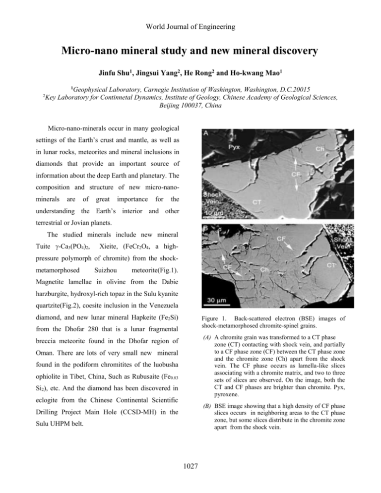
World Journal of Engineering Micro-nano mineral study and new mineral discovery Jinfu Shu1, Jingsui Yang2, He Rong2 and Ho-kwang Mao1 1 Geophysical Laboratory, Carnegie Institution of Washington, Washington, D.C.20015 Key Laboratory for Continnetal Dynamics, Institute of Geology, Chinese Academy of Geological Sciences, Beijing 100037, China 2 Micro-nano-minerals occur in many geological settings of the Earth’s crust and mantle, as well as in lunar rocks, meteorites and mineral inclusions in diamonds that provide an important source of information about the deep Earth and planetary. The composition and structure of new micro-nanominerals are of great importance for the understanding the Earth’s interior and other terrestrial or Jovian planets. The studied minerals include new mineral Tuite -Ca3(PO4)2, Xieite, (FeCr2O4, a high- pressure polymorph of chromite) from the shockmetamorphosed Suizhou meteorite(Fig.1). Magnetite lamellae in olivine from the Dabie harzburgite, hydroxyl-rich topaz in the Sulu kyanite quartzite(Fig.2), coesite inclusion in the Venezuela diamond, and new lunar mineral Hapkeite (Fe2Si) Figure 1. Back-scattered electron (BSE) images of shock-metamorphosed chromite-spinel grains. from the Dhofar 280 that is a lunar fragmental (A) A chromite grain was transformed to a CT phase zone (CT) contacting with shock vein, and partially to a CF phase zone (CF) between the CT phase zone and the chromite zone (Ch) apart from the shock vein. The CF phase occurs as lamella-like slices associating with a chromite matrix, and two to three sets of slices are observed. On the image, both the CT and CF phases are brighter than chromite. Pyx, pyroxene. breccia meteorite found in the Dhofar region of Oman. There are lots of very small new mineral found in the podiform chromitites of the luobusha ophiolite in Tibet, China, Such as Rubusaite (Fe0.83 Si2), etc. And the diamond has been discovered in eclogite from the Chinese Continental Scientific (B) BSE image showing that a high density of CF phase slices occurs in neighboring areas to the CT phase zone, but some slices distribute in the chromite zone apart from the shock vein. Drilling Project Main Hole (CCSD-MH) in the Sulu UHPM belt. 1027 World Journal of Engineering brilliant (105-7 times conventional x-ray), and can be focused to a couple of micron size spots by mirror. Since the system at APS HPCAT Beam 16 and at BNLNSLS X17C beam are built for highpressure experiments, the angle and energydipersive X-ray diffraction system combined with the Ge detector provides the state-of-the-art technique to investigate the micro-mineral experiment. In summary these results obtained by the Synchrotron x-ray diffraction technique combining with other petrological and mineralogical data will provide important information of origin and evolution of these rocks containing micro-nano mineral and develop a new micro-nano-mineralogy. Reference (1). Ru Y. Zhang, Jin F. Shu, H.K.Mao, and Juhn G. Liou (1999) Magnetite lamellae in olivine and clinohumite from Dabie UHF ultramafic rocks, Central China. American Mineralogist, 84, 564-569. (2). Ming Chen, Jinfu Shu Xiande Xie, and Hokwang Mao (2003) Natural CaTi2O4structured FeCr2O4 polymorph in Suizhou meteorite and its significance in mantle mineralogy Geochimica et Cosmochimica Acta Vol. 67, No.20, pp3937-3942 (3). Jinfu Shu, Ho-kwang Mao, Russel J.Hemley (2002) Micromineral study by Synchrotron X-ray Diffraction. he 4th World Chinese Conference on Geological Science, Nanjing China Abstracts pp.227 Figure 2. (a) Magnetite (Mag) lamellae exsolved from harzburgitic olivine and crosscut by fractures filled with serpentine and minute grains of magnetite. Cr-bearing magnetite (Cr-Mag) lamellae also occur (plane polarized light, width of view = 0.39 mm). (b) Back-scattered electron image of a small area from (a) Some lamellae beneath the surface do not appear in this image. The rods marked by arrows are the same as those in Figure 2a, (scale bar = 10 m). Due to the study natural micro-nano mineral sample are very rare and small, we need to use a much stronger X-ray source as well as a smaller Xray spots. Synchrotron x-ray diffraction technique which has been successfully applied to study of very high-pressure experiments and micro- nano minerals in natural rock is powerful to identify new mineral and crystal structure of micro-nano minerals. Synchrotron light source x-ray is very 1028
