2nd February 2015 Lanchester (B7), Room 3031, Highfield Campus
advertisement
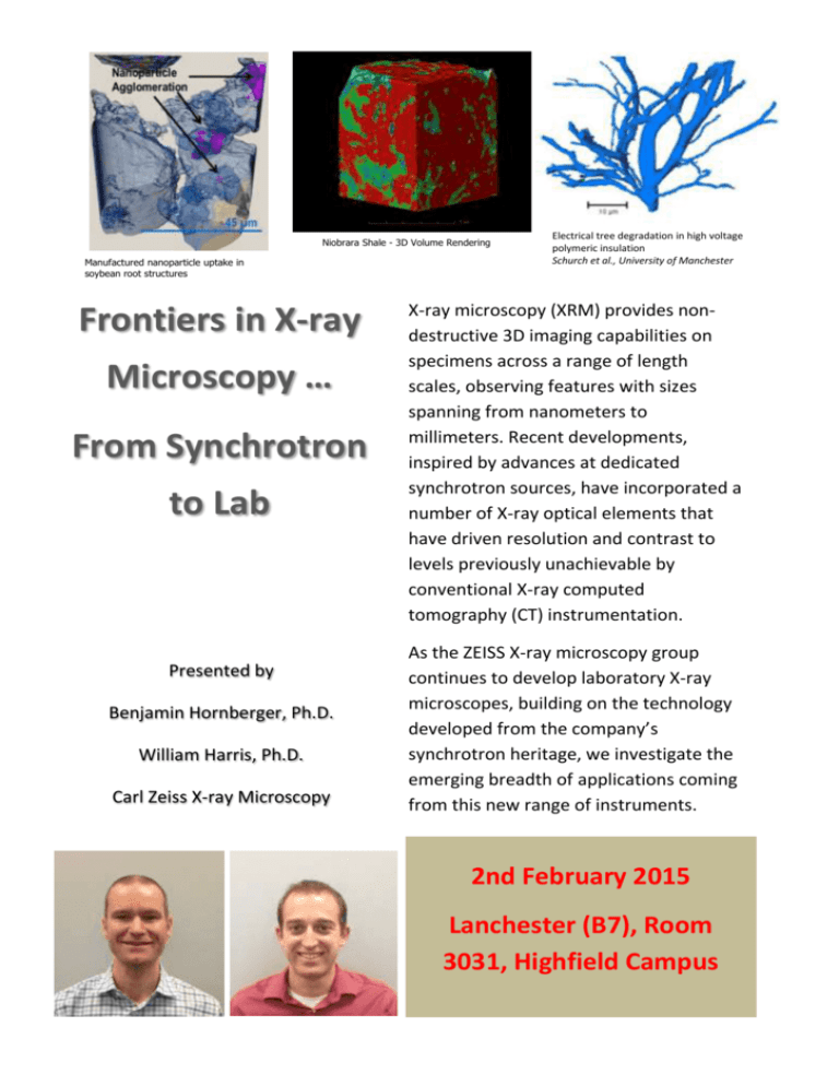
Niobrara Shale - 3D Volume Rendering Manufactured nanoparticle uptake in soybean root structures Frontiers in X-ray Microscopy … From Synchrotron to Lab Presented by Benjamin Hornberger, Ph.D. William Harris, Ph.D. Carl Zeiss X-ray Microscopy Electrical tree degradation in high voltage polymeric insulation Schurch et al., University of Manchester X-ray microscopy (XRM) provides nondestructive 3D imaging capabilities on specimens across a range of length scales, observing features with sizes spanning from nanometers to millimeters. Recent developments, inspired by advances at dedicated synchrotron sources, have incorporated a number of X-ray optical elements that have driven resolution and contrast to levels previously unachievable by conventional X-ray computed tomography (CT) instrumentation. As the ZEISS X-ray microscopy group continues to develop laboratory X-ray microscopes, building on the technology developed from the company’s synchrotron heritage, we investigate the emerging breadth of applications coming from this new range of instruments. 2nd February 2015 Lanchester (B7), Room 3031, Highfield Campus
