Vibration of eukariotic Cells in Suspension induced by a
advertisement
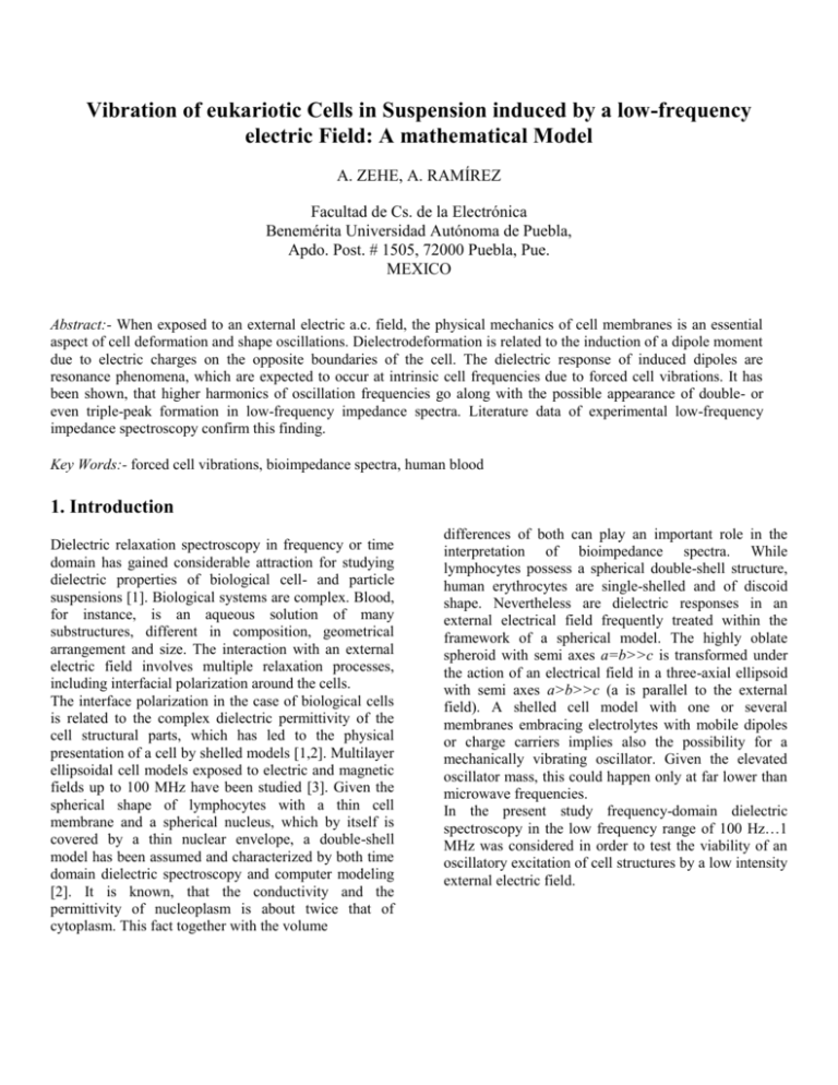
Vibration of eukariotic Cells in Suspension induced by a low-frequency electric Field: A mathematical Model A. ZEHE, A. RAMÍREZ Facultad de Cs. de la Electrónica Benemérita Universidad Autónoma de Puebla, Apdo. Post. # 1505, 72000 Puebla, Pue. MEXICO Abstract:- When exposed to an external electric a.c. field, the physical mechanics of cell membranes is an essential aspect of cell deformation and shape oscillations. Dielectrodeformation is related to the induction of a dipole moment due to electric charges on the opposite boundaries of the cell. The dielectric response of induced dipoles are resonance phenomena, which are expected to occur at intrinsic cell frequencies due to forced cell vibrations. It has been shown, that higher harmonics of oscillation frequencies go along with the possible appearance of double- or even triple-peak formation in low-frequency impedance spectra. Literature data of experimental low-frequency impedance spectroscopy confirm this finding. Key Words:- forced cell vibrations, bioimpedance spectra, human blood 1. Introduction Dielectric relaxation spectroscopy in frequency or time domain has gained considerable attraction for studying dielectric properties of biological cell- and particle suspensions [1]. Biological systems are complex. Blood, for instance, is an aqueous solution of many substructures, different in composition, geometrical arrangement and size. The interaction with an external electric field involves multiple relaxation processes, including interfacial polarization around the cells. The interface polarization in the case of biological cells is related to the complex dielectric permittivity of the cell structural parts, which has led to the physical presentation of a cell by shelled models [1,2]. Multilayer ellipsoidal cell models exposed to electric and magnetic fields up to 100 MHz have been studied [3]. Given the spherical shape of lymphocytes with a thin cell membrane and a spherical nucleus, which by itself is covered by a thin nuclear envelope, a double-shell model has been assumed and characterized by both time domain dielectric spectroscopy and computer modeling [2]. It is known, that the conductivity and the permittivity of nucleoplasm is about twice that of cytoplasm. This fact together with the volume differences of both can play an important role in the interpretation of bioimpedance spectra. While lymphocytes possess a spherical double-shell structure, human erythrocytes are single-shelled and of discoid shape. Nevertheless are dielectric responses in an external electrical field frequently treated within the framework of a spherical model. The highly oblate spheroid with semi axes a=b>>c is transformed under the action of an electrical field in a three-axial ellipsoid with semi axes a>b>>c (a is parallel to the external field). A shelled cell model with one or several membranes embracing electrolytes with mobile dipoles or charge carriers implies also the possibility for a mechanically vibrating oscillator. Given the elevated oscillator mass, this could happen only at far lower than microwave frequencies. In the present study frequency-domain dielectric spectroscopy in the low frequency range of 100 Hz…1 MHz was considered in order to test the viability of an oscillatory excitation of cell structures by a low intensity external electric field. z 2. Dielectric Model of Forced Cell Resonance 2.8 m y Forced vibrations of an oscillator result, when an external oscillatory force of frequency is applied to a particle subject to an electric field. The double-shell lymphocyte bears similarities in the physical concept of motion, where the external force acts on both, the inner (nucleoplasm) sphere and the outer (cytoplasm) sphere. Given the different geometrical and dielectric characteristics, the response of each might be different (Fig.1a). In the case of erythrocytes only one shell exists and the oscillator model could be simpler, although the more complex shape makes it more difficult in the experimental approach (Fig.1b). Let us suppose, that a shelled spherical cell containing a certain dipole charge density is exposed to an harmonically variable electric field. It is easy to imagine, that the elastic properties of the nuclear envelope and the cell membrane provide for a restoring force after a deformation due to the external field action on the mobile ions inside the sphere plasmas has occurred. nuclear envelope nucleoplasm = 60 = 0.5 S/m m2 = 120 = 1S/m m1 b a x 7.6 m Figure 1(b): Discoid shape of an erythrocyte of diameter 2R=7.6m and thickness at the rim 2L=2.8 m. a, b represent semi axes (a=b=R) in a spheroidal approach with small semi axis c in direction of z. half axis of the field-deformed ellipsoid of radius R (i.e. q=1 corresponds to a spheroid) characterizes both, the elongation (contraction) deformation and the forces, which determine the deformation [4]. The electric ponderomotive force is caused by the applied time-dependent field E0(t), and contains the dielectric properties of the cell: membrane Fpond ( , q ) V F ( ) Eloc ( , q) 2 cytoplasm 7 nm 40 nm 5.6m f L (q) . (1) q 7m Figure 1(a): Double-shelled lymphocyte cell as proposed by Ermolina et al. [2]; is the dielectric constant, specific electric conductivity, m1 - mass of the inner nucleus (m1=91011g), m2- mass of the outer shell region (m2=61011g), being mc= m1+m2 the total cell mass. The periodic deformation of cells in an electric field E0() with frequency is determined by the distribution of the electrical forces applied and the mechanical forces generated in the deformed membranes and in the adjacent layers of the cell. The mechanical forces include elastic and viscous shear stresses in the membranes. The axis relation q=R/L with L the longer Here fL(q) is the depolarizing factor in direction of the main axis L parallel to the external field vector E0. The local field Eloc(, q) is the value of the applied field inside the ellipsoid: Eloc ( , q) ( , q) Eo ; ( , q) m* ( ) m* ( ) [ *p ( ) m* ( )] f (q) (q ) m* ( ) (2) The factor (q) has been calculated previously for the general case of a dielectric object with arbitrary shape [5]. The frequency dependence of the ponderomotive electric field force is given by F() F ( ) p 2 2 p 0 m p 1) 2 ( e ) 2 ( ) 1 ( 4 m m m 1 ( ) 2 1 e e=0m/m. (3) Here p and m are the static relative permittivities of the cell and the suspending medium, p and m are the static conductivities, and 0 is the vacuum permittivity. The elastic force of the cell membrane is given by F (q ) ½ S (1 q ) 2 (4) with S the surface area of the object and the shear elasticity modulus. The friction force Ffr (q, dq/dt) between the cell and the surrounding medium is F (q , d q /d t ) S q 2 dq dt (5) with s the surface viscosity coefficient of the membrane. Due to the small mass of the considered object and low velocities of deformation dq/dt, it is safe to exclude the inertia term in the equation of cell motion. All forces acting on the cell will then fulfill the condition of instantaneous equilibrium; i.e., its sum must be equal to zero. Introducing 0=s/ as time of viscoelastic relaxation of the membrane, and summing up the forces given by ecs. (3,4,5), it follows 2 2 f ( q ) VE0 2 1 F ( ) ( , q) q q S . dq 1 2 0 dt (6) q(t ) ~ cos(t 1 ) 1 (E ) 2 1 2 2 o cos(2t 2 ) 1 2 (E ) 2 1 ; 2 o ( Eo ) o /qo 1 Bx02 /q02 ; xo2 VEo2 /S (7) 1 and 2 are phase angles of the harmonics, and related to tan n=n(Eo). 3. Comparison to Experimental Spectra If by any reason forced oscillations of cells give rise to a dissipative process at a peak frequency of the external field, one should expect peak repetitions at 2, and possibly at 3. Indeed would such a peak sequence in a spectrum be indicative, that a forced oscillation of cells has occurred. In contrast to the relaxation type response of dielectric objects with permanent dipoles, the dielectric response of induced dipoles are resonant phenomena, where energy resonance occurs at frequencies 0, which are easily calculated from the expressions for ’() and ’’(), and result in 0 [2Ne2 /0m']⅓. Here N is the number of induced dipoles per unit volume, m is the particle (oscillator) mass and e the elementary charge. (02-2)=(0+)(0-) was substituted by 20 as a first-order approximation for 0. c a 4 b log(dielectric loss/a.u.) This equation describes the time-dependent elongation (contraction) of the ellipsoid, depending on geometric (V, S), electrical (p, p) and mechanical (, s) cell parameters, as well as on experimental properties of the medium (m, m). As can be appreciated in ec. (1), the cell deformation is caused not only by the electric field amplitude E0, but by the dimensionless force parameters 3 2 1 1 2 (VEo2 / S ) . If we apply a time-dependent electric field Eo(t)=E0[1+δcosΩt] of frequency Ω, modulation depth δ, and consider the periodic changes of the axis relation q(t) as forced oscillations of the ellipsoid, a solution of ec. (1) is found, which contains such oscillations not only at the frequency but also at its higher harmonics. Details of the calculations are given in [4]. For small-signal modulation δ<<1 of the electric field amplitude, one gets 0 2 3 4 5 6 log(frequenc y/Hz) Figure 2: Spectra of dielectric loss D vs. frequency [1/s] of human blood, as published by Vázquez et al. [6]. The sample is described to be composed of (a) 0.1 ml blood + 20 ml H2O; (b) 0.1 ml blood + 20 ml H2O + 0.5 ml ethanol, introduced into a parallel-plate capacitor cell. (c) is a different donor. In order to arrive at numerical values, we apply for N 1024 m-3, m = 1.5 10-10 g, ' = 120, and = 200 s-1, which correspond to realistic experimental parameters of a certain type of human blood cells. Here is the width of the single resonance line, and 0 1.7 kHz is the experimentally determined [6] and by ec. (7) reproduced resonance frequency. A second peak could then be found at 3.4 kHz, which is actually seen in Fig. 2 of the mentioned spectra. Certainly, electrode polarization and thus the generation of electrode artifacts deserves attention. The measurements of conductive materials in the frequency range 1 Hz to 10 MHz are not so straight-forward as in higher frequency regions. Electrode polarization is a manifestation of molecular charge organization, which in the presence of water molecules and hydrated ions occur at the sample-electrolyte interface. As discussed previously [7], the effect increases with increasing sample conductivity, and its consequences are more pronounced on the capacitance of the ionic solutions and biological samples. It has been found that in the case of biological material the poorly conducting cells shield part of the electrodes from the ionic current and reduce polarization effects. 4. Discussion The low-frequency dispersion occurring below a few kHz is often assigned to counter-ion displacement about molecular structural parts or cell membranes. Moreover are biological tissues inhomogeneous and show considerable variability in structure and composition, and hence in the dielectric response. Although such variations are natural and may be due to physiological processes or other functional requirements, they make the meaningful interpretation of the measuring results an involved task. Additionally are relaxation processes of the sample material often obscured by electrode polarization. The implications of these effects deserve particular consideration. The appearance of pronounced peaks in low-frequency impedance spectra of human blood suspensions seems to support the forced oscillatory behavior of cells with the induction of dipole moments corresponding to the oscillation frequency of the external field. The dielectric responses of induced dipoles are resonant phenomena. The difference to other macromolecular structures arises from the fact, that the cell electrolytes are constrained in elastic membranes, which make them interact as a whole with the harmonically oscillating electric field. The elastic deformation of each single cell under the action of the slowly varying external field from spherical to oblate with harmonically forth and back changing the direction of the internal polarization vector makes the charges oscillate between two preferred localization sites, or the dipoles to assume one of two discrete orientations in space. Indeed would this picture represent an array of polarizable macrostructures with mutual interactions only by way of the counter-ions. The precise effect of the latter is not yet clear. Acknowledgements Financial support of SEP-SESIC, México, under contract #2003-21-001-023 is gratefully acknowledged. References [1] Irimajiri, I., Hanai, T., Inouye, A., A dielectric theory of ’Multi-Stratified Shell’ model with its applications to a lymphoma cell, J. Theor. Biol. 78, 1979, 251-269 [2] Ermolina, I., Polevaya, Yu., Feldman, Yu., Análisis of dielectric spectra of eukariotic cells by computer modeling, Eur. Biophys. J. 29, 2000, 141-145 [3] Sebastián, J.L., Muñoz, S., Sancho, M., Miranda, J.M., Analysis of cell geometry, orientation and cell proximity effects on the electric field distribution from direct R. F. exposure, Phys. Med. Biol. 46, 2001, 213225 [4] Kononenko, V.L., Kasymova, M.R., Biol. Membrany, 8, 1991, 297-313 [5] Zehe, A., Ramírez, A., The depolarization field in polarizable objets of arbitrary shape, Rev. Mex. Fís. 48, 2002, 427-431 [6] Vázquez, J., Starostenko, O., Martínez, J., Gutiérrez, F., Espectroscopia de bajas frecuencias para el monitoreo de cis-Platino en soluciones sanguíneas, Proceedings 1° Congreso Latino-Americano de Ingeniería Biomédica, México, 1998, pp. 686-688. [7] Schwan, H.P., Linear and nonlinear electrode polarization and biological materials, Ann. Biomed. Engineering 20, 1992, 269-288
