Structural RNA elements involved in internal initiation of translation
advertisement
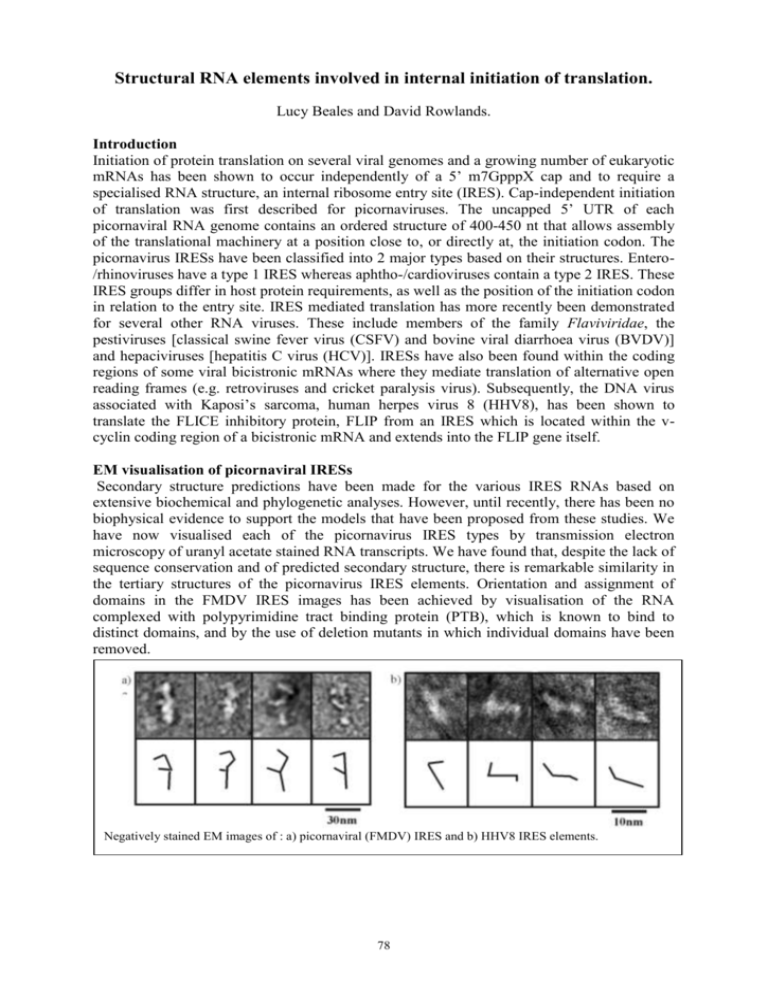
Structural RNA elements involved in internal initiation of translation. Lucy Beales and David Rowlands. Introduction Initiation of protein translation on several viral genomes and a growing number of eukaryotic mRNAs has been shown to occur independently of a 5’ m7GpppX cap and to require a specialised RNA structure, an internal ribosome entry site (IRES). Cap-independent initiation of translation was first described for picornaviruses. The uncapped 5’ UTR of each picornaviral RNA genome contains an ordered structure of 400-450 nt that allows assembly of the translational machinery at a position close to, or directly at, the initiation codon. The picornavirus IRESs have been classified into 2 major types based on their structures. Entero/rhinoviruses have a type 1 IRES whereas aphtho-/cardioviruses contain a type 2 IRES. These IRES groups differ in host protein requirements, as well as the position of the initiation codon in relation to the entry site. IRES mediated translation has more recently been demonstrated for several other RNA viruses. These include members of the family Flaviviridae, the pestiviruses [classical swine fever virus (CSFV) and bovine viral diarrhoea virus (BVDV)] and hepaciviruses [hepatitis C virus (HCV)]. IRESs have also been found within the coding regions of some viral bicistronic mRNAs where they mediate translation of alternative open reading frames (e.g. retroviruses and cricket paralysis virus). Subsequently, the DNA virus associated with Kaposi’s sarcoma, human herpes virus 8 (HHV8), has been shown to translate the FLICE inhibitory protein, FLIP from an IRES which is located within the vcyclin coding region of a bicistronic mRNA and extends into the FLIP gene itself. EM visualisation of picornaviral IRESs Secondary structure predictions have been made for the various IRES RNAs based on extensive biochemical and phylogenetic analyses. However, until recently, there has been no biophysical evidence to support the models that have been proposed from these studies. We have now visualised each of the picornavirus IRES types by transmission electron microscopy of uranyl acetate stained RNA transcripts. We have found that, despite the lack of sequence conservation and of predicted secondary structure, there is remarkable similarity in the tertiary structures of the picornavirus IRES elements. Orientation and assignment of domains in the FMDV IRES images has been achieved by visualisation of the RNA complexed with polypyrimidine tract binding protein (PTB), which is known to bind to distinct domains, and by the use of deletion mutants in which individual domains have been removed. a) a b) Negatively stained EM images of : a) picornaviral (FMDV) IRES and b) HHV8 IRES elements. 78 Other viral IRESs In contrast to the picornaviral IRESs, the HCV IRES is a relatively simple structure consisting of 2 major stems and a small spur. Colloidal gold labelling studies using biotinylated oligonucleotides have allowed assignment of the individual domains with regard to the proposed structure. The related viruses, CSFV and GBV-B have similar IRES structures to HCV and it is likely that they are assembled in the same way. The IRES found on one of the HHV8 mRNAs is the smallest IRES to be analysed by EM. In spite of the size difference, the overall appearance of this IRES seems to be similar to that of the flavivirus IRESs. Relating IRES structure to function The more elaborate nature of the picornavirus IRESs compared with the flavivirus IRESs may reflect the functional requirement for ancillary protein factors in the former. A major difference between these IRES groups is that picornavirus translation initiation requires the presence of the initiation factor eIF4G (or the virus protease cleaved form, p100) whereas translation from the HCV IRES is not dependent on this host factor. The smaller size and simplicity of the HHV8 IRES may reflect the fact that, unlike the picornavirus and flavivirus RNAs, the RNA in which it is located functions solely as a mRNA and is not involved in replication. There is, therefore, no requirement for the type of control elements found in positive sense RNA viruses that are thought to modulate the switch from translation to transcription during the growth cycle. In addition, the presence of coding sequence within the HHV8 IRES element may also constrain the size of this structure. References Beales, L. P., Rowlands, D. J. & Holzenburg, A. (2001) The internal ribosome entry site (IRES) of hepatitis C virus visualized by electron microscopy. RNA., 7, 661-670. Beales, L. P., Holzenburg, A. & Rowlands, D. J. (2002) Comparison of virally encoded internal ribosome entry sites by electron microscopy. RNA (submitted). Funding We thank the BBSRC for funding. 79


