Microscope lab
advertisement
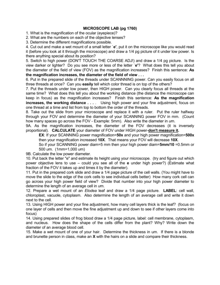
MICROSCOPE LAB (pg 1760) 1. What is the magnification of the ocular (eyepiece)? 2. What are the numbers on each of the objective lenses? 3. Determine the different magnifications possible. 4. Cut out and make a wet mount of a small letter `e', put it on the microscope like you would read it (before you look at it through the microscope) and draw a 1/4 pg picture of it under low power. Is there anything special about its position? 5. Switch to high power (DON'T TOUCH THE COARSE ADJ!) and draw a 1/4 pg picture. Is the view darker or lighter? Do you see more or less of the letter `e'? What does this tell you about the diameter of the field of view (FOV) as the magnification increases? Finish this sentence: As the magnification increases, the diameter of the field of view . . . 6. Put in the prepared slide of the threads under SCANNNING power. Can you easily focus on all three threads at once? Can you easily tell which color thread is on top of the others? 7. Put the threads under low power, then HIGH power. Can you clearly focus all threads at the same time? What does this tell you about the working distance (the distance the microscope can keep in focus) as the magnification increases? Finish this sentence: As the magnification increases, the working distance . . . . Using high power and your fine adjustment, focus on one thread at a time and list from top to bottom the order of the threads. 8. Take out the slide from your microscope and replace it with a ruler. Put the ruler halfway through your FOV and determine the diameter of your SCANNING power FOV in mm. (Count how many spaces go across the FOV - Example: 5mm). Also write the diameter in um. 9A. As the magnification increases, the diameter of the FOV decreases (it is inversely proportional). CALCULATE your diameter of FOV under HIGH power-don't measure it. EX: If your SCANNING power magnification=50x and your high power magnification=500x then your magnification increased 10X. That means your FOV will decrease 10X. So if your SCANNING power diam=5 mm then your high power diam=5mm/10 =0.5mm or 500 um. (1mm=1,000 um) 9B. Calculate the low power diameter. 10. Put back the letter "e" and estimate its height using your microscope. (try and figure out which power objective lens to use – could you see all of the e under high power?) (Estimate what fraction of the FOV it takes up and times it by the diameter). 11. Put in the prepared cork slide and draw a 1/4 page picture of the cell walls. (You might have to move the slide to the edge of the cork cells to see individual cells better) How many cork cell can go across your high power field of view? Divide that number into your high power diameter to determine the length of an average cell in um. 12. Prepare a wet mount of an Elodea leaf and draw a 1/4 page picture. LABEL: cell wall, chloroplast, vacuole, cytoplasm. Also determine the length of an average cell and write it down next to the cell. 13. Using HIGH power and your fine adjustment, how many cell layers thick is the leaf? (focus on one layer of cells and then move the fine adjustment up and down to see if other layers come into focus) 14. Using prepared slides of frog blood draw a 1/4 page picture, label: cell membrane, cytoplasm, and nucleus. How does the shape of the cells differ from the plant? Why? Write down the diameter of an average blood cell. 15. Make a wet mount of one of your hair. Determine the thickness in um. If there is a blonde and brunette person in class, make an X with the hairs on a slide and compare their thickness.
