Direct growth of graphene on hexagonal boron
advertisement
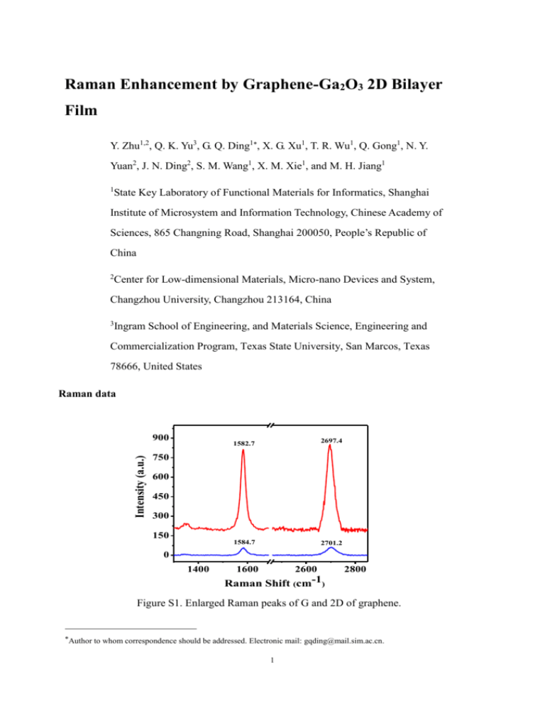
Raman Enhancement by Graphene-Ga2O3 2D Bilayer Film Y. Zhu1,2, Q. K. Yu3, G. Q. Ding1, X. G. Xu1, T. R. Wu1, Q. Gong1, N. Y. Yuan2, J. N. Ding2, S. M. Wang1, X. M. Xie1, and M. H. Jiang1 1 State Key Laboratory of Functional Materials for Informatics, Shanghai Institute of Microsystem and Information Technology, Chinese Academy of Sciences, 865 Changning Road, Shanghai 200050, People’s Republic of China 2 Center for Low-dimensional Materials, Micro-nano Devices and System, Changzhou University, Changzhou 213164, China 3 Ingram School of Engineering, and Materials Science, Engineering and Commercialization Program, Texas State University, San Marcos, Texas 78666, United States Raman data Intensity (a.u.) 900 1582.7 2697.4 1584.7 2701.2 750 600 450 300 150 0 1400 1600 2600 2800 Raman Shift (cm-1) Figure S1. Enlarged Raman peaks of G and 2D of graphene. Author to whom correspondence should be addressed. Electronic mail: gqding@mail.sim.ac.cn. 1 The graphene G-band downshifts from 1584.7 cm-1 to 1582.7 cm-1 when β-Ga2O3 sheet on the surface of graphene. At the same time, the 2D peak also has a downshift of 4 cm-1, 2 times of the G peak shift. 900 202 Intensity (a.u.) 750 600 450 300 417 171 146 653 348 474 319 630 769 150 0 200 400 600 800 Raman Shift (cm-1) Figure S2. Enlarged Raman peaks of β-G2O3. The Raman peaks range in 100-800 cm-1 can be assigned toβ-G2O3.Compared to the Raman peaks of G2O3 nanowires, the narrow full width at half maximum indicates the good crystalline fabricated G2O3 sheets on graphene. Table 1 shows the Raman peak positions from present work and the Ref. 14 and 17. It is clear that our samples can be well assigned to β-G2O3 since all the peaks can find its counterparts from bulk samples. It should be noted that 1-4 cm-1 upshift takes place for most peaks. This upshift may be induced by two effects: 1) the stain induced Raman shift. Ref. 17 has reported the Raman shift due to strains in the nanowires. Since Ga will expand during the 2 cooling while β-G2O3 will contract, the β-G2O3 sheet may undergo a compression stress at room temperature. 2) It has been confirmed by XPS that the O inβ-G2O3 has chemical bonding with C in grpahene, which affects the vibration of Ga-O inβ-G2O3. Table S1. The peak positions of G2O3 sheets and bulk G2O3 powders from Ref. 14 and 17. Bulk [14] 142 167 198 320 344 415 473 627 651 765 Bulk [17] 144 169 200 317 344 416 472 629 654 767 This work 146 171 202 319 348 417 474 630 653 769 C 1s Ga 2p Ga 2p 5 O KLL 1x10 (a) O 1s 5 Ga LMM Ga LMM 2x10 Ga 3d Ga 3p Ga 3s Counts / s XPS analysis 0 0 200 400 600 800 1000 1200 Binding Eenrgy(eV) Figure S3. the XPS survey spectrum of Ga2O3-graphene structure. The XPS spectra, scan range from 0 to 1200 eV, shows the peaks of the core levels of Ga2p, 3 Ga3s, Ga3p, Ga3d, Ga LMM peaks, as well as the O1s, OKLL and C1s. These peaks confirm the formation of graphene and Ga2O3. The growth mode Graphene (a) Ga Quartz Graphene (b) Ga2O3 Ga Quartz Figure S4. Schematic illustration of feasible growth mechanism (a) continuous graphene film covers the liquid Ga surface (d) separated Ga2O3 sheets deposited on graphene during the sample cooling process. 4
