objective
advertisement
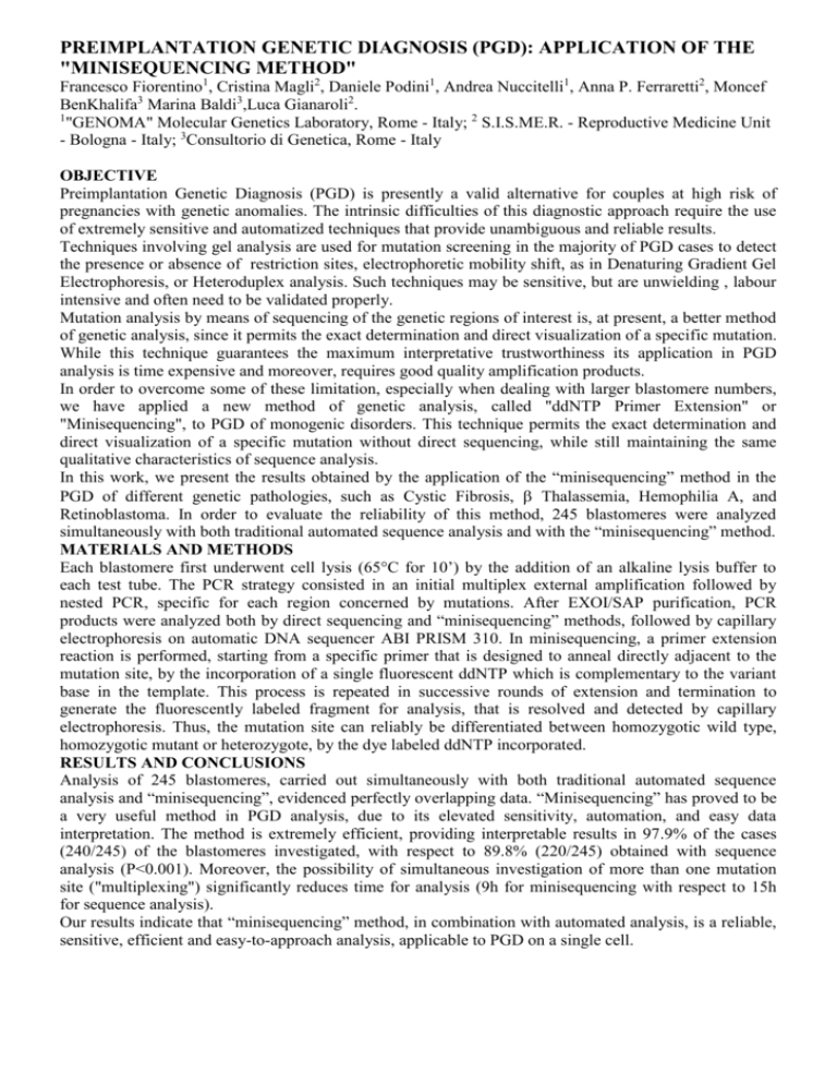
PREIMPLANTATION GENETIC DIAGNOSIS (PGD): APPLICATION OF THE "MINISEQUENCING METHOD" Francesco Fiorentino1, Cristina Magli2, Daniele Podini1, Andrea Nuccitelli1, Anna P. Ferraretti2, Moncef BenKhalifa3 Marina Baldi3,Luca Gianaroli2. 1 "GENOMA" Molecular Genetics Laboratory, Rome - Italy; 2 S.I.S.ME.R. - Reproductive Medicine Unit - Bologna - Italy; 3Consultorio di Genetica, Rome - Italy OBJECTIVE Preimplantation Genetic Diagnosis (PGD) is presently a valid alternative for couples at high risk of pregnancies with genetic anomalies. The intrinsic difficulties of this diagnostic approach require the use of extremely sensitive and automatized techniques that provide unambiguous and reliable results. Techniques involving gel analysis are used for mutation screening in the majority of PGD cases to detect the presence or absence of restriction sites, electrophoretic mobility shift, as in Denaturing Gradient Gel Electrophoresis, or Heteroduplex analysis. Such techniques may be sensitive, but are unwielding , labour intensive and often need to be validated properly. Mutation analysis by means of sequencing of the genetic regions of interest is, at present, a better method of genetic analysis, since it permits the exact determination and direct visualization of a specific mutation. While this technique guarantees the maximum interpretative trustworthiness its application in PGD analysis is time expensive and moreover, requires good quality amplification products. In order to overcome some of these limitation, especially when dealing with larger blastomere numbers, we have applied a new method of genetic analysis, called "ddNTP Primer Extension" or "Minisequencing", to PGD of monogenic disorders. This technique permits the exact determination and direct visualization of a specific mutation without direct sequencing, while still maintaining the same qualitative characteristics of sequence analysis. In this work, we present the results obtained by the application of the “minisequencing” method in the PGD of different genetic pathologies, such as Cystic Fibrosis, Thalassemia, Hemophilia A, and Retinoblastoma. In order to evaluate the reliability of this method, 245 blastomeres were analyzed simultaneously with both traditional automated sequence analysis and with the “minisequencing” method. MATERIALS AND METHODS Each blastomere first underwent cell lysis (65°C for 10’) by the addition of an alkaline lysis buffer to each test tube. The PCR strategy consisted in an initial multiplex external amplification followed by nested PCR, specific for each region concerned by mutations. After EXOI/SAP purification, PCR products were analyzed both by direct sequencing and “minisequencing” methods, followed by capillary electrophoresis on automatic DNA sequencer ABI PRISM 310. In minisequencing, a primer extension reaction is performed, starting from a specific primer that is designed to anneal directly adjacent to the mutation site, by the incorporation of a single fluorescent ddNTP which is complementary to the variant base in the template. This process is repeated in successive rounds of extension and termination to generate the fluorescently labeled fragment for analysis, that is resolved and detected by capillary electrophoresis. Thus, the mutation site can reliably be differentiated between homozygotic wild type, homozygotic mutant or heterozygote, by the dye labeled ddNTP incorporated. RESULTS AND CONCLUSIONS Analysis of 245 blastomeres, carried out simultaneously with both traditional automated sequence analysis and “minisequencing”, evidenced perfectly overlapping data. “Minisequencing” has proved to be a very useful method in PGD analysis, due to its elevated sensitivity, automation, and easy data interpretation. The method is extremely efficient, providing interpretable results in 97.9% of the cases (240/245) of the blastomeres investigated, with respect to 89.8% (220/245) obtained with sequence analysis (P<0.001). Moreover, the possibility of simultaneous investigation of more than one mutation site ("multiplexing") significantly reduces time for analysis (9h for minisequencing with respect to 15h for sequence analysis). Our results indicate that “minisequencing” method, in combination with automated analysis, is a reliable, sensitive, efficient and easy-to-approach analysis, applicable to PGD on a single cell.
