Hello Ashraf:
advertisement
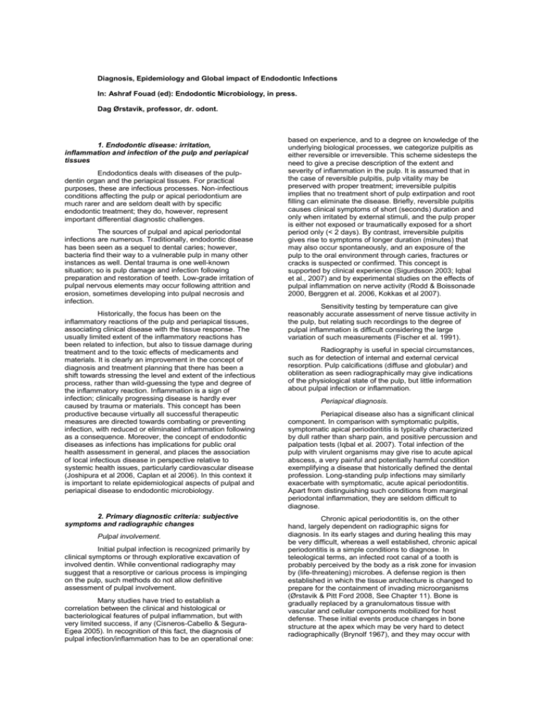
Diagnosis, Epidemiology and Global impact of Endodontic Infections In: Ashraf Fouad (ed): Endodontic Microbiology, in press. Dag Ørstavik, professor, dr. odont. 1. Endodontic disease: irritation, inflammation and infection of the pulp and periapical tissues Endodontics deals with diseases of the pulpdentin organ and the periapical tissues. For practical purposes, these are infectious processes. Non-infectious conditions affecting the pulp or apical periodontium are much rarer and are seldom dealt with by specific endodontic treatment; they do, however, represent important differential diagnostic challenges. The sources of pulpal and apical periodontal infections are numerous. Traditionally, endodontic disease has been seen as a sequel to dental caries; however, bacteria find their way to a vulnerable pulp in many other instances as well. Dental trauma is one well-known situation; so is pulp damage and infection following preparation and restoration of teeth. Low-grade irritation of pulpal nervous elements may occur following attrition and erosion, sometimes developing into pulpal necrosis and infection. Historically, the focus has been on the inflammatory reactions of the pulp and periapical tissues, associating clinical disease with the tissue response. The usually limited extent of the inflammatory reactions has been related to infection, but also to tissue damage during treatment and to the toxic effects of medicaments and materials. It is clearly an improvement in the concept of diagnosis and treatment planning that there has been a shift towards stressing the level and extent of the infectious process, rather than wild-guessing the type and degree of the inflammatory reaction. Inflammation is a sign of infection; clinically progressing disease is hardly ever caused by trauma or materials. This concept has been productive because virtually all successful therapeutic measures are directed towards combating or preventing infection, with reduced or eliminated inflammation following as a consequence. Moreover, the concept of endodontic diseases as infections has implications for public oral health assessment in general, and places the association of local infectious disease in perspective relative to systemic health issues, particularly cardiovascular disease (Joshipura et al 2006, Caplan et al 2006). In this context it is important to relate epidemiological aspects of pulpal and periapical disease to endodontic microbiology. 2. Primary diagnostic criteria: subjective symptoms and radiographic changes Pulpal involvement. Initial pulpal infection is recognized primarily by clinical symptoms or through explorative excavation of involved dentin. While conventional radiography may suggest that a resorptive or carious process is impinging on the pulp, such methods do not allow definitive assessment of pulpal involvement. Many studies have tried to establish a correlation between the clinical and histological or bacteriological features of pulpal inflammation, but with very limited success, if any (Cisneros-Cabello & SeguraEgea 2005). In recognition of this fact, the diagnosis of pulpal infection/inflammation has to be an operational one: based on experience, and to a degree on knowledge of the underlying biological processes, we categorize pulpitis as either reversible or irreversible. This scheme sidesteps the need to give a precise description of the extent and severity of inflammation in the pulp. It is assumed that in the case of reversible pulpitis, pulp vitality may be preserved with proper treatment; irreversible pulpitis implies that no treatment short of pulp extirpation and root filling can eliminate the disease. Briefly, reversible pulpitis causes clinical symptoms of short (seconds) duration and only when irritated by external stimuli, and the pulp proper is either not exposed or traumatically exposed for a short period only (< 2 days). By contrast, irreversible pulpitis gives rise to symptoms of longer duration (minutes) that may also occur spontaneously, and an exposure of the pulp to the oral environment through caries, fractures or cracks is suspected or confirmed. This concept is supported by clinical experience (Sigurdsson 2003; Iqbal et al., 2007) and by experimental studies on the effects of pulpal inflammation on nerve activity (Rodd & Boissonade 2000, Berggren et al. 2006, Kokkas et al 2007). Sensitivity testing by temperature can give reasonably accurate assessment of nerve tissue activity in the pulp, but relating such recordings to the degree of pulpal inflammation is difficult considering the large variation of such measurements (Fischer et al. 1991). Radiography is useful in special circumstances, such as for detection of internal and external cervical resorption. Pulp calcifications (diffuse and globular) and obliteration as seen radiographically may give indications of the physiological state of the pulp, but little information about pulpal infection or inflammation. Periapical diagnosis. Periapical disease also has a significant clinical component. In comparison with symptomatic pulpitis, symptomatic apical periodontitis is typically characterized by dull rather than sharp pain, and positive percussion and palpation tests (Iqbal et al. 2007). Total infection of the pulp with virulent organisms may give rise to acute apical abscess, a very painful and potentially harmful condition exemplifying a disease that historically defined the dental profession. Long-standing pulp infections may similarly exacerbate with symptomatic, acute apical periodontitis. Apart from distinguishing such conditions from marginal periodontal inflammation, they are seldom difficult to diagnose. Chronic apical periodontitis is, on the other hand, largely dependent on radiographic signs for diagnosis. In its early stages and during healing this may be very difficult, whereas a well established, chronic apical periodontitis is a simple conditions to diagnose. In teleological terms, an infected root canal of a tooth is probably perceived by the body as a risk zone for invasion by (life-threatening) microbes. A defense region is then established in which the tissue architecture is changed to prepare for the containment of invading microorganisms (Ørstavik & Pitt Ford 2008, See Chapter 11). Bone is gradually replaced by a granulomatous tissue with vascular and cellular components mobilized for host defense. These initial events produce changes in bone structure at the apex which may be very hard to detect radiographically (Brynolf 1967), and they may occur with teeth that still has vitality or at least nervous activity in the pulp (Fig. 1). A B periodontitis dominate as sources for acute dental pain in children and adults (Zeng et al 1994, Lygidakis et at 1998) which may be debilitating to the patient and lead to absence from work and involvement of costly health services. While we know that emergency dental services are in great demand in most countries, in urban as well as rural areas, there is very scant information on the actual incidence and prevalence of acute pulpal and apical periodontal disease. Therefore, one can only speculate that there is still, even in communities with well-developed dental services, a significant impact on the general wellbeing by acute pulpal and periodontal conditions (SindetPedersen et al 1985, Richardsson 2005). A frequently overlooked situation is the association of pulpal and apical disease with tooth loss in the elderly. Whereas marginal periodontal disease is generally accepted as a significant cause of tooth loss, pulpal and apical diseases are important causes for extraction (Eckerbom et al 1992) and may dominate after the age of approximately 50 years (Eriksen et al 1991). Fig. 1. Minimal bone structural changes at the apex in conjunction with chronic pulpitis (A), necessitating endodontic treatment (B). When periapical tissue remodeling has reached a state of complete granulomatous transformation, the lesion is very characteristic and easily diagnosed in the radiograph (Fig. 2). Treatment decision is then easy if the tooth does not respond to sensitivity testing. On the other hand, there may be total pulp necrosis and no infection or inflammation at the apex, such as when the pulp is devitalized by traumatic injury (Sundqvist 1976). The tooth with pulpitis is obviously in danger of becoming infected and developing apical periodontitis. Correct and prompt treatment of the acute situation is therefore important not only to curb the pain and to reestablish a functional tooth, but also to reduce or eliminate the risk for the insidious spreading of the infection and the emergence of a periapical lesion. It has been known for a very long time that the prognosis for treatment of apical periodontitis is much poorer than expected treatment outcome after vital pulpectomy (See Chapter 17). Early detection and root canal treatment of teeth at definitive risk of developing root canal infection is therefore essential. Failure to provide adequate treatment early will facilitate the development of an infection (Fig. 3), which would reduce the prognosis. Fig.3. Chronic apical periodontitis developing in 6 months after inadequate emergency treatment. The prognosis is reduced from >95% to <85%. Fig. 2. Chronic apical periodontitis: incipient at mesial root, established at distal root of mandibular left first molar. 3. Pulpal inflammation vs infection: public health consequences The clinical aspects of endodontic diseases may be serious and with some consequences for individual and public health. Symptomatic, acute pulpitis and apical 4. Epidemiology of pulpal inflammation Information in the literature about the incidence of dental and oral pain is scarce in itself (Lipton et al.1993, Pau et al 2003), and the separation of the pulpal or periapical component from the inclusive diagnosis is difficult if at all possible. What little there is of targeted epidemiological data, point to a limited, but significant occurrence of acute pain of pulpal origin (Sindet-Pedersen et al 1985, Zeng et al 1994, Lygidakis et at 1998). This is an area in need of continued and extensive research. The incidence and prevalence of symptomatic pulpitis and apical periodontitis are obviously important for the targeting of dental services, and forms important background knowledge for the design of dental curricula and for public health measures. 5. Criteria for assessing periapical health: presence/absence, PAI score Asymptomatic, chronic apical periodontitis poses a different challenge from pulpitis and symptomatic, acute apical periodontitis. The insidious nature and frequently pain-free course of this disease makes it evasive to detection outside of the dental treatment situation. The fact that asymptomatic, chronic apical periodontitis relies on radiography for detection poses limitations on the possibilities for screenings and population surveys. Moreover, when radiographic data have been made available for analysis, lack of standardization in scoring makes comparisons across studies difficult. The radiographic technique may also influence the ability to detect with certainty the occurrence of asymptomatic, chronic apical periodontitis. For population surveys, panoramic radiography provides information at far less radiation dosage than full-mouth periapical examinations, but the possibilities of detection of apical lesions may be diminished. Comparisons of panoramic and periapical films for diagnosis of apical periodontitis suggest that there is some, but not a dramatic, reduction in the detectability of periapical lesions (Sameshima & Asgarifar 2001, Ridao-Sacie et al 2007). Newer methods, such as tomography (Tammisalo et al. 1996) and computed tomography (Huumonen et al. 2006) and conebeam tomography (Lofthag-Hansen et al. 2007, Patel et al.2007, Estrela et al. 2008), are more sensitive and probably more specific than periapical radiographs, but the radiation dose strongly limits their application for use in surveys. Radiographic characteristics of chronic apical periodontitis Verbal descriptors of the radiographic characteristics of asymptomatic, chronic apical periodontitis have included a widened periodontal space; interruptions of the lamina dura, and/or the presence of a radiolucent area at the site of exit of the pulp to the periodontal membrane (Ørstavik & Larheim 2008). Only when there is an overt radiolucency associated with the root tip and a concomitant finding of a necrotic pulp, are the signs pathognomonic (Ørstavik & Larheim 2008). While it is possible to make assumptions from different studies with similar descriptions of the criteria used for detection of asymptomatic, chronic apical periodontitis, it is not possible to draw conclusions with any certainty. The periapical index The periapical index was developed with the aim of overcoming this difficulty (Fig. 4). Fig. 4. The periapical index. The periapical condition is scored by comparison with a series of reference radiographs of teeth with known histology. Reproduced with permission from Ørstavik et al (1986). It makes use of an ordinal scale with 5 steps indicating increasing severity of apical periodontitis (Ørstavik et al 1986). The steps are represented by radiographs that have histological verification from an extensive study on human cadavers (Brynolf 1967). This makes possible a visual reference scale that reduces the risk of personal bias otherwise associated with subjective radiographic assessments. Also the system is used after extensive and standardized calibration of the observers, which facilitates comparisons of different studies and pooling of data. While developed for clinical, follow-up studies of endodontic treatment in prospective studies, the PAI scoring system is easily modified for use in epidemiological surveys (Eriksen 1991). A general principle in epidemiology is to avoid scoring a healthy condition wrongly as disease. This is accomplished by restricting the categorization as “diseased” (i.e., with apical periodontitis) to teeth with scores 3-5 (Fig. 5). In this way, some cases of asymptomatic, chronic apical periodontitis will go undetected, but only a minimal number of healthy teeth will be scored as diseased. 100 Scored as healthy Scored as diseased 80 60 40 20 0 1 2 Healthy 3 4 5 Diseased Fig. 5. Dichotomization of PAI scores applied to epidemiology. Blue line, teeth without apical periodontitis; red line, teeth with apical periodontitis. A minimum of false positives (healthy apical periodontium scored as diseased; blue cases in red sector) is acceptable at the expense of some false negatives (diseased teeth registered as healthy; red cases in blue sector) Irrespective of the radiographic method of detection, it is apparent that radiographic assessments of apical periodontitis on the whole will underestimate its true incidence or prevalence (Brynolf 1967). Even with all these provisos, it may still be prudent to review and compare results from different areas and cohorts, as long as the shortcomings of the radiographic methods are kept in mind. 6. Results of epidemiological surveys of asymptomatic, chronic apical periodontitis When periapical disease was seen only as an extension of caries, epidemiological studies paid little if any attention to the incidence and prevalence of apical periodontitis. Based on numerous institutional studies on the outcome of endodontic treatment, the notion that endodontic treatment was predictable and generally successful was accepted (Strindberg 1956, Grossman 1964, Kerekes & Tronstad 1979, Ørstavik et al 1987), and the extent and importance of apical periodontitis in the general population was largely overlooked. In a series of studies, Eriksen and co-workers (Eriksen 1991, Eriksen et al 1991, 1995, Marques et al 1998, Sidaravicius et al 1999, Aleksejuniene et al 2000, Skudyte-Rysstad et al 2006) examined the general prevalence of apical periodontitis and placed in its proper perspective. The PAI scoring system was used together with simple criteria for the assessment of root filling quality. A primary aim was to reassess the association of the quality of the root filling as seen on the radiograph with the periapical status of the teeth. Similar to what had been documented in the institutional, follow-up studies, there was a clear association between poor root filling quality and the presence of apical periodontitis, emphasizing the need for focus on high-quality technical performance during the endodontic procedures. However, there was also an unexpectedly high prevalence of apical periodontitis in most populations and age groups. This was a source of concern and had to be considered in oral health assessments in general. Moreover, the finding that pulpal and periapical disease were a major reason for extractions in adults, surpassing marginal periodontitis around the 5th decade of life, emphasized the impact of periapical health for retention of the dentition into old age (Eriksen et al 1991, Eckerbom et al 1988). These studies have later been supplemented by several others from almost all corners of the globe, and with few exceptions, the results are quite disheartening in different countries and populations, regardless of the degree and perceived quality of the dental services offered. Many studies have made use of the PAI scoring system; others rely on a simple assessment on the presence or absence of a radiolucent area indicating periodontitis. Fig. 6 shows the prevalence of apical periodontitis in populations from 19 different populations in different countries. Apical periodontitis occurs with a prevalence of 30 to 80 percent in different populations, generally increasing in older age groups (Chen et al 2007) and in populations at high risk of infectious disease (diabetes) (Britto et al 2003). 100 Prevalence, per cent r s 80 l 60 n o p q j k 40 20 0 Fig. 6. The prevalence of apical periodontitis in different populations. a, Dugas et al 2003; b, Marques et al 1998; c, Frisk & Hakeberg 2005; d, Loftus et al 2005; e, Buckley & Spangberg 1995; f, DeCleen et al 1993; g, Eriksen et al 1991; h, Dugas et al 2003; i, Kirkevang et al 1991; j, Frisk & Hakeberg 2005; k, Chen et al 2007; l, JiménezPinzón et al 2004; n, De Moor et al 2000; o, Saunders et al 1997; p, Sidaravicius et al 1999; q, Tsuneishi et al 2005; r, Kabak & Abbott 2005; s, Segura-Egea et al 2005. Figures produced by this kind of surveys generally do not account for alternative ways of dealing with apical periodontitis in different environments. It is tempting to speculate that populations with low prevalence have had teeth with apical periodontitis extracted: indeed, for the Portuguese population studied by Marques et al (1998) which showed the lowest prevalence, it was found that they had a lower mean number of remaining teeth than a comparable Norwegian population with higher prevalence of apical periodontitis (Eriksen et al 1991). 6. Quality of root canal treatment and the development and persistence of apical periodontitis Institutional follow-up studies and epidemiological surveys have all documented that there is a very clear correlation between presence of apical periodontitis and inadequate technical quality of the root filling as it appears in the radiograph. The association is strongest for teeth that are diagnosed with apical periodontitis at the start of treatment, and far less dominant when the root filling is placed in teeth with no lesion prior to treatment (Sjögren et al 1990). In the latter situation, typically less than 10 per cent of treated cases develop apical periodontitis; contrarily, teeth treated for primary apical periodontitis show persistence of lesions in 20-25% in institutional studies. In all likelihood, there is a poorer outcome for both preoperative diagnoses in practice compared to the institutional setting. By inference, when epidemiological surveys indicate that 30-40% of root filled teeth have apical periodontitis, it seems fair to assume that less than 50 per cent of teeth with apical periodontitis are cured in the average treatment setting in practice. This should not be placed in a context to advocate more radical treatment or prophylaxis of apical periodontitis. The preservation of teeth by endodontic procedures is, after all, a clinically very successful and predictable procedure. The sequels to extractions and various prosthetic procedures, as alternative treatments, are numerous and often of greater consequence. However, these epidemiological findings clearly point to a need for improvements in the quality of endodontic care. 7. General oral health, oral health strategies, and tooth preservation as risk factors for oral infections The infectious aspects of endodontic diseases pose the question if the nature of the organisms and their activities in the pulp space and beyond are a source of concern for the general health of the individual. Marginal periodontitis seems to have a definitive, albeit limited association with cardiovascular disease, and there is data emerging in the literature indicating that this is the case also for apical periodontitis (Caplan et al 2006, Olsen et al 2007); however, others have failed to establish such an association (Frisk et al. 2003). The concept of endodontic diseases primarily as infections with the potential to spread and thereby to affect organs at distant sites may be important for patients’ systemic health, particularly the risk of cardiovascular events (Ørstavik & Pitt Ford 2008, see also Chapter 16). On the one hand, this affects the decision whether to provide antibiotic coverage prior to surgery in patients at risk of infective endocarditis or infection of vascular implants; on the other, the possible association of pulp and periapical infection with the risk of developing cardiovascular disease has a major impact on the rationale and case selection for endodontic treatment, and especially on prophylactic efforts to prevent pulpal infection in the first place. The notion that granulomas may be “sterile” or caused by medicaments or materials has been abandoned. Periapical lesions are virtually all apical periodontitis, and apical periodontitis is caused by microbial infection of the root canal system. Imminent or established infections of the pulp and periapical tissues need to be contained or eliminated. Early and appropriate endodontic intervention is necessary in such cases, with emphasis on proper case selection and highly skilled technical performance of treatment. The provision of high-quality endodontic care at all levels of dental service to the individual patient as well as to populations is therefore crucial for optimum, longterm oral health. The goals for these services are several: to prevent pulpal infection by effective caries prevention, by protection against dental trauma, and by appropriate dentin treatment under restorations; to limit pulpal pain as a source of discomfort and loss of work; and to eliminate dental infection and prevent its recurrence by root filling and surgical endodontic procedures. References Aleksejuniene J, Eriksen HM, Sidaravicius B, Haapasalo M. Apical periodontitis and related factors in an adult Lithuanian population. Oral Surg Oral Med Oral Pathol Oral Radiol Endod. 2000 Jul;90(1):95-101. Bletsa A, Berggreen E, Fristad I, Tenstad O, Wiig H. Cytokine signalling in rat pulp interstitial fluid and transcapillary fluid exchange during lipopolysaccharideinduced acute inflammation. J Physiol. 2006 May 15;573(Pt 1):225-36. Epub 2006 Mar 9. Britto LR, Katz J, Guelmann M, Heft M. Periradicular radiographic assessment in diabetic and control individuals. Oral Surg Oral Med Oral Pathol Oral Radiol Endod. 2003 Oct;96(4):449-52. Brynolf L. Histological and roentgenological study of periapical region of human upper incisors. Odontologisk Revy 1967;18: suppl.11. Buckley M, Spangberg LS. The prevalence and technical quality of endodontic treatment in an American subpopulation. Oral Surg Oral Med Oral Pathol Oral Radiol Endod. 1995 Jan;79(1):92-100. Caplan DJ, Chasen JB, Krall EA, Cai J, Kang S, Garcia RI, Offenbacher S, Beck JD. Lesions of endodontic origin and risk of coronary heart disease. J Dent Res. 2006 Nov;85(11):996-1000. Chen CY, Hasselgren G, Serman N, Elkind MS, Desvarieux M, Engebretson SP. Prevalence and quality of endodontic treatment in the Northern Manhattan elderly. J Endod. 2007 Mar;33(3):230-4. Cisneros-Cabello R, Segura-Egea JJ. Relationship of patient complaints and signs to histopathologic diagnosis of pulpal condition. Aust Endod J. 2005 Apr;31(1):24-7. Cisneros-Cabello R, Segura-Egea JJ. Relationship of patient complaints and signs to histopathologic diagnosis of pulpal condition. Aust Endod J. 2005 Apr;31(1):24-7. Csillag M, Berggreen E, Fristad I, Haug SR, Bletsa A, Heyeraas KJ. Effect of electrical tooth stimulation on blood flow and immunocompetent cells in rat dental pulp after sympathectomy. Acta Odontol Scand. 2004 Dec;62(6):305-12. De Cleen MJ, Schuurs AH, Wesselink PR, Wu MK. Periapical status and prevalence of endodontic treatment in an adult Dutch population. Int Endod J. 1993 Mar;26(2):112-9. De Moor RJ, Hommez GM, De Boever JG, Delme KI, Martens GE. Periapical health related to the quality of root canal treatment in a Belgian population. Int Endod J. 2000 Mar;33(2):113-20. Dugas NN, Lawrence HP, Teplitsky PE, Pharoah MJ, Friedman S. Periapical health and treatment quality assessment of root-filled teeth in two Canadian populations. Int Endod J. 2003 Mar;36(3):181-92. Eckerbom M, Magnusson T, Martinsson T. Reasons for and incidence of tooth mortality in a Swedish population. Endod Dent Traumatol. 1992 Dec;8(6):230-4. Eriksen HM, Berset GP, Hansen BF, Bjertness E. Changes in endodontic status 1973-1993 among 35-year-olds in Oslo, Norway. Int Endod J. 1995 May;28(3):129-32 Eriksen HM, Bjertness E. Prevalence of apical periodontitis and results of endodontic treatment in middle-aged adults in Norway. Endod Dent Traumatol. 1991 Feb;7(1):1-4 Eriksen HM. Endodontology--epidemiologic considerations. Endod Dent Traumatol. 1991 Oct;7(5):189-95 Estrela C, Bueno MR, Leles CR, Azevedo B, Azevedo JR. Accuracy of cone beam computed tomography and panoramic and periapical radiography for detection of apical periodontitis. J Endod. 2008 Mar;34(3):273-9. Fischer C, Wennberg A, Fischer RG, Attström R.Clinical evaluation of pulp and dentine sensitivity after supragingival and subgingival scaling.Endod Dent Traumatol. 1991 Dec;7(6):259-65 Frisk F, Hakeberg M. A 24-year follow-up of root filled teeth and periapical health amongst middle aged and elderly women in Goteborg, Sweden. Int Endod J. 2005 Apr;38(4):246-54. Fristad I, Berggreen E, Haug SR. Delta (delta) opioid receptors in small and medium-sized trigeminal neurons supporting the dental pulp of rats. Arch Oral Biol. 2006 Apr;51(4):273-81. Epub 2005 Nov 2. Grossman LI, Shepard LI, Pearson LA. Roentgenologic and clinical evaluation of endodontically treated teeth. Oral Surgery, Oral Medicine, Oral Pathology 1964;17:368-374. Huumonen S, Kvist T, Grondahl K, Molander A. Diagnostic value of computed tomography in re-treatment of root fillings in maxillary molars. Int Endod J. 2006 Oct;39(10):827-33. Iqbal M, Kim S, Yoon F. An investigation into differential diagnosis of pulp and periapical pain: a PennEndo database study. J Endod. 2007 May;33(5):548-51. Epub 2007 Mar 21. Jiménez-Pinzón A, Segura-Egea JJ, Poyato-Ferrera M, Velasco-Ortega E, Ríos-Santos JV. Prevalence of apical periodontitis and frequency of root-filled teeth in an adult Spanish population. Int Endod J. 2004 Mar;37(3):16773. Joshipura KJ, Pitiphat W, Hung HC, Willett WC, Colditz GA, Douglass CW. Pulpal inflammation and incidence of coronary heart disease. J Endod. 2006 Feb;32(2):99103. Kabak Y, Abbott PV. Prevalence of apical periodontitis and the quality of endodontic treatment in an adult Belarusian population. Int Endod J. 2005 Apr;38(4):23845. Kerekes K, Tronstad L. Long-term results of endodontic treatment performed with a standardized technique.J Endod. 1979 Mar;5(3):83-90. Kirkevang LL, Horsted-Bindslev P, Ørstavik D, Wenzel A. Frequency and distribution of endodontically treated teeth and apical periodontitis in an urban Danish population. Int Endod J. 2001 Apr;34(3):198-205. Kokkas AB, Goulas A, Varsamidis K, Mirtsou V, Tziafas D. Irreversible but not reversible pulpitis is associated with up-regulation of tumour necrosis factor-alpha gene expression in human pulp. Int Endod J. 2007 Mar;40(3):198-203. Lipton JA, Ship JA, Larach-Robinson D. Estimated prevalence and distribution of reported orofacial pain in the United States. J Am Dent Assoc. 1993 Oct;124(10):115-21. Locker D, Grushka M. Prevalence of oral and facial pain and discomfort: preliminary results of a mail survey. Community Dent Oral Epidemiol. 1987 Jun;15(3):16972. Lofthag-Hansen S, Huumonen S, Grondahl K, Grondahl HG. Limited cone-beam CT and intraoral radiography for the diagnosis of periapical pathology. Oral Surg Oral Med Oral Pathol Oral Radiol Endod. 2007 Jan;103(1):114-9. Epub 2006 Apr 24. Loftus JJ, Keating AP, McCartan BE. Periapical status and quality of endodontic treatment in an adult Irish population. Int Endod J. 2005 Feb;38(2):81-6. Lygidakis NA, Marinou D, Katsaris N. Analysis of dental emergencies presenting to a community paediatric dentistry centre. Int J Paediatr Dent. 1998 Sep;8(3):18190. Marques MD, Moreira B, Eriksen HM. Prevalence of apical periodontitis and results of endodontic treatment in an adult, Portuguese population. Int Endod J. 1998 May;31(3):161-5. Olsen I, Nafstad P, Schwarze P, Rønningen KS, Håheim LL. Tooth Extractions, Bacterial Antibodies, C-reactive Protein and Myocardial Infarction Abstract # 2291, 85th General Session & Exhibition of the IADR, New Orleans, Louisiana. Ørstavik D, Kerekes K, Eriksen HM. Clinical performance of three endodontic sealers. Endod Dent Traumatol. 1987;3,178-186. Ørstavik D, Kerekes K, Eriksen HM. The periapical index: A scoring system for radiographic assessment of apical periodontitis. Endod Dent Traumatol. 1986;2,20-34. Ørstavik D, Larheim TA. Radiology of Apical Periodontitis. In: Ørstavik D, Pitt Ford TR. (eds.) Essential Endodontology: Prevention and Treatment of Apical Periodontitis. Blackwell, Second Edition, 2008 Ørstavik D, Pitt Ford TR. Apical Periodontitis: Microbial Infection and Host Responses. In: Ørstavik D, Pitt Ford TR. (eds.) Essential Endodontology: Prevention and Treatment of Apical Periodontitis. Blackwell, Second Edition, in press2008 Patel S, Dawood A, Ford TP, Whaites E. The potential applications of cone beam computed tomography in the management of endodontic problems. Int Endod J. 2007 Oct;40(10):818-30. Pau AK, Croucher R, Marcenes W. Prevalence estimates and associated factors for dental pain: a review. Oral Health Prev Dent. 2003;1(3):209-20. Richardsson PS. Dental morbidity in United Kingdom Armed Forces, Iraq 2003. Mil Med 2005 Jun;170(6):53641. Ridao-Sacie C, Segura-Egea JJ, Fernández-Palacín A, Bullón-Fernández P, Ríos-Santos JV. Radiological assessment of periapical status using the periapical index: comparison of periapical radiography and digital panoramic radiography. Int Endod J. 2007 Jun;40(6):433-40. Epub 2007 Apr 19. Sameshima & Asgarifar 2001 Rodd HD, Boissonade FM. Substance P expression in human tooth pulp in relation to caries and pain experience. Eur J Oral Sci. 2000 Dec;108(6):467-74. Sameshima GT Asgarifar KO. Assessment of root resorption and root shape: periapical vs panoramic films. Angle Orthod. 2001 Jun;71(3):185-9. 2 Saunders WP, Saunders EM, Sadiq J, Cruickshank E. Technical standard of root canal treatment in an adult Scottish sub-population. Br Dent J 1997;182:382-386. Segura-Egea JJ, Jiménez-Pinzón A, Ríos-Santos JV, Velasco-Ortega E, Cisneros-Cabello R, Poyato-Ferrera M. High prevalence of apical periodontitis amongst type 2 diabetic patients. Int Endod J. 2005 Aug;38(8):564-9. Sidaravicius B, Aleksejuniene J, Eriksen HM. Endodontic treatment and prevalence of apical periodontitis in an adult population of Vilnius, Lithuania. Endod Dent Traumatol. 1999 Oct;15(5):210-5. Sigurdsson A. Pulpal diagnosis. Endodontic Topics 2003;5:12-23 Sindet-Pedersen S, Petersen JK, Götzsche PC. Incidence of pain conditions in dental practice in a Danish county. Community Dent Oral Epidemiol. 1985 Aug;13(4):244-6. Sjögren U, Hägglund B, Sundqvist G, Wing K. Factors affecting the long-term results of endodontic treatment.J Endod. 1990 Oct;16(10):498-504. Skudutyte-Rysstad R, Eriksen HM. Endodontic status amongst 35-year-old Oslo citizens and changes over a 30-year period. Int Endod J. 2006 Aug;39(8):637-42. Strindberg LZ. The dependence of the results of pulp therapy on certain factors. An analytic study based on radiographic and clinical follow-up examination. Acta Odontologica Scandinavica 1956;14; suppl. 21. Sundqvist G. Bacteriological studies of necrotic dental pulps. Umeå, Sweden: Umeå University Odontological Dissertations, 1976. Tammisalo T, Luostarinen T, Vahatalo K, Neva M. Detailed tomography of periapical and periodontal lesions. Diagnostic accuracy compared with periapical radiography. Dentomaxillofac Radiol. 1996 Apr;25(2):89-96. Tsuneishi M, Yamamoto T, Yamanaka R, Tamaki N, Sakamoto T, Tsuji K, Watanabe T. Radiographic evaluation of periapical status and prevalence of endodontic treatment in an adult Japanese population. Oral Surg Oral Med Oral Pathol Oral Radiol Endod. 2005 Nov;100(5):631-5. Zeng Y, Sheller B, Milgrom P. Epidemiology of dental emergency visits to an urban children's hospital. Pediatr Dent. 1994 Nov-Dec;16(6):419-23.
