BBSI Final Report - Virginia Commonwealth University
advertisement
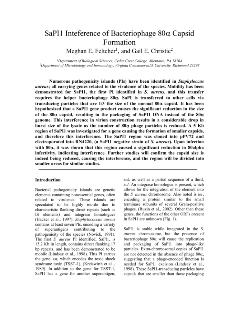
SaPI1 Inteference of Bacteriophage 80 Capsid Formation Meghan E. Feltcher1, and Gail E. Christie2 1 Department of Biological Sciences, Cedar Crest College, Allentown, PA 18104 Department of Microbiology and Immunology, Virginia Commonwealth University, Richmond 23298 2 Numerous pathogenicity islands (PIs) have been identified in Staphyloccus aureus; all carrying genes related to the virulence of the species. Mobility has been demonstrated for SaPI1, the first PI identified in S. aureus, and this transfer requires the helper bacteriophage 80. SaPI is transferred to other cells via transducing particles that are 1/3 the size of the normal 80 capsid. It has been hypothesized that a SaPI1 gene product causes the significant reduction in the size of the 80 capsid, resulting in the packaging of SaPI1 DNA instead of the 80 genome. This interference in virion construction results in a considerable drop in burst size of the lysate as the number of 80 phage particles is reduced. A 5 Kb region of SaPI1 was investigated for a gene causing the formation of smaller capsids, and therefore this interference. The SaPI1 region was cloned into pPV72 and electroporated into RN4220, (a SaPI1 negative strain of S. aureus). Upon infection with 80, it was shown that this region caused a significant reduction in 80alpha infectivity, indicating interference. Further studies will confirm the capsid size is indeed being reduced, causing the interference, and the region will be divided into smaller areas for similar studies. Introduction Bacterial pathogenicity islands are genetic elements containing nonessential genes, often related to virulence. These islands are speculated to be highly motile due to characteristic flanking direct repeats (such as IS elements) and integrase homologues (Hacker et al., 1997). Staphylococcus aureus contains at least seven PIs, encoding a variety of superantigens contributing to the pathogenicity of the species (Novick, 1991). The first S. aureus PI identified, SaPI1, is 15.2 Kb in length, contains direct flanking 17 bp repeats, and has been demonstrated to be mobile (Lindsay et al., 1998). This PI carries the gene, tst, which encodes the toxic shock syndrome toxin (TSST-1), (Kreiswirth et al ., 1989). In addition to the gene for TSST-1, SaPI1 has a gene for another superantigen, sek, as well as a partial sequence of a third, sel. An integrase homologue is present, which allows for the integration of the element into the S. aureus chromosome. Also noted is ter, encoding a protein similar to the small terminase subunits of several Gram-positive phages. (Ruzin et al., 2002). Other than these genes, the functions of the other ORFs present in SaPI1 are unknown (Fig. 1). SaPI1 is stable while integrated in the S. aureus chromosome, but the presence of bacteriophage 80 will cause the replication and packaging of SaPI1 into phage-like particles. Extra-chromosomal copies of SaPI1 are not detected in the absence of phage 80, suggesting that a phage-encoded function is needed for SaPI1 excision (Lindsay et al., 1998). These SaPI1-transducing particles have capsids that are smaller than those packaging 80 DNA, and the transduction frequency of SaPI1 through these elements is extremely high (Lindsay et al., 1998). The observed interference of SaPI1 in the lytic growth and packaging of 80 is speculated to be controlled by the PI itself. Although the mechanism for interference by SaPI1 is unknown, one model is provided by previous studies with the P4/P2 coliphages. Without the presence of P2, P4 exists in E. coli as a plasmid or prophage. Superinfection with P2 allows lytic growth of P4. P4 capsid particles are composed of the same protein monomers as those of P2, but P4 is able to control the assemblage of these proteins by way of the protein Sid. This results in a smaller head only capable of packaging the smaller P4 genome (Lindqvist et al., 1993). The smaller SaPI1 transducing capsids contain the same protein composition of bacteriophage 80 capsids. Because of similarities between the SaPI1-80 and P4-P2 systems, it is worth investigating the unknown ORFs of SaPI1 for a gene responsible for this packaging interference. FIG. 1. The genes and suspected ORFs of SaPI1 identified thus far. Materials and Methods for plasmid selection in both organisms when appropriate. Bacterial Cultures and Strains. E. coli strain DH5 was used as an intermediate host for plasmid construction. RN4220, a restrictiondefective derivative of S. aureus strain RN450 was used as host for infection with bacteriophage 80. DH5 was routinely grown in Luria-Bertani medium at 37C while shaking (200rpm) or on LB agar plates. RN4220 was grown either in trypic soy broth (200rpm) or brain heart infusion plates at 30C. Prior to 80 infection, RN4220 strains were grown overnight in CYGL broth and combined with top agar with 5mM CaCl2. The top agar was poured onto phage agar plates with 5mM CaCl2. All cultures were supplemented with 10 μg/mL of tetracycline PCR Reaction. Amplification of the SaPI1 region was accomplished using the following primers: 5’ – TGCGCTGCAGAATGACGGA TCCACATATCAAAGTG – 3’ (forward) and 5’ – TCCGCTGCAGCAATATGGCT TTAGTGTAGATTTAGCTTC – 3’ (reverse), both designed to include Pst I sites upstream and downstream of the amplified product. A 50 μL reaction with LATaq polymerase (TaKaRa) and SaPI1 lysate template was performed in a Biometra Tgradient thermocycler under the following conditions: 5 min at 95C; 30 cycles of 1 min at 95C, 1.5 min at 50C, and 72C for 5.5 min; 10 min at 72C, and held at 4C. General DNA Procedures. Qiagen kits were used for isolation of plasmid DNA, and extraction of DNA from agarose gels for purification of digests. Lysostaphin was supplimented for minipreps from RN4220. Sureclean (Bioline) was used for PCR cleanup and further purification of plasmid DNA. Restricted plasmid DNA was treated with Antarctic Phosphatase (New England Biolabs) prior to ligation reations. The 5KB region of SaPI1 was successfully amplified and inserted into the MCS of vector pPV72 to yield pMEF2. The pPV72 plasmid has been designed specifically for growth in RN4220 and has been used for allelic recombination in this organism. It has a temperature sensitive origin for replication in S. aureus which allows for either Plasmid Construction. The SaPI1 region and pPV72 (Fig. 2) were digested with Pst I (New England Biolabs) and purified. Ligation was carried out using T4 DNA ligase at room temperature for 1 hour. The ligation was electroporated into DH5. The new plasmid construct, pMEF2, was purified and checked using restriction digests and PCR. pMEF2 was electroporated into RN4220. Electroporations were carried out using the Micropulser Electroporation Apparatus (BIORAD) according to manufactorer’s instructions. Bacteriophage Spot Infections. A purified 80 lysate with a concentration of 2 x 10¹⁰ was used to create serial dilutions of 10-2, 10-4, 10-6, and 10-8 in phage buffer. Two hundred microliters of overnight CYGL cultures were combined with 3 mL of top agar with 5mM CaCl2. Five microliters of each phage dilution were dropped in series on each of the three plates. Results SaPI1 was investigated for the most probable location of a gene encoding a protein interfereing with bacteriophage capsid assembly. The most likely location for a gene involved in capsid assembly would be in a late-gene cluster. Due to the modular structure of phage genomes, the fact that orf15 is similar in sequence to a phage helicase (an early bacteriophage gene), and terminase is involved in late transcription, this likely location would be from orf15 to ter. Therefore primers were designed from the 5’ edge of orf15 to the 3’ edge of tst. FIG. 2. The vector, pPV72, used to transport the SaPI1 region into S. aureus strain RN4220. chromosomal recombination or propagation of the plasmid. RN4220 with pMEF2 was grown at 30C to allow plasmid replication. Although this plasmid contains no promoter region, the large SaPI1 region was selected with hopes of carrying along necessary promoter/termination regions. Overnight growths in CYGL broth of RN4220, RN4220 with pPV72, and RN4220 with pMEF2 were combined with top agar to create bacterial lawns on phage agar plates. Interestingly, pMEF2 cultures exhibited a distinct color difference from RN4220 (alone and with pPV72). This color was a deeper orange compared to the normal pale yellow of RN4220 (Fig. 3). Serial dilutions of a 2 x 10¹⁰ bacteriophage 80 stock were created and spot dropped onto the plates. After overnight incubation, the plates were observed for plaque formation. Table 1 shows the phage concentrations used and the plaque observations. Strain RN4220 and pPV72 show no siginificant changes in plaque formation while pMEF2 caused a drop in the infectivity of the 2 x 10⁴ and 2 x 10⁴ dilutions. performed in this study. However, some regions may require cloning into an expression plasmid considering the possibility of many of these ORFs being under control of one promoter, which may not be included in some of the smaller regions. The intense color change in this study is a good indication of the expression of SaPI1 region. This, combined with the plaque counts, highly suggests a gene product in the region is causing capsid size reduction. Unfortunately, a control with a SaPI1 positive strain of RN4220 was not run alongside the pMEF2 strain to see if the interference is reduced at all when compared to wild type SaPI1. This test will be performed in the near future, but it is expected the interference will be no different, given the modular nature of the PI and the fact that the excluded region contains and early gene homologues. FIG. 3. CY-GL broth overnight cultures of A) RN4220, B) pPV72, and C) pMEF2. pMEF2 contains the SaPI1 region and the culture exhibits a distinct color difference compared to the other two cultures (both free of SaPI1 influence). Discussion Preliminary results indicate that this 5 KB region of SaPI1 is enough to cause a significant inference in bacteriophage 80 infection. The presence of the plasmid carrying the region (pMEF2) caused a reduction in plaques from the 2 x 10⁴ dilution and complete absence of any plaques from the 2 x 10² dilution of phage. This supports the notion that a gene product from the orf15 to terminase region is interfering with capsid formation. However, conformation through electron microscopy or density gradient centrifugation is needed. Before capsid size difference is confirmed, however, the area will be divided up further to narrow down the gene or genes responsible for capsid size difference between transducing and phage particles. These smaller regions will be cloned into pPV72 as well and interference tested through spot infection as Interestingly, the cloning of this large SaPI1 region has introduced an additional Pst I cut site, in the middle of orf11. This mutation was detected after purified pMEF2 was tested by restriction with Pst I. The two pieces created by this cut will be inserted into pPV72 and the resultant plasmids used to test interference. The exact nature of this mutation will have to be determined using sequencing, though it is suspected that it was introduced during replication in DH5, since the two fragments were only obtainable after electroporating in to the E. coli intermediate. Further interference studies of SaPI1 will hopefully reveal the gene product or products which are essential in the smaller construction of the 80 capsid. Additionally, by breaking apart SaPI1 into smaller regions, this approach will shed light on the expression profile of the PI, indicating which ORFs are under the control of a single promoter. This study has so far indicated the significant value of such an easy approach to determining the function of unknown genes in a system like SaPI1 and its helper bacteriophage 80. Table 1: Plaque observations after spot infection with bacteriophage 80 dilutions. +++ indicates complete clearing in spot area, ++ indicates an incomplete clearing, + indicates individual plaques, and – indicates no plaques. Phage Dilution 2 x 10¹⁰ 2 x 10⁸ 2 x 10⁶ 2 x 10⁴ 2 x 10² RN4220 +++ +++ +++ +++ + +++ +++ +++ +++ + +++ +++ +++ + – pPV72 (SaPI1 -) pMEF2 (SaPI1 +) Acknowledgments This work was supported by grant EEC0234104 from the NSF/NIH Bioinformatics and Bioengineering Summer Institute program. REFERENCES 1. Hacker, J., Blum-Oehler, G., Muhldorfer, I., and Tschape, H. (1997) Pathogenicity islands of virulent bacteria: structure, function, and impact on microbial evolution. Molecular Microbiology 23: 1089-1097. 2. Kreiswirth, B.N., Projan, S.J., Schlievert, P.M., and Novick, R.P. (1989) Toxic shock syndrom toxin-1 is encoded by a variable genetic element. Review of Infectious Diseases. 11: S75-S82. 3. Lindqvist B.H., Deho, G., Calenda,r R., 1993. Mechanisms of genome propagation and helper exploitation by satellite phage P4. Microbiology Review. 57: 683-702. 4. Novick, R.P. (2003) Mobile genetic elements and bacterial toxinoses: the superantigen-encoding pathogenicity islands of Staphyloccus aureus. Plasmid. 49: 93-105. 5. Ruzin, A., Lindsay J., and Novick R.P. (2002) Molecular genetics of SaPI1 - a mobile pathogenicity island in Staphylococcus aureus. Molecular Microbiology 41(2): 365377.
