Lab - ID Biological Molecules
advertisement
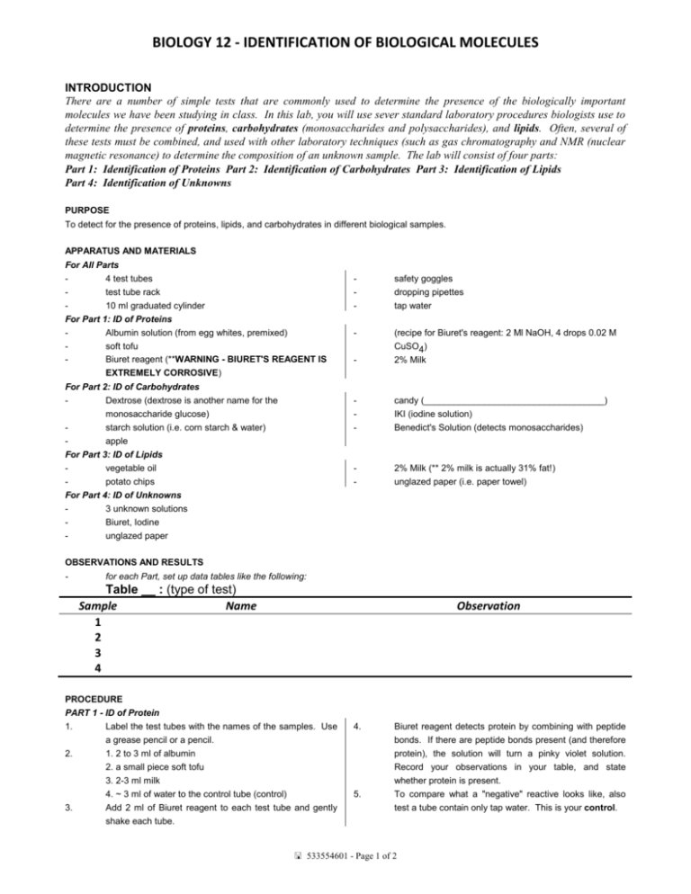
BIOLOGY 12 - IDENTIFICATION OF BIOLOGICAL MOLECULES INTRODUCTION There are a number of simple tests that are commonly used to determine the presence of the biologically important molecules we have been studying in class. In this lab, you will use sever standard laboratory procedures biologists use to determine the presence of proteins, carbohydrates (monosaccharides and polysaccharides), and lipids. Often, several of these tests must be combined, and used with other laboratory techniques (such as gas chromatography and NMR (nuclear magnetic resonance) to determine the composition of an unknown sample. The lab will consist of four parts: Part 1: Identification of Proteins Part 2: Identification of Carbohydrates Part 3: Identification of Lipids Part 4: Identification of Unknowns PURPOSE To detect for the presence of proteins, lipids, and carbohydrates in different biological samples. APPARATUS AND MATERIALS For All Parts 4 test tubes test tube rack 10 ml graduated cylinder For Part 1: ID of Proteins Albumin solution (from egg whites, premixed) soft tofu Biuret reagent (**WARNING - BIURET'S REAGENT IS EXTREMELY CORROSIVE) For Part 2: ID of Carbohydrates Dextrose (dextrose is another name for the monosaccharide glucose) starch solution (i.e. corn starch & water) apple For Part 3: ID of Lipids vegetable oil potato chips For Part 4: ID of Unknowns 3 unknown solutions Biuret, Iodine unglazed paper - safety goggles dropping pipettes tap water - (recipe for Biuret's reagent: 2 Ml NaOH, 4 drops 0.02 M CuSO4) 2% Milk - candy (____________________________________) IKI (iodine solution) Benedict's Solution (detects monosaccharides) - 2% Milk (** 2% milk is actually 31% fat!) unglazed paper (i.e. paper towel) OBSERVATIONS AND RESULTS - for each Part, set up data tables like the following: Table __ : (type of test) Sample Name 1 2 3 4 Observation PROCEDURE PART 1 - ID of Protein 1. Label the test tubes with the names of the samples. Use a grease pencil or a pencil. 2. 1. 2 to 3 ml of albumin 2. a small piece soft tofu 3. 2-3 ml milk 4. ~ 3 ml of water to the control tube (control) 3. Add 2 ml of Biuret reagent to each test tube and gently shake each tube. 4. 5. Biuret reagent detects protein by combining with peptide bonds. If there are peptide bonds present (and therefore protein), the solution will turn a pinky violet solution. Record your observations in your table, and state whether protein is present. To compare what a "negative" reactive looks like, also test a tube contain only tap water. This is your control. 533554601 - Page 1 of 2 BIOLOGY 12 - IDENTIFICATION OF BIOLOGICAL MOLECULES PART 2 - ID of Carbohydrate You will need 2 tables (test for monosaccharides and test for polysachararides) each with spaces for observations of 4 samples. 1. We will first test for monosaccharides. Label test tubes 1-4. Add the following to the separate test tubes: 1. 2 to 3 ml of dextrose 2. a small piece of apple <.5cm 3 with 2ml of water 3. candy pieces to the first tube with 2ml water 4. ~ 3 ml of water to the control tube (control) Add 5 drops of Benedict's solution to the tubes. Have one group member place the test tubes in a hot water bath (there will be one set up for you for all 2. 3. 4. students to use). Leave in for 2 to three minutes and observe any colour changes. Other group members should work on the IKI test. 5. ** 6. 7. 7. 8. If a coloured precipitate forms, then there has been a positive reaction for monosaccharides. NOTE: Do not touch the water bath with your hands. Use TONGS to carefully remove your test tube from the bath, and let it cool before touching it! IKI test for starch. Label 4 test tubes 1-4. Add the following to the separate test tubes: 1. 2 ml of starch solution 2. a small piece of apple <.5cm 3 with 2ml of water 3. candy pieces to the first tube with 2ml water 4. ~ 3 ml of water to the control tube (control) Add 3 or 4 drops of IKI solution to each tube, and observe any changes. A positive reaction will occurs if a colour change of blueblack occurs. Part 3: ID of Lipids 1. First do a "Grease Spot/Translucence). Place a small 2. After 5 minutes, hold the paper up to the light and look for amount of the sample to be tested on apiece of unglazed any translucent spots. paper. Label each sample right on the paper and spread 3. If a translucent spot is visible, then the presence of lipid it as thinly as possible (use a stirring rod or plastic knife has been verified. Record your observations. to do this). Use tap water as a control. Part IV: ID of Unknown You will need 4 separate tables, one for each of the tests that you are going to conduct. (Proteins, monosacharides, polysaccarides and lipids) 1. You will obtain samples of 3 unknowns. Label your test your test tube and get more of the unknown for each test. tubes 1, 2, 3, and add about 2 ml of each solution for Your kind and good-looking teacher may or may not ask each test. that you skip one or more of the tests. 2. Using the techniques learned in parts 1-3, test for the 3. Clean everything up as conscientiously as if the eternal presence of lipids (do only the paper-towel test for lipids). future of your immortal soul depended on it. Amen. carbohydrates, and protein. You will need to clean out *** TO CLEAN UP, RETURN EVERYTHING TO ITS ORIGINAL PLACE, CLEAN AND DRY ALL GLASSWARE, CLEAN ALL PIPETTES, STIR RODS, GRADUATED CYLINDER ETC., AND MAKE SURE YOUR WORK AREA IS CLEAN AND DRY. QUESTIONS 1. 2. 3. 4. 5. 6. 7. What happens to proteins when they are exposed to very high temperatures? Contrast carbohydrates and proteins in relation to their chemical structure and their functions in the organism. Name some foodstuffs high in carbohydrate content. Although carbohydrates are a rich source of energy, can people survive on a diet consisting only of carbohydrates? Explain. What biological process provides all of the carbohydrates consumed by man? In what form is excess carbohydrate stored in an animal? In a plant? Did your sample test positive fro more than one test? Explain why this might occur. 533554601 - Page 2 of 2
