Genomic DNA isolation Method
advertisement
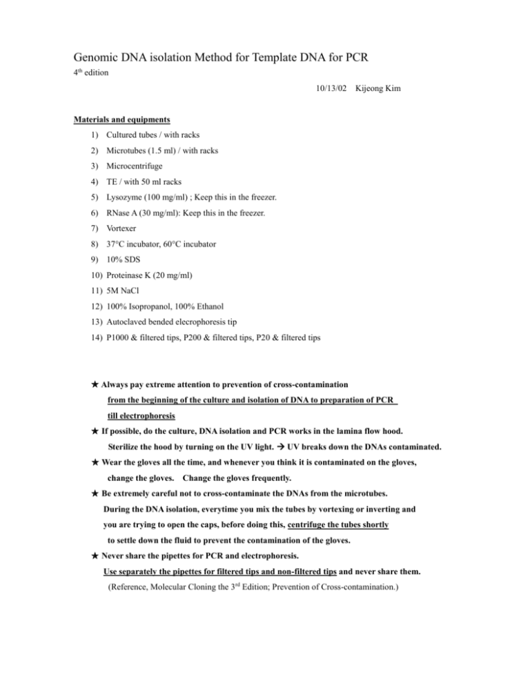
Genomic DNA isolation Method for Template DNA for PCR 4th edition 10/13/02 Kijeong Kim Materials and equipments 1) Cultured tubes / with racks 2) Microtubes (1.5 ml) / with racks 3) Microcentrifuge 4) TE / with 50 ml racks 5) Lysozyme (100 mg/ml) ; Keep this in the freezer. 6) RNase A (30 mg/ml): Keep this in the freezer. 7) Vortexer 8) 37C incubator, 60C incubator 9) 10% SDS 10) Proteinase K (20 mg/ml) 11) 5M NaCl 12) 100% Isopropanol, 100% Ethanol 13) Autoclaved bended elecrophoresis tip 14) P1000 & filtered tips, P200 & filtered tips, P20 & filtered tips Always pay extreme attention to prevention of cross-contamination from the beginning of the culture and isolation of DNA to preparation of PCR till electrophoresis If possible, do the culture, DNA isolation and PCR works in the lamina flow hood. Sterilize the hood by turning on the UV light. UV breaks down the DNAs contaminated. Wear the gloves all the time, and whenever you think it is contaminated on the gloves, change the gloves. Change the gloves frequently. Be extremely careful not to cross-contaminate the DNAs from the microtubes. During the DNA isolation, everytime you mix the tubes by vortexing or inverting and you are trying to open the caps, before doing this, centrifuge the tubes shortly to settle down the fluid to prevent the contamination of the gloves. Never share the pipettes for PCR and electrophoresis. Use separately the pipettes for filtered tips and non-filtered tips and never share them. (Reference, Molecular Cloning the 3rd Edition; Prevention of Cross-contamination.) 1. Grow bacteria shaking vigorously at 35C, 5 hrs 2. Centrifuge 1 ml of culture in 1.5 ml microtubes for 2 min. 3. Discard the supernatant by using pipette and add 420 l TE. 4. Add 15 l lysozyme (100 mg/ml) and 10 l RNase A(30mg/ml) and vortex until suspension is homogenous. (Lysozyme final 3 mg/ml, RNase A final conc. 600 ug/ml; standard = 100 ug/ml) 5. Incubate at 37C, 1 hr 6. Shortly centrifuge, and add 25 l of 10% SDS and invert it 20 times gently (Final SDS % should be 0.5%) incubate at 60 C, 30 min (The suspension will be clear by lysis.) 7. Shortly centrifuge, and add 10 l of proteinase K (20mg/ml) and invert it 20 times gently incubate at 60 C, 30 min. (Final conc. > 100 ug/ml; standard = 50 ug/ml) 8. Shortly centrifuge, and add 20 l of 5 M NaCl and invert it 20 times gently (Final NaCl should be 0.2 M in 0.5% SDS) 9. Centrifuge for 5 ~ 10 min. Transfer a clear volume of supernatant to a new tube by using 1 ml sterile tip. If the transfer is difficult, centrifuge it again. 10. Add 0.7 volume of isopropanol and invert very gently 10 times until precipitated DNA is visible. If DNA is so dense then invert more up to 20 ~ 40 times to precipitate it or add more isopropanol (Alternatively you can use 2 V of ethanol instead of isoproanol) 11. Centrifuge all the samples for 10 min. (Alternatively, if DNA strands are visible: transfer them, using a sterile bended tip at the end of tip into 100 ~ 300 l of TE) 12. Remove the supernatant using 1ml sterile microtip and remove the remnant of supernatant fluid completely using 20 l sterile tip. 13. (Option) Rinse with 0.5 ml of 95% ethanol. Repeat centrifugation for 5 min. Do the step 12. 14. Stay the tubes cap open for five minutes to dry. 15. Add 100 l or 50 ul (if pellet is too small) of TE to dissolve. To help dissolving the highly concentrated DNA, incubate it at 60 C, for 15~30 min. and vortex it gently. 16. Estimate the Concentration of DNA using spectrophotometer at 260 nM [ DNA conc. (ng/l) = O.D at 260 nM x 50 g/ml x dilution factor ] The ratio of O.D. at 260/280 should be higher than 1.7. The ratio lower than 1.7 means protein contamination. However, even if the ratio is lower than 1.7, that’s OK for the template for PCR. 17. By this method, proteins could not be completely removed. The ratio is usually lower than 1.6 If you want to remove proteins, extract with phenol-choloroform-isoamlyalcohol, wash with chloroform, and add 0.1 V 3M Sod. Acetate and then precipitate with 2 V of ethanol or 0.7 V of isopropanol.
