Proc
advertisement
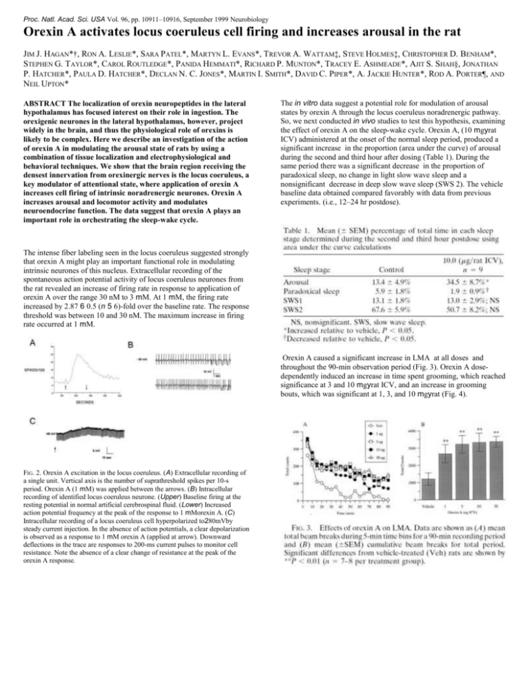
Proc. Natl. Acad. Sci. USA Vol. 96, pp. 10911–10916, September 1999 Neurobiology Orexin A activates locus coeruleus cell firing and increases arousal in the rat JIM J. HAGAN*†, RON A. LESLIE*, SARA PATEL*, MARTYN L. EVANS*, TREVOR A. WATTAM‡, STEVE HOLMES‡, CHRISTOPHER D. BENHAM*, STEPHEN G. TAYLOR*, CAROL ROUTLEDGE*, PANIDA HEMMATI*, RICHARD P. MUNTON*, TRACEY E. ASHMEADE*, AJIT S. SHAH§, JONATHAN P. HATCHER*, PAULA D. HATCHER*, DECLAN N. C. JONES*, MARTIN I. SMITH*, DAVID C. PIPER*, A. JACKIE HUNTER*, ROD A. PORTER¶, AND NEIL UPTON* ABSTRACT The localization of orexin neuropeptides in the lateral hypothalamus has focused interest on their role in ingestion. The orexigenic neurones in the lateral hypothalamus, however, project widely in the brain, and thus the physiological role of orexins is likely to be complex. Here we describe an investigation of the action of orexin A in modulating the arousal state of rats by using a combination of tissue localization and electrophysiological and behavioral techniques. We show that the brain region receiving the densest innervation from orexinergic nerves is the locus coeruleus, a key modulator of attentional state, where application of orexin A increases cell firing of intrinsic noradrenergic neurones. Orexin A increases arousal and locomotor activity and modulates neuroendocrine function. The data suggest that orexin A plays an important role in orchestrating the sleep-wake cycle. The in vitro data suggest a potential role for modulation of arousal states by orexin A through the locus coeruleus noradrenergic pathway. So, we next conducted in vivo studies to test this hypothesis, examining the effect of orexin A on the sleep-wake cycle. Orexin A, (10 mgyrat ICV) administered at the onset of the normal sleep period, produced a significant increase in the proportion (area under the curve) of arousal during the second and third hour after dosing (Table 1). During the same period there was a significant decrease in the proportion of paradoxical sleep, no change in light slow wave sleep and a nonsignificant decrease in deep slow wave sleep (SWS 2). The vehicle baseline data obtained compared favorably with data from previous experiments. (i.e., 12–24 hr postdose). The intense fiber labeling seen in the locus coeruleus suggested strongly that orexin A might play an important functional role in modulating intrinsic neurones of this nucleus. Extracellular recording of the spontaneous action potential activity of locus coeruleus neurones from the rat revealed an increase of firing rate in response to application of orexin A over the range 30 nM to 3 mM. At 1 mM, the firing rate increased by 2.87 6 0.5 (n 5 6)-fold over the baseline rate. The response threshold was between 10 and 30 nM. The maximum increase in firing rate occurred at 1 mM. Orexin A caused a significant increase in LMA at all doses and throughout the 90-min observation period (Fig. 3). Orexin A dosedependently induced an increase in time spent grooming, which reached significance at 3 and 10 mgyrat ICV, and an increase in grooming bouts, which was significant at 1, 3, and 10 mgyrat (Fig. 4). FIG. 2. Orexin A excitation in the locus coeruleus. (A) Extracellular recording of a single unit. Vertical axis is the number of suprathreshold spikes per 10-s period. Orexin A (1 mM) was applied between the arrows. (B) Intracellular recording of identified locus coeruleus neurone. (Upper) Baseline firing at the resting potential in normal artificial cerebrospinal fluid. (Lower) Increased action potential frequency at the peak of the response to 1 mMorexin A. (C) Intracellular recording of a locus coeruleus cell hyperpolarized to280mVby steady current injection. In the absence of action potentials, a clear depolarization is observed as a response to 1 mM orexin A (applied at arrow). Downward deflections in the trace are responses to 200-ms current pulses to monitor cell resistance. Note the absence of a clear change of resistance at the peak of the orexin A response.
