Muscle Testing Guide: Neck, Shoulder, Elbow, Wrist
advertisement
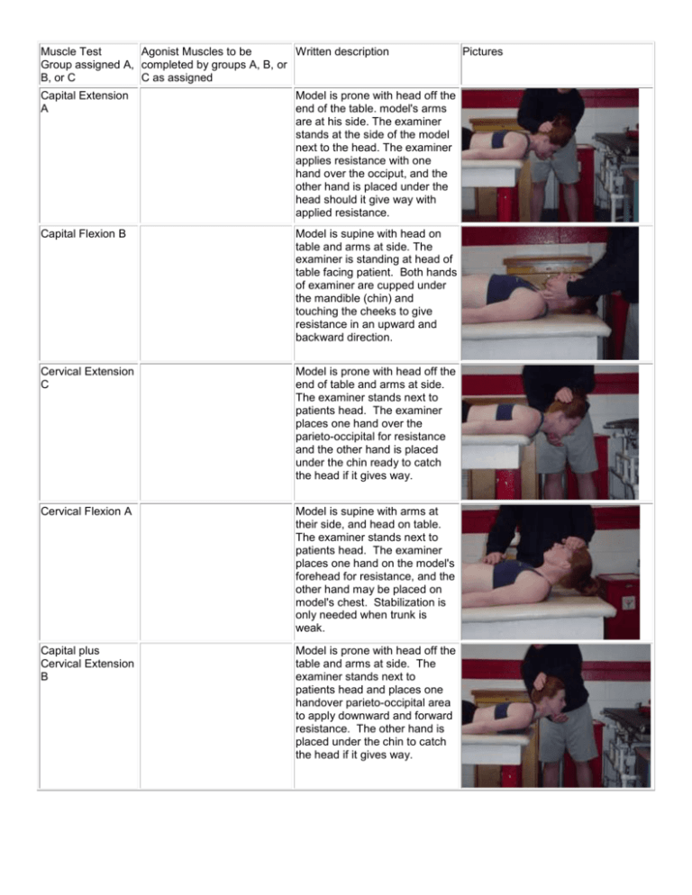
Muscle Test Agonist Muscles to be Written description Group assigned A, completed by groups A, B, or B, or C C as assigned Capital Extension A Model is prone with head off the end of the table. model's arms are at his side. The examiner stands at the side of the model next to the head. The examiner applies resistance with one hand over the occiput, and the other hand is placed under the head should it give way with applied resistance. Capital Flexion B Model is supine with head on table and arms at side. The examiner is standing at head of table facing patient. Both hands of examiner are cupped under the mandible (chin) and touching the cheeks to give resistance in an upward and backward direction. Cervical Extension C Model is prone with head off the end of table and arms at side. The examiner stands next to patients head. The examiner places one hand over the parieto-occipital for resistance and the other hand is placed under the chin ready to catch the head if it gives way. Cervical Flexion A Model is supine with arms at their side, and head on table. The examiner stands next to patients head. The examiner places one hand on the model's forehead for resistance, and the other hand may be placed on model's chest. Stabilization is only needed when trunk is weak. Capital plus Cervical Extension B Model is prone with head off the table and arms at side. The examiner stands next to patients head and places one handover parieto-occipital area to apply downward and forward resistance. The other hand is placed under the chin to catch the head if it gives way. Pictures Capital plus Cervical Flexion C Model is supine with head on table and arms at side. The examiner stands at the side of the table at the level of the model's shoulder. One hand is placed on the forehead for resistance and the other hand may be used for stabilization of the trunk if it is weak. Neck Flexion with Rotation A Patient is supine with head supported on table and turned to the right to test the left sternocleidomastoid. Therapist faces the patient with one hand placed on the temporal area above the ear for resistance. Patient raises head from table against resistance, keeping head turned throughout the movement. Glenohumeral Abduction B Patient is short sitting with arm at side and elbow slightly flexed. Examiner is standing behind patient giving resistance over arm just above elbow. Patient abducts arm to 90 degrees against maximal downward pressure. Glenohumeral Flexion C Patient is short sitting with arms at sides, elbow slightly flexed, and forearm supinated. Examiner stands at test side with one hand giving maximal resistance over distal humerus and other hand stabilizing shoulder. Glenohumeral Extension A Patient is prone with arms at sides and shoulder internally rotated. Examiner stands at test side and applies maximal resistance over posterior arm just above elbow. Patient raises arm off table keeping elbow straight. Shoulder Extension to Isolate Latissimus Dorsi B Patient is short sitting with hands flat on table next to hips. If the patient's arm are too short, blocks may be used under each hand. Therapist stands behind patient to observe or palpate the fibers of the Latissimus dorsi on the lateral aspects of the thoracic wall (bilaterally) just above the waist. Patient pushes down on hands to lift buttocks from table. Glenohumeral Internal Rotation C Patient is prone with shoulder abducted to 90 degrees with folded towel placed under distal arm and forearm hanging vertically over edge of table. Examiner stands at test side giving resistance over volar side of forearm just above the wrist in a downward and forward direction. The other hand provides counterforce at the elbow in a backward and slightly upward direction. Glenohumeral External Rotation A Patient is prone with shoulder abducted to 90 degrees and forearm hanging vertically over edge of table. Examiner stands at test side at level of patient's waist. One hand gives resistance at wrist while other hand supports and provides counter pressure at the elbow. Patient moves forearm upward through range of external rotation. Glenohumeral Horizontal Abduction B Patient is prone with shoulder abducted to 90 degrees and forearm off edge of table with elbow flexed. Examiner stands at test side giving resistance over posterior arm just above elbow with other hand stabilizing the trunk. Patient lifts elbow toward ceiling against resistance. Glenohumeral Scaption C Patient is short sitting with arms at sides. Examiner stands slightly to the test side of the patient. Resistance hand contours over the arm just above the elbow, while other hand stabilizes the upper body. Patient elevates arm halfway between flexion and abduction against resistance. Elbow Flexion A The patient should be short sitting with arms at side. The therapist stands in front of patient toward the test side. The hand giving resistance is contoured over the flexor surface of the forearm proximal to the wrist, and the other hand applies a counterforce by cupping the palm over the anterior superior surface of the shoulder. Elbow Extension B The patient should be prone on a table with the arm abducted 90 degrees, and the forearm flexed and hanging vertically over the side of the table. The examiner should provide support just above the elbow with one hand, and with the other hand he should apply a downward resistance on the dorsal side of the wrist. Forearm Supination C The patient sits on a table with arms at side and elbow bent at 90 degrees on test arm. The forearm should be in neutral. The examiner stands at side or in front of patient. One hand supports the elbow of the patient and the other hand grasps the forearm on the volar surface at the wrist, for resistance. Forearm Pronation A The patient sits on a table with arms at side and elbow bent at 90 degrees on test arm. The forearm should be in neutral. The examiner stands at side or in front of patient. One hand supports the elbow of the patient and the other hand grasps the forearm on the dorsal surface at the wrist, for resistance. Wrist Abduction B The patient sits with forearm in neutral (thumb side up) with hand hanging off table. The therapist stabilizes the forearm against the table with one hand and uses the other hand to apply downward resistance toward wrist adduction. The patient actively abducts the wrist. Wrist Adduction C The patient lies prone with forearm and wrist in neutral (thumb side down). The test arm should slightly hang off the edge of the table. The therapist stabilizes the forearm against the table with one hand and uses other hand to apply downward resistance toward wrist abduction. The patient actively adducts the wrist. Wrist Flexion A The patient sits with forearm in supination and wrist in neutral. The therapist stabilizes the patient's forearm against table with one hand and the other hand grasps the patient's hand in a handshake position. Resistance is given on the palmar surface of the hand in the direction of extension. The patient actively flexes the wrist. Wrist Extension B The patient sits with forearm in pronation and wrist in neutral. The therapist stabilizes the patient's forearm against table with one hand and the other hand is placed on the dorsal aspect of the patient's hand . Resistance is given on the dorsal surface of the hand in the direction of flexion. The patient actively extends the wrist. MCP Flexion C The patient is sitting or supine with forearm in supination. The wrist is in neutral with the MCP joints fully extended. The therapist stabilizes the metacarpals just proximal to the MCP joint, and applies resistance on the palmer surface of the proximal row of phalanges in the direction of MCP extension while the patient flexes at the MCP joint. MCP Extension A The patient's forearm is in pronation with the wrist in neutral. MP joints and IP joints are in relaxed flexion posture. Therapist stabilizes the wrist and places the index finger of the resistance hand across the dorsum of all proximal phalanges just distal to the MCP joints. Give resistance in the direction of flexion. PIP Flexion B The patient's forearm is in supination with the wrist in neutral. The finger to be tested is in slight flexion at the MCP joint. The therapist holds all fingers, except the test finger, in extension at all joints. The therapist applies resistance at the head of the middle phalanx in the direction of extension while the patient actively flexes the PIP joint. PIP Extension C The patient's forearm is in pronation with the wrist in neutral. The finger being tested should be in slight extension at the MCP joint. The patient's other fingers are flexed against the table, except the test finger. The therapist stabilizes the test finger at the proximal phalanx. The therapist applies resistance distal to PIP joint in the direction of flexion, while the patient extends the PIP joint. DIP Flexion A The patient's forearm is in supination with the wrist in neutral, with the PIP joint in extension. The therapist stabilizes the middle phalanx by grasping it on either side, and resistance is applied at the distal phalanx in the direction of extension while the patient actively flexes the DIP joint. DIP Extension B The patient's forearm is in pronation with the wrist in neutral. The finger being tested should be in slight extension at the PIP joint. The patient's other fingers are flexed against the table, except the test finger. The therapist stabilizes the test finger at the middle phalanx. The therapist applies resistance distal to DIP joint in the direction of flexion, while the patient extends the DIP joint. Finger Abduction C The patient's forearm is pronated with the wrist in neutral. Fingers start in extension and adduction. MCP joints are in neutral while avoiding hyperextension. Therapist supports wrist in neutral. The fingers of the other hand are used to give resistance on the distal phalanx, on the radial side of one finger and the ulnar side of the adjacent finger. The direction of resistance is toward adduction while patient actively abducts. Finger Adduction A The patient's forearm is pronated, wrist in neutral, and fingers extended and abducted. The therapist supports the wrist in neutral. The fingers of the other hand are used to give resistance on the distal phalanx, on the radial side of one finger and the ulnar side of the adjacent finger. The direction of resistance is toward abduction while the patient actively adducts. Thumb Abduction B Patient sits with the wrist in neutral, and thumb relaxed in adduction. Therapist stabilizes metacarpals by maintaining wrist in neutral in somewhat of a handshake position. Resistance is applied to the lateral aspect of proximal phalanx in the direction of adduction. Patient lifts thumb toward ceiling against resistance. Thumb Adduction C The patient sits with wrist in neutral, and thumb relaxed and hanging down in abduction. Therapist stabilizes metacarpals by grasping the patient's hand around the ulnar side. Resistance is given on medial side of proximal phalanx in the direction of abduction. Patient adducts against resistance. Thumb IP Extension A The patient sits with wrist in neutral, and the ulnar side of the hand resting on the table. The thumb should be relaxed and in a flexed position. The therapist uses the table to support the ulnar side of the hand and stabilize the proximal phalanx of the thumb. Resistance is applied over the dorsal surface of the distal phalanx of the thumb in the direction of flexion. The patient actively extends the IP joint. Thumb IP Flexion B The patient sits with wrist in neutral, and the MP joint of the thumb in extension. The therapist stabilizes MP joint in extension then gives resistance with the other hand against the palmar surface of the distal phalanx in the direction of extension. Patient actively flexes IP joint. Thumb MP Extension C The patient sits with wrist in neutral, with the CMC and IP joints relaxed in slight flexion. The MP joint is in abduction and flexion. The therapist stabilizes the first metacarpal allowing motion to occur only in the MP joint. Resistance is provided with the other hand on the dorsal surface of the proximal phalanx in the direction of flexion. The patient actively extends MP joint. Thumb MP Flexion A The patient sits with the wrist in neutral, with the CMC and IP joints in extension. The MP joint is in abduction and in extension. The therapist stabilizes the first metacarpal allowing motion to occur only in the MP joint. Resistance is provided with the other hand on the palmar surface of the proximal phalanx in the direction of extension. The patient actively flexes MP joint. Thumb Opposition B The patient sits with forearm in supination, and wrist in neutral. The therapist stabilizes the hand by placing the dorsal aspect of his fingers on the palmar aspect of the patient's fingers, and the same with the thumb. Resistance is given on the palmar side of the thumb in the direction of extension. The patient actively flexes the thumb toward the little finger. Lumbar Extension A Patient lies prone with hands clasped behind head. Examiner stands so as to stabilize the lower extremities just above the ankles if the patient has normal hip strength. Patient extends lumbar spine until thorax is raised above table level. Thoracic Extension B The patient lies prone with head and upper trunk extending off the table from about the nipple line. The examiner stands so that he can stabilize the lower limbs at the ankles. The patient then extends thoracic spine to the horizontal. Trunk Flexion C The patient lies supine with hands clasped behind head. Examiner stands at the side of the table level with the patient's chest, so he can determine if patient's scapulae clear the table. The patient flexes trunk through range of motion. Trunk Rotation A Patient is supine with hands clasped behind head. The examiner stands at patient's waist level. Patient flexes trunk and rotates to one side. Pelvic Elevation B Patient is supine with hip and lumbar spine in extension. Patient grasps edge of table for stabilization during resistance. The examiner stands at foot of table facing the patient. Examiner grasps test limb with both hands and pulls evenly and smoothly. The patient hikes that pelvis, approximating the pelvic rim to the inferior margin of the rib cage. Hip Abduction C The patient is side lying with test leg uppermost. The therapist stands behind the patient and stabilizes with one hand at the hip. This hand is proximal to the greater trochanter. The other hand applies resistance across the lateral surface of the knee. Patient abducts hip against downward resistance. Hip Abduction Flexed A The patient is side lying with test leg uppermost, and hip flexed to 45 degrees. The therapist stands behind the patient and stabilizes with one hand at the hip. This hand is proximal to the greater trochanter. The other hand applies resistance across the lateral surface of the knee. Patient abducts hip against downward resistance. Hip Adduction B The patient is side lying with the test leg lowermost and resting on the table. The uppermost leg is abducted to 25 degrees and supported by the examiner. The therapist stands behind the patient at the knee level. The resistance hand is placed on the distal medial femur of the test leg. Resistance is applied in a downward motion while the patient actively adducts. Hip Flexion C The patient is short sitting with thighs fully supported and legs hanging over the edge. The therapist stands next to the test leg. The therapist places one hand on the distal thigh and proximal knee, and applies resistance in a downward direction as the patient actively flexes at the hip Hip Extension A The patient lies prone on the table. The therapist stands on the side of the test leg, at pelvis level. One hand stabilizes the pelvis, and the other hand is placed on the distal calf. The hand on the distal calf applies resistance in a downward direction ad the patient actively extends at the hip. Hip Internal Rotation B The patient is short sitting. The therapist sits on a stool or kneels beside patient. The therapist places one hand at the medial aspect of the distal thigh and applies resistance in a lateral direction. The other hand grasps the lateral ankle just above the malleolus, and applies resistance in a medial direction. The patient is actively internally rotating at the hip. Hip External Rotation C The patient is short sitting. The therapist sits on a stool or kneels beside patient. The therapist places one hand at the lateral aspect of the distal thigh and applies resistance in a medial direction. The other hand grasps the medial ankle just above the malleolus, and applies resistance in a lateral direction. The patient is actively externally rotating at the hip. Hip Flexion Abduction Ext. Rotation A The patient is short sitting with thighs supported on table and legs hanging over side. The therapist stands lateral to the test leg while placing one hand on the lateral side of the knee and using the other hand to grasp the medial anterior surface of the distal leg. Hand at knee gives downward and inward resistance. Hand at ankle gives upward and outward resistance. Patient flexes, abducts, and externally rotates the hip and flexes the knee. Knee Flexion B The patient is prone with legs straight and toes hanging over the edge of the table. The therapist stands next to the test leg. The therapist places one hand on the posterior thigh and the other hand applies resistance at the distal calf or just above the ankle. The resistance is applied in downward direction as the patient actively flexes the knee. Knee Extension C The patient is short sitting, place a wedge or pad under the distal thigh to maintain the femur in the horizontal position. The therapist stands at the side of the leg being tested. The hand giving resistance is contoured over the anterior surface of the distal leg just above the ankle. Resistance is applied in a downward direction as the patient actively extends the knee. Knee Flexion with External Rotation A Patient is prone with knee flexed to less than 90 degrees and the leg in external rotation. Therapist stabilizes the thigh with one hand and gives downward and inward resistance at the ankle with the other hand. Patient flexes the knee, maintaining the leg in external rotation. Knee Flexion with Internal Rotation B Patient is prone with knee flexed to less than 90 degrees and the leg in internal rotation. Therapist stabilizes the thigh with one hand and gives downward and outward resistance at the ankle with the other hand. Patient flexes the knee, maintaining the leg in internal rotation. Plantarflexion (Gastrocnemius and Soleus in standing position) C Patient stands on test limb with knee extended. Patient may place one or two fingers on a table or other external surface to assist with balance. Therapist should stand or sit with a lateral view of test limb. Patient actively raises heel from floor 20 consecutive times without rest or fatigue through a full range of plantarflexion. Plantarflexion (Soleus only in standing position) A Patient stands on test limb with knee slightly flexed. One or two fingers may be used to assist with balance. Therapist stands or sits with a lateral view of test limb. Patient actively raises heel from floor 20 consecutive times without rest or great fatigue through full range of plantarflexion. Neutral Dorsiflexion (Tibialis Anterior, Extensor Digitorum Longus, Peroneus Tertius, Extensor Hallicus Longus) B Patient is short sitting with ankle plantarflexed. Therapist sits on stool in front of patient and uses one hand to stabilize the leg just above the malleoli. The other hand is used for resistance by placing it on the dorsal aspect of the foot. Patient actively dorsiflexes against resistance. Inversion C The patient is short sitting with ankle in slight plantar flexion. Therapist sits in front or on side of test limb and uses one hand to stabilize the ankle just above the malleoli. The other hand provides resistance by contouring over the dorsum and medial side of the foot at the level of the metatarsal heads. Resistance is directed toward eversion and slight dorsiflexion while patient actively inverts foot. Inversion with dorsiflexion A Patient may be short sitting or supine. The therapist sits on a stool in front of the patient with patient's heel resting on thigh. One hand stabilizes posterior leg just above the malleoli while other hand provides resistance over the dorsomedial aspect of foot. Patient actively dorsiflexes ankle and inverts foot, keeping toes relaxed. Eversion B The patient is short sitting with ankle in slight plantar flexion. Therapist sits in front or on side of test limb and uses one hand to stabilize the ankle just above the malleoli. The other hand provides resistance by contouring over the dorsum and medial side of the foot at the level of the metatarsal heads. Resistance is directed toward inversion and slight dorsiflexion while patient actively everts foot. Eversion with plantar flexion C Patient is short sitting with ankle in neutral position. Therapist is sitting on a stool in front of patient. One hand stabilizes the ankle just above the malleoli while other hand provides resistance around the dorsum and lateral border of the forefoot. Resistance is directed toward inversion and slight dorsiflexion. Patient actively turns the foot down and out. Scapular Abduction A Patient is short sitting with hands on lap. Examiner stands at test side with one hand giving resistance on the arm just above elbow. The other hand uses thumb, index finger, and web space in between to palpate inferior angle of scapula. Patient raises arm to about 130 degrees of flexion against resistance. Scapula should abduct without winging. Scapular Adduction B Patient is prone with shoulder at edge of table. Shoulder is abducted to 90 degrees and externally rotated. Elbow may be flexed to a right angle. Examiner stands at test side and stabilizes the contralateral scapular area. Resistance hand is placed over distal humerus. Patient lifts elbow towards ceiling which adducts scapula. Scapular Adduction and Downward Rotation C Patient is prone with shoulder internally rotated, elbow flexed, and arm adducted across the back. Examiner stands at test side with resistance hand placed on humerus just above the elbow. Resistance is given in a downward and outward direction. Palpation hand is placed on vertebral border of scapula. Patient lifts hand off the back against resistance which adducts scapula. Scapular Elevation A Patient is short sitting with hands relaxed in lap. Examiner stands behind patient and places hands over the top of both shoulders to give resistance in a downward direction. Patient shrugs shoulders simultaneously against resistance. MP Flexion of Great Toe B The patient is short sitting with legs hanging over the edge of the table. The ankle is in a neutral position. The therapist is seated on a stool in front of the patient. The test foot rests on the examiner's lap. The therapist stabilizes the dorsum of the foot just below the ankle with one hand, and uses the index finger of the other hand to resist beneath the proximal phalanx of the great toe. Then patient actively flexes the great toe. IP Flexion of Great Toe C The patient is short sitting with legs hanging over the edge of the table. The ankle is in a neutral position. The therapist is seated on a stool in front of the patient. The test foot rests on the examiner's lap. The therapist stabilizes the dorsum of the foot just below the ankle with one hand, and uses the index finger and thumb of the other hand to grasp the distal phalanx of the great toe for resistance. Then patient actively flexes the great toe at the IP joint. MP Flexion of Lesser Toes A The patient is short sitting with legs hanging over the edge of the table. The ankle is in a neutral position. The therapist is seated on a stool in front of the patient. The test foot rests on the examiner's lap. The therapist stabilizes the dorsum of the foot just below the ankle with one hand, and uses the index finger of the other hand to resist beneath the MP joints of the four lesser toes. Then patient actively flexes the toes at the MP joints, keeping the IP joints neutral. PIP Flexion of the Lesser Toes B The patient is short sitting with legs hanging over the edge of the table. The ankle is in a neutral position. The therapist is seated on a stool in front of the patient. The test foot rests on the examiner's lap. The therapist uses both hands to grasp the anterior foot with the fingers across the dorsum of the foot and the thumbs under the proximal phalanges for resistance. Then patient actively flexes the toes at the PIP joints. DIP Flexion of the Lesser Toes C The patient is short sitting with legs hanging over the edge of the table. The ankle is in a neutral position. The therapist is seated on a stool in front of the patient. The test foot rests on the examiner's lap. The therapist uses both hands to grasp the anterior foot with the fingers across the dorsum of the foot and the thumbs under the distal phalanges for resistance. Then patient actively flexes the toes at the DIP joints. MP Extension of Great Toe A The patient is short sitting with legs hanging over the edge of the table. The ankle is in a neutral position. The therapist is seated on a stool in front of the patient. The test foot rests on the examiner's lap. The therapist stabilizes the metatarsal area by contouring hand around the plantar surface of the foot with the thumb curving around the base of the great toe. For resistance, place thumb over the MP joint. The other hand stabilizes the foot at heel. The patient actively extends MP joint. IP Extension of Great Toe B The patient is short sitting with legs hanging over the edge of the table. The ankle is in a neutral position. The therapist is seated on a stool in front of the patient. The test foot rests on the examiner's lap. The therapist stabilizes the metatarsal area by contouring hand around the plantar surface of the foot with the thumb curving around the base of the great toe. For resistance, place thumb over the IP joint. The other hand stabilizes the foot at heel. The patient actively extends IP joint. MP Extension of Lesser Toes C The patient is short sitting with legs hanging over the edge of the table. The ankle is in a neutral position. The therapist is seated on a stool in front of the patient. The test foot rests on the examiner's lap. The therapist uses both hands to stabilize the metatarsals with the fingers on the plantar surface and the thumbs on the dorsum of the foot. The patient actively extends MP joints of lesser toes. PIP Extension of Lesser Toes A The patient is short sitting with legs hanging over the edge of the table. The ankle is in a neutral position. The therapist is seated on a stool in front of the patient. The test foot rests on the examiner's lap. The therapist uses one hand to stabilize the metatarsals with the fingers on the plantar surface and the thumb on the dorsum of the foot. The other hand is used to give resistance with the thumb placed over the dorsal surface of the proximal phalanges of toes. The patient actively extends PIP joints. DIP Extension of Lesser Toes B The patient is short sitting with legs hanging over the edge of the table. The ankle is in a neutral position. The therapist is seated on a stool in front of the patient. The test foot rests on the examiner's lap. The therapist uses one hand to stabilize the metatarsals with the fingers on the plantar surface and the thumb on the dorsum of the foot. The other hand is used to give resistance with the thumb placed over the dorsal surface of the distal phalanges of toes. the patient actively extends DIP joints.

