ABCs of dermatology (bearded Hamzavi)
advertisement
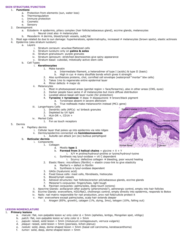
SKIN STRUCTURE/FUNCTION 1. Functions a. Protection from elements (sun, water loss) b. Thermoregulation c. Immune protection d. Cosmetic e. Sensory 2. Development a. Ectoderm epidermis, piliary complex (hair follicle/sebaceous gland), eccrine glands, melanocytes i. Neural crest also melanocytes b. Mesoderm dermis, blood/lymph vessels, subQ fat 3. Most age-related Δs due to sun damage: hyperkeratosis, epidermatrophy, increased # melanocytes (brown spots), elastic actinosis 4. Epidermis (aka stratum lucidum) a. Layers i. Stratum corneum: anuclear/flattened cells ii. Stratum lucidum: only on palms & soles iii. Stratum granulosum: purple granules iv. Stratum spinosum: stretched desmosomes give spiny appearance v. Stratum basal: cuboidal, mitotically-active stem cells b. Cell Types i. Keratinocytes 1. Make keratin a. Intermediate filament, a heterodimer of type I (acidic) & type II (basic) b. High in cys many disulfide bonds which gives it strength 2. Also synthesizes proteins, chol, cornified cell envelope (waterproof “mortar” btw cells) 3. Takes 1mo to regenerate entire epidermal layer 4. Minor defects major dz ii. Melanocytes 1. Most in photoexposed areas (genital region > face/forearms), also in other areas (CNS, eyes) 2. Darker people have same # of melanocytes but more diffuse distribution 3. Located above basal cell layer nuclei (for protection) 4. Tyrosine + tyrosinase dopa dopaquinone brown/black pigment a. Tyrosinase absent in severe albinoism b. True redheads make melanocortin instead (MC1 gene) iii. Langerhaans Cells 1. Dendritic cells (APCs) w/ birbeck granules 2. Depleted by UV light 3. HLA-DR +, CD1A + iv. Merkel Cells 1. Fxn as touch receptors 5. Dermis a. Papillary dermis i. Cellular layer that pokes up into epidermis via rete ridges ii. Dermis/epidermis connected via hemidesmosomes 1. AutoAb can attack jxn (ex) bullous pemphigoid b. Reticular dermis i. Components 1. Collagen a. Mostly type 1 b. Formed from 3 helical chains = glycine + X + Y i. X/Y = proline/hydroxyl-proline or lysine/hydroxyl-lysine c. Synthesis req lysyl-oxidase = vit C dependent i. Scurvy: defective collagen bleeding, poor wound healing 2. Elastic fibers: microfibers (fibrillin) + elastin cross-link to give elasticity a. Marfan’s = defect in fibrillin b. Synthesis is lysyl-oxidase dependent 3. GAGs (hyaluronic acid) 4. Fixed tissue cells: mast cells, fibroblasts, histiocytes 5. Blood/lymph vessels 6. Adnexel structures: hair follicles/erector pili/sebaceous glands, eccrine glands 7. Meissner’s corpuscles: fingers/toes, light tough 8. Pacinian corpuscles: palms/soles, deep touch (onions) ii. Apocrine Glands: axilla/groin after puberty (pheromones?), adrenergic control, empty into hair follicles iii. Eccrine Glands: everywhere, H2O/NaCl, cholinergic control, empty directly into epidermis, responds to Botox iv. Nails: nail matrix responsible for nail production, prox nail fold/cuticle protect it v. Hair: everywhere except palms/soles, scalp hair extends deeper 1. Anagen (85%, growth), catagen (1%, dying, 3mo), telogen (10%, falling out) LESION NOMENCLATURE 1. Primary lesions a. macule: flat, non-palpable lesion w/ only color Δ < 5mm (ephelies, lentigo, Mongolian spot, vitiligo) b. patch: flat, non-palpable lesion w/ only color Δ > 5mm c. papule: raised, solid lesion < 5mm (moluscum contageousum, verruca vulgaris) d. plaque: raised, solid lesion > 5mm (psoriasis, lichen planus) e. nodule: solid, deep, dome-shaped lesion < 5mm (basal cell carcinoma, keratoacanthoma) f. tumor: solid, deep, dome-shaped lesion > 5mm 2. 3. 4. 5. 6. 7. g. vesicle: raised, fluid-filled lesion < 5mm (herpes, eczema) h. bulla: raised, fluid-filled lesion > 5mm (bullous pemphigoid, pemphigus vulgaris) i. pustule: vesicle filled with purulent fluid (acne, folliculitis) j. wheal: smooth, superficial, flat-topped lesion, due to dermal edema (urticaria) Secondary lesions a. atrophy: thinned, depressed skin (aging) b. excoriation: depression in skin caused by scratching c. induration: dermal thickening d. lichenification: thickening of skin w/accentuation of skin lines (lichen planus) e. erosion: loss of epidermis, heals w/o scarring (herpes) f. ulcer: loss of epidermis + dermis, heals w/ scarring (decubitus ulcer) g. fissure: linear erosion or ulcer (chapping) h. crust: dried exudates (impetigo) i. scale: excess dead stratum corneum, white (psoriasis) j. desquamation: peeling of sheets of stratum cornum/epidermis k. comedo: plug of sebaceous/keratinous material in hair follicle, closed or open (acne) By color a. erythematous: red b. blanching: color fades with pressure c. telangiectasia: dilated superficial blood vessels d. petechia: fine speckled, non-blanching color <5 mm e. purpura: petechia > 5mm f. hypo/hyper/depigmentation: obvi By shape a. annular: forming a ring b. arcuate: forming half a circle c. nummular: coin-like d. serpiginous: curving irregularly like a snake By configuration a. linear: in a line b. grouped: clustered c. confluent: smaller lesions joining to form larger ones d. reticulated: net-like By distribution a. acral: affecting extremities, ears, nose b. dermatomal: along linear bands of skin innervation c. blaschkonian: along linear bands of skin migration d. generalized: involving most of skin surface e. photodistributed: sun-exposed areas By morphology a. papulosquamous: well-defined lesions with scale i. psoriasis (ext), lichen planus, tinea infections, secondary syphilis, mycosis fungoides b. eczematous: itchy processes of scaly lesions with indistinct borders i. eczema, atopic dermatitis (flex), contact dermatitis c. granulomatous: dermal inflammation without scale i. granuloma annulare, sarcoidosis, mycobacterial & deep fungal infections d. vesiculobullous: blisters (epi only or at epi/dermis jxn) and erosions i. pemphigus vulgaris (flaccid), bullous pemphigoid (tense), herpes, bullous impetigo e. pigmentary: varying amounts of melanin in the skin i. melasma/post-inflammatory(hyper), tuberous sclerosis/pityriasis alba (hypo), vitiligo/piebaldism (de) f. poikiloderma: triad of lacy hyper/hypopigmentation, epidermal atrophy, telangiectasias i. dermatomyositis, pokiloderma of Civatte (cheeks & neck, middle-aged ♀) g. alopecia: hair loss i. androgenic alopecia (diffuse/no scar), alopecia areata/trichotillomania/traction (focal/no scar), lupus (scarring) ERYTHEMA & PURPURA 1. Erythema: redness that blanches w/ pressure a. Urticaria: mast cell degranulation histamine release increased vessel permeability leakage of plasma into skin i. Smooth lesions w/ central pallor (tho no depression) that are very pruritic, transient, and move around ii. In kids, trigger is often infxn/new meds iii. Tx is antihistamines and avoidance of triggers b. Erythema multiforme: immunologic rxn fixed & symmetric papules/plaques w/ a central duskiness i. In kids, assoc w/ drug rxn (ex) penicillin vs in adults, assoc w/ HSV or mycoplamsa ii. Tx is tx of underlying cause 1. Can progress to toxic epidermal necrolysis = life-threatening sloughing of skin c. Erythema migrans: Lyme dz (tick bite) erythematous annular plaques on trunk & extremities w/ bulls-eye appearance i. Can cause neuro/cardio complications ii. Tx is antibiotics 2. Purpura: purplish macules/papules that DO NOT blanch w/ pressure a. Non-palpable i. Thrombocytopenic purpura: due to decreased plts, occurs first in extremities ii. DIC: due to excessive clotting bleeding, assoc w/ fevers + systemic sx b. Palpable (vasculitis) i. Henoch-Schonlein purpura: inflamm of venules acute onset of skin lesions in butt/feet + GI pain/hematuria 1. Affects Caucasian kids 4-7y, possibly infectious etiology since recurrent cases are post-streptococcal 2. Tx is topical steroids, if kidneys are involved, oral steroids ACNE & ROSACEA 1. Disorder of pilosebaceous unit: increased cohesiveness of shed corneocytes occlusion of follicular ostium w/ buildup of cells/sebum a. Non-inflammatory forms open or closed comedones vs. inflammatory forms erythematous papules, pustules b. Clinical features: hyperpigmentation, persistent erythema, +/- scarring c. P acnes: G+ anaerobic rod produces mediators which convert sebum to chemotactic FFAcids neutrophils/inflamm i. [] of bacteria does NOT correlate to dz severity d. Sebum levels increase in puberty i. Adrenarche assoc w/ increase in DHEA-S (precursor to androgens) which stimulate sebaceous gland ii. Estrogens can oppose effects of androgens 2. Variants a. Acne fulminans: most severe form, common in young men i. Acute onset pustules hemorrhagic crusts/ulcers severe scarring ii. Assoc w/ osteolytic bone lesions, elevated ESR, leukocytosis b. Acne conglobata: above w/o systemic sx i. Triad = trunk + scalp (dissecting cellulitis) + axilla/inguinal nodules (hidradenitis suppurativa) c. Acne mechanica: friction acne (ex) chinstraps, suspenders comedone formation d. Acne excoriee: young women w/ OCD pick at comedones, linear configuration e. Drug-induced: monomorphic papules from steroids/Li/phenytoin f. Neonatal: appears 1w after birth, disappears by 3m, possibly due to yeast Topical tx (more for DRUG Benzoyl peroxide Salicylic acid Antibiotics Retinoids non-inflamm acne) MOA bacteriocidal (↓ P. acne) dries up active lesions bacteriocidal (↓ P. acne) alters keratinization Azelaic acid above + ↓ P. acne Oral tx (more for inflamm acne) DRUG MOA Tetracyclines Binds 30s ribo subunit Erythromycin Hormonal Binds 50s ribo subunit Increases estrogen levels Spironolactone Isoretinoin Androgen R antag 3. PEARLS SE: bleaching, irritation, erythema combine w/ peroxide to decrease bacterial resistance SE: irritation, erythema, scaling tretinoin inactivated by sun tazarotene most potent, contra in pregnancy also good for lightening hyperpigmentation, from cereal grains PEARLS SE: esophagitis, binding of divalent cations Tetra stains growing teeth, contra in pregnancy Doxy phototox Mino vertigo, pseudomotor cerebri, pigmentation of acne scars SE: nausea SE: nausea, wt gain, irregular menses severe SE: thrombophlebitis, PE, HTN SE: hyperK SE: dry mouth, ↑ triglycerides, depression/suicide contra in pregnancy Rosacea: idiopathic, chronic inflamm dz of blood vessels/sebaceous glands a. Erythema/telangiectasias/papules/pustules but comedones are not present b. Affects the face in “sunburn” pattern, most common in 30-40yo c. Complications: edema, conjunctivitis, rhinophyma d. Triggered by sun, caffeine, EtOH, spicy foods, stress e. Tx: topical metronidazole, azelaic acid or oral tetracycline, isotretinoin + avoid triggers/steroids ECZEMOUS RASHES/WHITE SPOTS/INFLAMM PAPULES 1. Eczema/dermatitis = intracellular edema (aka spongiosis) a. Acute forms = papulovesicular w/ spongiosis, subacute = more inflamm/less spongiosis, chronic = lichenification b. Irritant contact: due to chemical exposure, most common on dorsal hand, tx is irritant avoidance c. Allergic contact: due to type IV rxn red/scaly papules w/ sharp margins @ site of contact ~1-2d after i. Most common – poison ivy, cosmetics, nickel, rubber, meds, perfumes ii. Tx is allergen avoidance + antihistamines/steroids d. Nummular: coin-shaped lesions on lower extremities, older men, tx is antihistamines/topicals e. Seborrheic: overgrowth of yeast on scalp/intertriginous w/ greasy yellow scaling, infants/adults, tx is topical antifungals/Zn i. Also assoc w/ AIDS, Parkinson’s f. Stasis: venous HTN serum leakage/inflamm on med/ant shin, tx is better blood flow, compression hose, topical steroids g. Lichen Simplex: repetitive rubbing lichenified plaque on neck/ankles/genitalia, tx is topical steroids/Codran/no more itching i. Also assoc w/ neurological/psych issues, tx is much more difficult 2. White spots = areas of hypo/depigmentation a. Tinea versicolor: due to Malassezia yeast, scaly round macules/patches on oily areas (esp trunk), tx w/ topical antifungals/Zn i. Lesions can be pruritic or asx ii. Assoc w/ higher temps/humidity b. Pityriasis Alba: asx hypopigmented patches, common on face/in kids, seen when surrounding skin tans, tx is topical steroids c. Post-inflamm hypopigmentation: similar to vitiligo but assoc w/ inflamm dz (ex) dermatitis, seborrheic, psoriasis d. Vitiligo: autoimmune, no melanocytes, common on bony surfaces & around eyes/mouth, gradually enlarge but totally asx i. Assoc w/ thyroid dz, pernicious anemia, cement workers ii. Tx is long and ineffective, steroids/immunomodulators/phototherapy 3. Inflamm papules a. Scabies: mite lays eggs in s. corneum severe itching (worse at night) + scattered macules/papules/pustules i. Nodular form in genitalia of kids, crusted form in immunocomprimised ii. Tx is topical permethrin x1w, wash bedding, tx all close contacts (as this is how it’s spread) b. c. d. Insect bites: rxn to injected chemicals acute urticaria/pain, tx all w/ steroids/antihistamines i. Brown recluse spider: necrosis w/ red/white/blue sign ii. Pubic lice: attach to hairs & produce itchy papules + blue-gray macules, STI iii. Head lice: “dandruff” that won’t detach + papular rash on post neck Lichen Planus: idiopathic, chronic, 4 Ps (purple/polygonal/pruritic/papules) on wrists/ankles, tx is steroids/antihistamines i. High assoc w/ hep C, ALL pts should get a hep C screening Miliaria: heat rash due to occlusion of sweat ducts, pruritic/small/uniform papules (also “dewdrop” form), tx is cooling skin VESICULOBULLOUS DZ 1. Intraepidermal vesicles/bulla (flaccid) w/ +Nikolsky, steroid responsive a. Bullous impetigo: most common b. Pemphigous vulgaris: usually scalp and oral lesions c. Epidermolysis bullosa: genetic dz in which blisters are caused by minor trauma 2. Subepidermal vesicles/bulla (tense) a. Bullous pemphigoid: most common, often caused by meds (ex) furosemide, HCTZ b. Herpes gestantionis: increased risk of fetal mortality, C3+ c. Porphyria cutaneous tarda: EtOH induced, photosensitivity w/ metabolic changes, erosive hand lesions, dark urine d. Linear IgA: childhood, can be caused by Vanco e. Dermatitis herpetiformis: itchy, causes gluten-sensitive enteropathy TYPES Pemphigus vulgaris Bullous Pemphigoid Erythema multiforme PCT/SLE Dermatitis Herpetiformis Epidermolysis bullosa INDIRECT Intercellular IgG Basement membrane IgG +ANA - DIRECT Intercellular IgG Basement membrane IgG Jxn IgG Papilla IgA Jxn IgG SKIN Intercellular IgG Basement membrane IgG Jxn IgG Jxn IgA - PAPULOSQUAMOUS DZ 1. Common presentation: scaling aka hyperkeratosis + sharp margination (epidermal), increased thickness + erythema (dermal) 2. Drug rxns: can be anything 3. Psoriasis vulgaris (most common) a. H&P: scaly, thick red plaques symmetrically on elbows/knees/gluts, assoc w/ arthritis, nail Δs, +Auspitz’s (punctuate bleeds after scale removal), Koebner phenom (plaques appear near areas of trama), Guttate type = rain-drops, post streptococcal b. Path: keratinocyte transit time 10 days, acanthosis, thin suprapapillary plate Auspitz’s, parakeratosis, Monro’s microbabcesses (neutrophils) c. Tx: NO ORAL STEROIDS, topicals, UV light (prevents it), systemic MTX 4. Lichen planus a. H&P: 5 Ps (polygonal, purple, pruritic, planar, papules), asymmetric dist’b on wrists/mucosal/Koebner, assoc w/ HepC b. Path: orthokeratosis, band like infiltrate c. Tx: oral or topical steroids, UV light, HepC screen 5. Pityriasis Rosea a. H&P: oval Herald patches in xmas tree pattern on trunk, spring/fall b. Path: spongiosis, parakeratosis c. Tx: self-limiting but must do VDRL to rule out secondary syphilis 6. Secondary syphilis a. H&P: similar to above but no Herald patch, palms/soles, persistant rash post-chancre b. Path: T Palladium spirochetes c. Tx: penicillin 7. Tinea corporis a. H&P: ringworm lesions, worsened by steroids b. Path: hyphae in s. corneum c. Tx: antifungals 8. Tinea versicolor: see Eczema lecture, path is spaghetti and meatballs 9. Mycoises fungoides (T cell lymphoma) a. H&P: itchy dermatitis in non-sun exposed areas, patchplaquetumorSezary syndrome, white/young/females b. Path: must test to prove monoclonal c. Tx: steroids, UV light, MTX, chemo ONLY if progressed to tumor/Sezary 10. DLE a. H&P: similar to lichen planus but no mouth involvement, +ANA, follicular something in ears, AA/females b. Tx: steroids, heals w/ depigmented scars VIRAL INFXNS VIRUS HPV non-env dsDNA INFXN cutaneous warts genital warts oral warts CLINICAL Direct contact, autoinnoulation Verruca vulgaris: hyperkeratotic, black dots, disrupt skin lines, hands/fingers Palmar/plantar warts: thick papules, painful, toes/feet Flat warts: flat papules, smooth, linear dist’b on dorsal hands/face 1/3 of sex active women, often subclinical HPV 16/18/31 cervical cancer Condylomata acuminate: external genitalia/periannally, can be confluent Bowenoid Papulosis; red-brown warts, resemble genital warts but are high grade squamous dysplasias, see in sexually active young adults Tx Spont regression Cryotherapy Cantharidin Salicylic acid Visible only Condoms do not prevent Reg pap smears Imiquimod Vaccine Digital or oral-genital transmission, soft & white in mucosa As above VIRUS HSV1 HSV2 dsDNA INFXN Cold sores Genital herpes HSV3 (VZV) dsDNA Eczema herpeticum Herpes whitlow Neonatal herpes Varicella Zoster/shingles HHV 6 dsRNA HHV 8 dsRNA Roseola infantum Aka 6th disease Classic Kaposi’s Aids Kaposi’s Coxsackie A16 Paramyxoviurs Hand/foot/mouth Measles Togavirus env ss RNA Rubella Parvovirus B19 ssRNA Variola virus dsDNA Poxvirus Erythema Infect Aka 5th disease Small pox Molluscum contagiosum CLINICAL 90% of adults positive to HSV1, 30% positive to HSV2 Initial infect, virus up nerve into DRG, latency, reactivation Triggers: stress, UV, light, fever, immunosuppression, trauma Oral: prodrome (lymphadenopathy, malaise) vesicles ulcerative pustules Genital: urinary retention/aseptic meningitis lesions, can look like syphilis Freq of recurrences correlates directly with severity of primary infection Infants & children w/ atopic dermatitis disseminated eruption of HSV Medical personnel who don’t use gloves lesions on digits Esp if women has primary herpes infection close to delivery lesions/systemic Vaccine has significantly reduced # infxns Varicella: very contagious, droplet transmission, travels up nerves into DRG, prodrome lesions are in all stages of development Zoster: eruption w/in dermatome, very painful post-herp neuralgia Disseminated: immunocomprimised All kids get it by 3y, most is subclinical Rapid onset of high fever (104+), rose-red macules w/ white halos Vascular endothelial malignancy Classic: old Mediterranean men, plaques on LL, NO oral mucosa or GI AIDS: commonly involves genital mucosa, lungs, oral mucosa, GI tract Vesicular eruption of palms and soles + erosive stomatitis + fever/malaise Prodrome w/ 3Cs: cough, coryza, conjunctivitis Koplik Spots (papules on buccal mucosa) + exanthem on head Erythematous macules/papules from facebody, lympahdenopathy Forchheimer spots = macules on soft palate Complications: miscarriage/stillbirth, severe congenital head/heart problems Bright red macular erythema of cheeks “slapped cheek dz” Complications: arthritis, fetal issues Lesions all same stage of development [] on face/limbs Firm, umbillicated pearly papules with waxy surface Common in children, considered STD in adults Tx Acyclovir Don’t rid the virus ImmunoΦ must tx completely Asx shedding w/ meds Vaccine Acyclovir Anti varicella Ig Zoster vaccine if >60y HAART therapy i.e. treat the AIDS Biopsy Vaccine Vaccine Spont resolve Curettage EPIDERMAL GROWTHS 1. Warts: covered in Viral Infxns lecture 2. Corns: localized thickening of the epidermis, secondary to friction/pressure a. H&P: pain w/ pressure, NO interruption of skin lines, NO pinpoint vessels b. Tx: scraper, salicylic acid, preventing source of friction 3. Seborrheic keratosis: dark “stuck on” plaques w/ visible keratin pits (i.e. raised age spots) a. H&P: no pain, crumble off but return, middle age, NO sign of Leser-Trelat (rapid size/# + pruritis = internal malignancy) b. Tx: none, unless very bothersome 4. Skin tags: benign, pedunculated, fleshy papule in areas of friction (ex) neck, axilla, inframammary a. H&P: more common in overweight, older pts b. Tx: cut or shave off 5. Molluscum contagiosum: covered in Viral Infxns lecture 6. Actinic keratosis: precancerous epidermal growth due to UV-induced mutation of p53 tumor suppressor gene a. H&P: red scaly patches/papules on sun-exposed skin b. Tx: since untx’ed SCC, ALL PTS are tx w/ cryotherapy, chemical peel, sunscreen prophylaxis 7. Bowen’s dz: SCC in situ a. H&P: older, fair-skinned, red scaly plaque on sun-exposed skin b. Path: atypical keratinocytes w/ full thickness atypia c. Tx: excision w/ 4mm margins 8. Squamous cell carcinoma: malignant + metastasize, 2nd most common, due to chronic sun exposure a. Includes keratoacanthomas (rapidly growing nodules w/ hyperkeratinized center) b. H&P: older male, fair-skinned, plaque/nodules on head/neck or lower lip (esp smokers) c. Path: atypical cells that have invaded the dermis d. Tx: excision w/ 5mm (well-diff) or 6-7mm margins (poorly diff), possibly Mohs surgery (covered in Surgery lecture) 9. Basal cell carcinoma: malignant but DO NOT metastasize, most common, due to intermittent sun exposure a. 3 types i. Nodular = pearly w/ telangiectasias and central crater ii. Pigmented = blue-black and shiny iii. Superficial = red and scaly b. H&P: older, fair-skinned, bleeding/crusted papule on sun-exposed skin c. Path: uniform cells w/ peripheral palisading + retraction d. Tx: excision w/ 4mm margins, possibly Mohs surgery CLINICAL MELANOMA/PIGMENTED LESIONS 1. Epidemiology: incidence rising, RF = UV light, sunburns, family hx, fair skin 2. Nevi: acquired benign prolif of melanocytes, sun exposure/genetics determine how many, begin in kids & stop by 60s a. Junctional: melanocytes only at jxn = flat, brown macule b. Compound: melanocytes in both jxn and dermis = raised c. Intradermal: melanocytes only in dermis = dome shaped 3. Atypical nevi: dysplastic melanocytic nevus w/ ABCDs, most are benign but can be a risk toward progression into melanoma 4. 5. Melanoma a. ABCDEs of melanoma i. Asymmetry, Border irregularity, Color variation, Diameter >6mm, Evolution (changes like bleeding, crusting) b. Types i. Melanoma in situ: dysplastic melanoma that is pre-malignant, has not yet spread beyond the skin ii. Superficial Spreading: invasive form, radial growth > vertical growth iii. Nodular: vertical growth > radial growth, don’t fit ABCD well but do fit E iv. Lentigo Maligna: slow radial growth from sunspot, highly assoc with sun exposure v. Acral Lentiginous: least common, seen in palms/soles/nails, seen in AA/asians c. Dx i. Self skin checks, full skin exam by dermatologist ii. Biopsy if needed – want appropriate depth but w/o disrupting lymphatics (may need to map sentinel LN for removal) iii. Full body photography, dermoscopy Benign pigmented lesions a. Freckles (ephelides): appear in first 3y w/ sun-exposure, disappear w/o exposure, = in melanin, NOT melanocytes b. Solar lentignes (liver spot): permanent freckles that appear later in life c. Lentigo simplex: focal area of increased melanocytes d. Café au lait macules: light tan, discrete, oval, completely macular, assoc w/ NF1 e. Becker’s nevus: adolescent males, onset of pigment after intense sun exposure which persists hypertrichosis f. Benign nevi: covered above g. Mongolian spot: blue-gray colored lumbosacral macules, AA & Asians h. Nevus of Ito/Ota: melanocytes in upper 1/3 of dermis, trigeminal nerve distribution (Ota) or shoulder (Ito) i. Congenital Nevus: babies born with moles, pretty rare i. Large congential nevi have inc risk for malignant transformation LESION Lentignes Café au lait macules Posterior Axis Congential Nevi Nevus of Ota GENETIC ASSOCIATION Peutz-Jeghers: AD, triad of circum-oral lentigines, intestinal polyposis, tumors Neurofibromatosis I: neurofibromin defect, dx by >5 café-au-laits, neurofibromas, optic gliomas, Lisch nodules, osseous lesions, 1st degree affected relative Neurocutaneous Melanosis: increased ICP, hydrocephalus, leptomeningeal melanoma, HI mortality Glaucoma PATHOPHYS OF MELANOMA 1. Etiology (mechanism that transforms melanocytes into malignant cells is still unknown) a. UV light i. Only lentigo maligna melanoma (~5-10%) is clearly due to UV exposure ii. Rest thought to be due to intermittent exposure, 5+ sunburns in adolescence, decreasing latitude 1. Cons: MM in sun-protected areas, MM in babies, no distinct UVB-induced mutation, recall bias b. Familial i. 10% attributed to genetic factors ii. Predisposition must be considered if tumors in uvea/pancreas/CNS are in relatives of melanoma pt 2. Pathophysiology a. Radial Growth: upward spreading of melanocytes into epidermis, melanoma in situ b. Vertical Growth: grows down into dermis, now able to metastasize c. Metastasis: lymphatic > vascular, most get trapped in most proximal lymph node (sentinel LN) i. Seed and Soil – melanoma must both reach & establish itself, due to cytokines 1. Melanoma cells express C4, C7, C10 2. Lungs/LN/skin express C12, C21, C27 (think 4*3=12, 7*3=21, 10*3 almost=27) 3. Genetics a. CDKN2A i. Codes p16, which normally (-) cyclin D hypoP of Rb (-) E2F transcription factor cell cycle blocked at G1 ii. Codes p14, which normally (-) degradation of p53 by HDM2, thus maintaining regulation of apoptosis 1. Mutations in p16/p14 loss of this regulation and uncontrolled cell growth iii. Tests for gene available 1. Increased likelihood of +test if: a. High # of infected family members b. Certain ethnicities, geographic locations c. Presence of familial uveal, pancreatic, CNS tumors d. NOT dependent on # of dysplastic nevi or age of onset 2. Not necessarily recommended due to false sense of security w/ negative result (?false negative) b. B-RAF i. B-RAF (proto-oncogene) mutations constituent RAS activation of cell cycle ii. Altered in majority of sporadic melanoma but no correlation btw activation & Breslow thickness, stage, or px c. MCR1: assoc w/ red hair/fair skin/sun sensitivity & may be a susceptibility gene for melanoma (variants risk x2) 4. Staging and px a. Most important px factor is regional LN involvement (if no identified distal metastases, obvi) b. If no identified lymph-node involvement, key px indicator is Breslow thickness i. Stage 0: only in epidermis (melanoma in situ) ii. Stage 1a: <1mm + invades papillary dermis iii. Stage 1b: <1mm + invades reticular dermis iv. Stage 2: <4mm v. Stage 3: mets to LN only vi. Stage 4: mets to distant organ vii. Ulcerations = jump one stage down 5. Tx: surgery best, otherwise try chemo PHOTODERMATOLOGY 1. Basic principles a. Longer the wavelength, the deeper the penetration i. UVC: 200 -290nm, used only a germicidal lamp ii. UVB: 290-320nm (upper dermis), sunburn, phototherapy iii. UVA2: 320-340nm (deep dermis), drug-photosensitivity, PUVA iv. UVA1: 340-400nm (dermis/subQ jxn), drug-photosensitivity, PUVA v. Visible: 400-780nm, general light vi. Infrared: 780-1100nm, heat b. UV light is the most biologically important due to induced DNA damage i. UVA indirect damage via ROS, forms 8-oxo-guanosine (mutagenic) a. Non-bulky pdts allow base excision repair by DNA glycosylase b. Deep penetration, thus basal cell layer/dermis are damaged BCC ii. UVB direct damage, forms thymine dimers a. Bulky pdts mean nucleotide exicision repair by DNA ligase b. Superficial penetration, thus suprabasal damage 2. Acute effects a. Erythema: primarily UVB, peaks in 24h (vs UVA which resolves before 24h) b. Pigment darkening: primarily UVA, secondary to oxidation of pre-existing melanin i. Immediate (resolves w/in min) vs persistant (lasts 2-24h) c. Delayed tanning: due to synth of new melanin d. Hyperplasia: occurs hr to days after, mildly photoprotective e. Vit D synth: UVB converts 7-dehydro to cholecalciferol in skin, converted to active form in kidney 3. Chronic effects a. Photoaging: wrinkled skin due to up-regulation of matrix metalloproteinases dermal degradation + imperfect repair b. Photocarcinogenesis i. SCC: due to chronic sun exposure ii. BCC: due to intermittent sun exposure iii. MM: not completely clear (see melanoma lecture) 4. Photodermatosis a. Polymorphous light eruption: most common, adults <30y, idiopathic erythematous papules in early summer, w/ hardening b. Photoallergy: delayed hypersensitivity rxn in sensitized pt, erythema/edema, usually topical agents (sunscreen, creams) c. Phototox: immediate non-immune rxn, vesicles/hyperpigmentation, usually oral meds (thiazides, tetracyclines) 5. Photoprotection a. Sunscreen can significantly decrease risk of skin cancer b. Broad spectrum contains both UVA/UVB filters c. Inorganic types include zinc oxide, titanium dioxide DRUG RXNS 1. Drug rxns should be included in every ddx w/ a skin-related problem a. Most common are analgesics, antibiotics, and diuretics b. Temporal relationship is important – new drug? old drug? 2. Four groups of rxns can occur a. SJS/TEN: generalized erythroderma + exfoliative dermatitis, think penicillin/allopurinol/sulfas/phenytoin/barbs i. Type IVc rxn skin peels off, this is an emergency! Major risk of gram negative sepsis. ii. SJS has progressed to TEN if >30% epidermal attachment 1. Begins w/ severe mucosal erosions diffuse detachment 2. Atypical target lesions w/ +Nikolsky sign iii. Tx w/ systemic steroids b. Lupus-like syndromes: will see high ANA titer, think minocycline/hydralazine/procaineamide c. Acute generalized exanthematous pustulosis: req hospitalization, think NSAIDS d. DRESS: drug reaction with eosinophils and systemic symptoms, get ESR, think all of the above HYPERSENSITIVITY RXN Type 1 IgE, mast cell degeneration Type II IgG, FcR-mediatd cell destruction Type III immune complex vasculitis Type IVa Th1, monocyte activation Type IVb Th2, eosinophil activation Type IVc CD4/8 mediated killing Type IVd CD8 mediated killing DRUG INTERACTION/SE urticaria and anaphylaxis (ex) lots of stuff pemphigoid (ex) furosemide vasculitis eczema (ex) statins maculopapular & bullous exanthema SJS/TEN (ex) penicillin/allopurinol generalized erythroderma, exfoliative dermatitis, EM (ex) vancomycin pustules (ex) phenytoin DERMATOLOGIC SURGERY 1. Principles a. Heal by primary closure or secondary intention (let heal on own, low risk of infxn since in persistent inflammatory state b. Minimal scarring if incision is w/in a wrinkle (aesthetics only, no medical purpose) 2. Mohs surgery a. Developed to minimize removal of tissue peripheral to a lesion in aesthetically sensitive areas, esp face i. First - thin layers of tissue are removed w/ minimal margins ii. Second - examined pathologically in “pie slice” sparing of unaffected tissue b. c. 3. 4. 5. Highest cure rate of all treatment methods, essentially 100% Indications: recurrent tumors, aggressive tumors, high risk areas where tissue sparing is paramount (periorbital, perinasal, periauricular, fingers, genitals, etc) BCC/SCC tx a. Primary excision: cut it out, 4mm margins, 5y cure rate = 97% b. ED & C: burning and scraping, non cosmetic areas like trunk, economical, uncomplicated cancers, 5y cure rate = 90% c. Mohs: excision w/ 1mm margins + histological exam so only dysplastic part is removed, expensive, 5y cure rate = 100% d. Photodynamic therapy: topical porphyrins + UV light ROS that kill tumor cells, 5y cure rate = 85% e. Cryotherapy: freezing, works well but leaves significant scar f. Imiquimod: toll-like R7 enhances immune response Melanoma tx: wide excision + possible SLN biopsy/excision Cosmetic tx a. Laser & Light therapy: different wavelengths destroy specific tissue while sparing surrounding, targets water/Hgb/melanin b. Botox: some medical indications but in derm mostly used to decrease wrinkles, prevents reuptake of Ach in muscle paralysis c. Cutaneous resurfacing and dermal fillers: used for age spots, acne scars, etc DERMPATH (look at notes for pics, he has really good ones) 1. Biopsy guidelines a. Good clinical hx & description b. Type selection i. Punch – most common, (ex) psoriasis ii. Shave – razor-blade the top, doesn’t not require suture closure (ex) rash iii. Excision – big hunk of skin including fat, requires suture closure (ex) melanoma c. Site selection – important to biopsy an area that hasn’t been disturbed (they haven’t scratched) BENIGN NEOPLASM Circumscribed Symmetric Well differentiated Uniform nuclei Few mitoses No inflammation 2. 3. MALIGNANT NEOPLASM Poorly circumscribed Asymmetric Undifferentiated Pleomorphism Many mitoses Inflammation Histological pattern of neoplasms a. Basal Cell Carcinoma i. Indolent and non-aggressive, no metastases ii. Peripheral palisading of nuclei iii. Clefting between islands of cells and stroma b. Squamous cell i. Actinic keratosis are precursor lesions ii. Islands of pale staining squamous cells iii. Atypical keratinocytes (Nuclei remain, very thick stratum corneum) c. Malignant Melanoma i. Pagetoid growth of atypical melanocytes, both singularly and nests ii. Breslow thickness is key for Px, so excision is most important biopsy d. Melanocytic Nevus: symmetric, uniform nests of normal cells e. Dysplastic nevus: nests of non-uniform size/shape f. Verruca Vulgaris (warts): epidermal hyperplasia, koilocytes in s. granulosum, black dots i. Koilo = vacuolated cells containing virions g. Lentigo Senilis (liver spots): elongated rete ridges, basal layer pigmentation, “dirty feet,” LOOK IN HANDOUT FOR PIC Histological patterns of inflammation a. Spongiosis: edema & inflammatory cells in epidermis (ex) ezcema b. Lichenoid: hydropic change of basal cell layer, cytoid bodies, band-like infiltrate (ex) lichen planus c. Psoriasiform: exocytosis of neutrophils (Munro microabscesses), acanthosis, suprapap thinning (ex) psoriasis d. DLE: focal lichenoid changes + atrophy epidermis/hair follicles e. Urticaria: dermal edema, no epidermal changes, mainly lymphocytes with some PMNs DERM THERAPY 1. Topical: pros – reduces SE/toxicity ; cons – time-consuming, req large amts meds, must be absorbed well, compliance a. Absorption: main diffusion barrier is s. corneum i. Slower on hands/feet where thicker, faster on face/ears/eyelids/genitalia ii. Increased absorption w/ anything that increases permeability (skin breakdown, moisture), increased [], occlusive form b. Vehicle matters i. Ointments: greasy, occlusive, and good if allergies, best for dryness/scale/lichenification ii. Creams: water-based but due to preservatives can cause allergic rxn, best for blisters/oozing iii. Lotions: best for blisters/oozing iv. Alcohols: evaporate leaving meds, never on open wound v. Gels/foams: best for hairy areas c. Location matters i. Body: desonide, triaminolone, or clobetasol (very resistant lesions) ii. Face: hydrocortisone 2.5% d. Type of wound matters i. Clean: non-adherent dressings ii. Moist w/ debridement: adherent dressings iii. Acute inflamm: wet dressings, removes crusts/exudates iv. Chronic ulcers: occlusive (semiperm plastic), allows migration of new keratinocytes + auto-digestion of necrotic tissue v. Crusting wounds: baths, soothing, used esp for chickenpox e. 2. 3. 4. Steroids: used w/ localized lesion, little SE/tox i. Strength lo-hi: hydrocortisone < desonide < triamcinolone/memetasone (safe in preg) < fluocinonide < clobetasol ii. SE: irreversible atrophy, perioral dermatitis (acneform lesions), impaired wound healing, increased risk of infxn (esp fungal – steroids feed fungus), allergic dermatitis, ocular lesions Phototherapy a. Psoriasis b. Vitiligo c. Eczema d. Mycosis fungoides e. Pityriasis rosea f. Pruritis Cryotherapy: used for warts, actinic keratoses, seborrheic keratoses, freezes skin which forms a blister and then falls off ED&C: used for BCC/SCC removal, scrap and cauterize, heals by secondary intention PEDIATRIC DERM Only studying the questions, since I hate derm more than embryo, immuno, and biochem combined 1. A 3 month old with a hemangioma - which of the following would require a work up for further analysis? A. at the tip of the nose B. at the right breast C. on the calf D. on the left ear E. asymmetric gluteal cleft 2. All are NF-1 signs, except? A. Ashleaf spots B. Plexiform lesions C. relative with NF-1 D. 2 or more nodule Ashleaf is for tuber sclerosis 3. Which of the following port-wine distributions has the highest risk of Sturg-Weber syndrome? A. over the shoulder/arms B. venous vascular C. bilateral V1, V2, V3 D. unilateral V1 E. unilateral over the upper and lower eyelid E is the second most common 4. In a newborn with a giant nevus that covers the majority of the back, what is the % risk for developing melanoma? He said that the answer was 3-5% and to just memorize it. 5. A 3 year old has isolated nevus depigmentation over the C-spine, what is it and how should it be treated? It is benign and found in 20% of the population. There is no treatment. 6. A 6 year old has black dot ringworm in the scalp. Which of the following are treatments? A. Oral antifungals B. Antifungal shampoo C. UV light D. Exfoliate all the hair E. X-ray Said it a hundred times in class. They used to use X-ray/exfoliation but no more.


